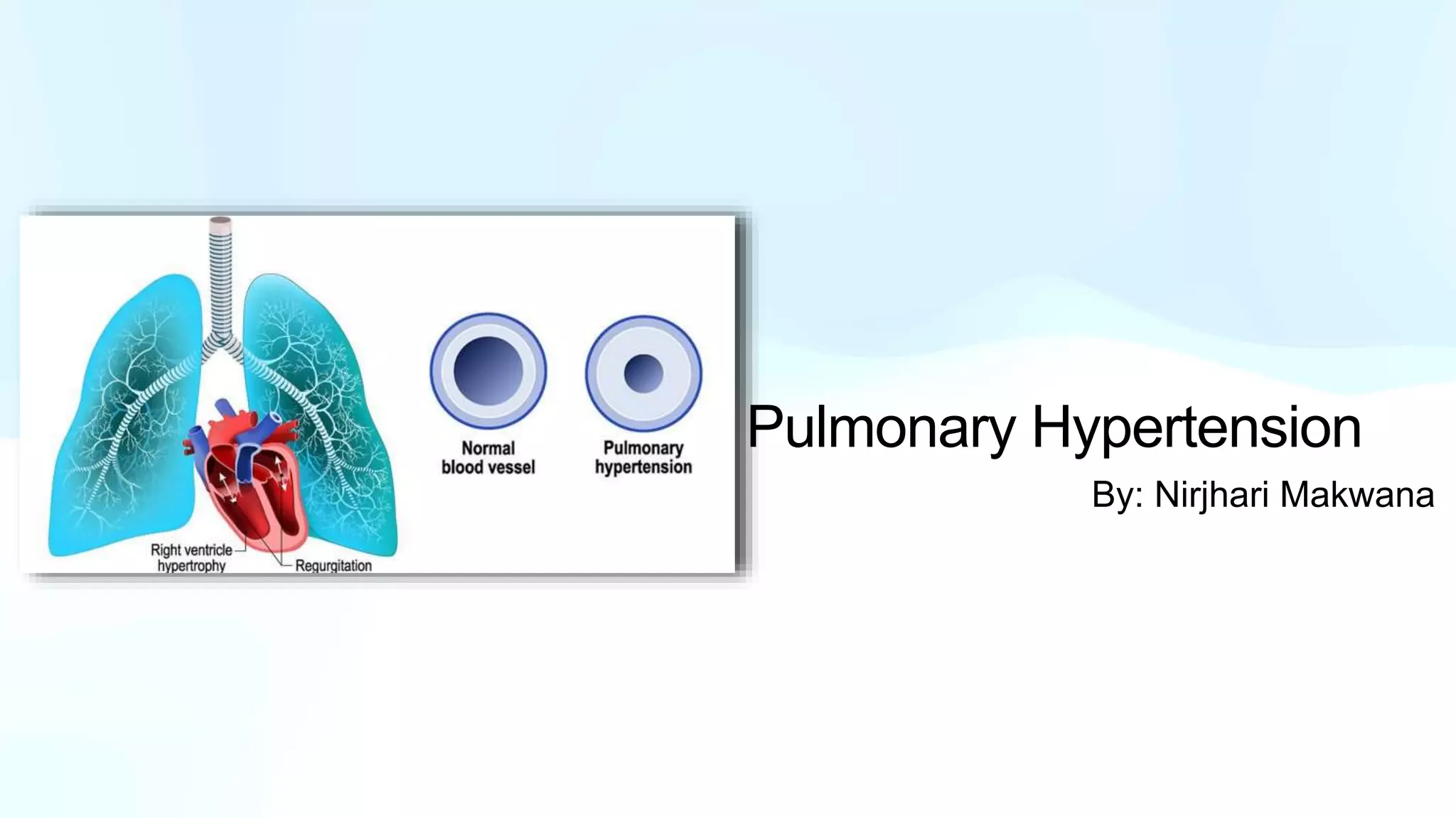
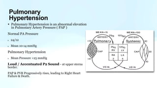
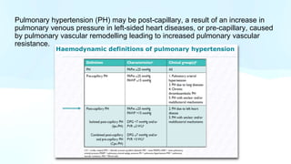
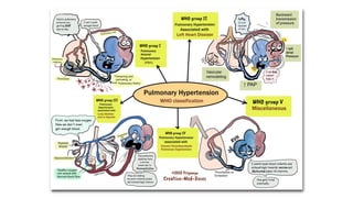
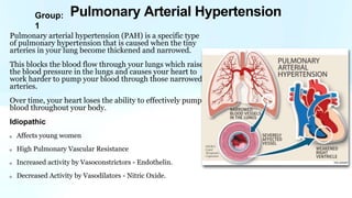
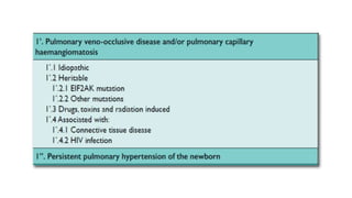
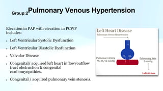
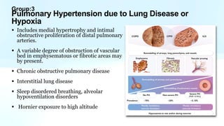
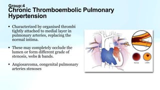
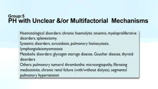
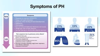
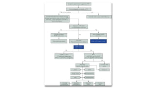
Pulmonary Hypertension
By: Nirjhari Makwana
Pulmonary Circulation
Low Pressure System
Systemic - 120/80
Pulmonary Artery - 24/12
Pulmonary artery has very thin walls
• Little smooth muscles
• Low resistance to flow
• Very distensible
Resistance to blood flow as blood moves to the lungs.
Two Types of Vessels
1. Alveolar Vessels - Capillaries ( surrounds alveoli )
2. Extra Alveolar Vessels - Arteries & Veins ( supplies to
capillaries )
Increased Lung Volume
• Crushes Alveolar Vessels —> High Resistance
• Pulls Extra Alveolar Vessels open —> Low Resistance
Resistance goes high & low while inhalation &
exhalation during respiratory cycle.
Pulmonary Vascular Resistance
• Pulmonary Hypertension is an abnormal elevation
in Pulmonary Artery Pressure ( PAP )
Normal PA Pressure
24/12
Mean 10-14 mmHg
Pulmonary Hypertension
Mean Pressure >25 mmHg
Loud / Accentuated P2 Sound - at upper sterna
border.
PAP & PVR Progressively rises, leading to Right Heart
Failure & Death.
Pulmonary
Hypertension
Pulmonary hypertension (PH) may be post-capillary, a result of an increase in
pulmonary venous pressure in left-sided heart diseases, or pre-capillary, caused
by pulmonary vascular remodelling leading to increased pulmonary vascular
resistance.
Group:
1
Pulmonary arterial hypertension (PAH) is a specific type
of pulmonary hypertension that is caused when the tiny
arteries in your lung become thickened and narrowed.
This blocks the blood flow through your lungs which raises
the blood pressure in the lungs and causes your heart to
work harder to pump your blood through those narrowed
arteries.
Over time, your heart loses the ability to effectively pump
blood throughout your body.
Idiopathic
Affects young women
High Pulmonary Vascular Resistance
Increased activity by Vasoconstrictors - Endothelin.
Decreased Activity by Vasodilators - Nitric Oxide.
Pulmonary Arterial Hypertension
Plexiform Lesions
Unique to Idiopathic PAH.
Endothelial proliferation forms
multiple lesions.
Small arteries branch points
from medium arteries.
BMPR2 Mutation
Bone morphogenetic protein receptor
type II
• Inhibits smooth muscle proliferation.
• Mutations —> abnormal growth (
endothelium, smooth muscle )
25% - idiopathic cases
80% - familial cases
Others : activin receptor kinase-like1
(ALK1), Endoglin (ENG), Smad8
Heritable
Associated with:
• Connective Tissue Disease
• Human immunodeficiency virus
(HIV) infection
• Portal hypertension
• Congenital Heart Disease
• Schistosomiasis
• Drugs - Amphetamines, Cocaine
• Epoprostenol: Prostacyclin (IV)
PGI2
Potentvasodilator
• Bosentan:
Antagonist endothelin-1 receptors (PO)
• Sildenafil:
Inhibits PDE-5 in smooth muscle of lungs
Treatment
Group:2
Elevation in PAP with elevation in PCWP
includes:
Left Ventricular Systolic Dysfunction
Left Ventricular Diastolic Dysfunction
Valvular Disease
Congenital/ acquired left heart inflow/outflow
tract obstruction & congenital
cardiomyopathies.
Congenital / acquired pulmonary vein stenosis.
Pulmonary Venous Hypertension
Group:3
• Includes medial hypertrophy and intimal
obstructive proliferation of distal pulmonary
arteries.
• A variable degree of obstruction of vascular
bed in emphysematous or fibrotic areas may
by present.
• Chronic obstructive pulmonary disease
• Interstitial lung disease
• Sleep disordered breathing, alveolar
hypoventilation disorders
• Hornier exposure to high altitude
Pulmonary Hypertension due to Lung Disease or
Hypoxia
Group:4
• Characterized by organised thrombi
tightly attached to medial layer in
pulmonary arteries, replacing the
normal intima.
• These may completely occlude the
lumen or form different grade of
stenosis, webs & bands.
• Angiosarcoma, congenital pulmonary
arteries stenoses
Chronic Thromboembolic Pulmonary
Hypertension
Group:5
PH with Unclear &/or Multifactorial Mechanisms
Symptoms of PH
Clinical Presentation
• The physical signs of PH include, left parasternal lift,
• an accentuated pulmonary component of the second heart sound,
• an RV third heart sound,
• a pan-systolic murmur of tricuspid regurgitation and,
• a diastolic murmur of pulmonary regurgitation.
• Elevated jugular venous pressure,
• hepatomegaly, ascites,
• peripheral oedema and
• cool extremities characterise patients with advanced disease.
• Wheeze and crackles are usually absent
Functional classification of PH according to WHO
Chest Radiograph
• In 90% of patients with IPAH the chest radiograph is abnormal at the time
of diagnoses
• patients with PAH include central pulmonary arterial dilatation, which
contrasts with ‘pruning’ (loss) of the peripheral blood vessels.
• Right atrium (RA) and RV enlargement may be seen in more advanced
cases
Pulmonary function tests and arterial blood
gases
• Pulmonary function tests and arterial blood gases identify the contribution
of underlying airway or parenchymal lung disease. Patients with PAH have
usually mild to moderate reduction of lung volumes related to disease
severity
• The prevalence of nocturnal hypoxaemia and central sleep ap- noeas are
high in PAH (70 – 80%).
• Echo is the most important screening tool for
determining the possibility of PH
• Transthoracic echocardiography is used to
image the effects of PH on the heart and
estimate PAP from continuous wave Doppler
measurement
• Evaluating RV function, including right atrial
area and RV ejection fraction (RVEF)
• Transthoracic Doppler echo can detect chronic
thromboembolic PH (CTEPH) in patients who
have had acute pulmonary embolism
Echocardiography
EPIDEMIOLOGY
• Pulmonary hypertension is a major
global health issue
• All age groups are affected
• Present estimates suggest a PH
prevalence of ∼1% of the global
population
• higher in individuals aged >65 years
• In UK, prevalence has doubled in the
last 10 years and is currently 125
cases/million inhabitants

Pulmonary Hypertension.pptx

  • 1.
  • 2.
    Pulmonary Circulation Low PressureSystem Systemic - 120/80 Pulmonary Artery - 24/12 Pulmonary artery has very thin walls • Little smooth muscles • Low resistance to flow • Very distensible
  • 3.
    Resistance to bloodflow as blood moves to the lungs. Two Types of Vessels 1. Alveolar Vessels - Capillaries ( surrounds alveoli ) 2. Extra Alveolar Vessels - Arteries & Veins ( supplies to capillaries ) Increased Lung Volume • Crushes Alveolar Vessels —> High Resistance • Pulls Extra Alveolar Vessels open —> Low Resistance Resistance goes high & low while inhalation & exhalation during respiratory cycle. Pulmonary Vascular Resistance
  • 5.
    • Pulmonary Hypertensionis an abnormal elevation in Pulmonary Artery Pressure ( PAP ) Normal PA Pressure 24/12 Mean 10-14 mmHg Pulmonary Hypertension Mean Pressure >25 mmHg Loud / Accentuated P2 Sound - at upper sterna border. PAP & PVR Progressively rises, leading to Right Heart Failure & Death. Pulmonary Hypertension
  • 6.
    Pulmonary hypertension (PH)may be post-capillary, a result of an increase in pulmonary venous pressure in left-sided heart diseases, or pre-capillary, caused by pulmonary vascular remodelling leading to increased pulmonary vascular resistance.
  • 8.
    Group: 1 Pulmonary arterial hypertension(PAH) is a specific type of pulmonary hypertension that is caused when the tiny arteries in your lung become thickened and narrowed. This blocks the blood flow through your lungs which raises the blood pressure in the lungs and causes your heart to work harder to pump your blood through those narrowed arteries. Over time, your heart loses the ability to effectively pump blood throughout your body. Idiopathic Affects young women High Pulmonary Vascular Resistance Increased activity by Vasoconstrictors - Endothelin. Decreased Activity by Vasodilators - Nitric Oxide. Pulmonary Arterial Hypertension
  • 9.
    Plexiform Lesions Unique toIdiopathic PAH. Endothelial proliferation forms multiple lesions. Small arteries branch points from medium arteries.
  • 10.
    BMPR2 Mutation Bone morphogeneticprotein receptor type II • Inhibits smooth muscle proliferation. • Mutations —> abnormal growth ( endothelium, smooth muscle ) 25% - idiopathic cases 80% - familial cases Others : activin receptor kinase-like1 (ALK1), Endoglin (ENG), Smad8 Heritable
  • 11.
    Associated with: • ConnectiveTissue Disease • Human immunodeficiency virus (HIV) infection • Portal hypertension • Congenital Heart Disease • Schistosomiasis • Drugs - Amphetamines, Cocaine
  • 12.
    • Epoprostenol: Prostacyclin(IV) PGI2 Potentvasodilator • Bosentan: Antagonist endothelin-1 receptors (PO) • Sildenafil: Inhibits PDE-5 in smooth muscle of lungs Treatment
  • 14.
    Group:2 Elevation in PAPwith elevation in PCWP includes: Left Ventricular Systolic Dysfunction Left Ventricular Diastolic Dysfunction Valvular Disease Congenital/ acquired left heart inflow/outflow tract obstruction & congenital cardiomyopathies. Congenital / acquired pulmonary vein stenosis. Pulmonary Venous Hypertension
  • 15.
    Group:3 • Includes medialhypertrophy and intimal obstructive proliferation of distal pulmonary arteries. • A variable degree of obstruction of vascular bed in emphysematous or fibrotic areas may by present. • Chronic obstructive pulmonary disease • Interstitial lung disease • Sleep disordered breathing, alveolar hypoventilation disorders • Hornier exposure to high altitude Pulmonary Hypertension due to Lung Disease or Hypoxia
  • 16.
    Group:4 • Characterized byorganised thrombi tightly attached to medial layer in pulmonary arteries, replacing the normal intima. • These may completely occlude the lumen or form different grade of stenosis, webs & bands. • Angiosarcoma, congenital pulmonary arteries stenoses Chronic Thromboembolic Pulmonary Hypertension
  • 17.
    Group:5 PH with Unclear&/or Multifactorial Mechanisms
  • 18.
  • 20.
    Clinical Presentation • Thephysical signs of PH include, left parasternal lift, • an accentuated pulmonary component of the second heart sound, • an RV third heart sound, • a pan-systolic murmur of tricuspid regurgitation and, • a diastolic murmur of pulmonary regurgitation. • Elevated jugular venous pressure, • hepatomegaly, ascites, • peripheral oedema and • cool extremities characterise patients with advanced disease. • Wheeze and crackles are usually absent
  • 22.
    Functional classification ofPH according to WHO
  • 23.
    Chest Radiograph • In90% of patients with IPAH the chest radiograph is abnormal at the time of diagnoses • patients with PAH include central pulmonary arterial dilatation, which contrasts with ‘pruning’ (loss) of the peripheral blood vessels. • Right atrium (RA) and RV enlargement may be seen in more advanced cases
  • 24.
    Pulmonary function testsand arterial blood gases • Pulmonary function tests and arterial blood gases identify the contribution of underlying airway or parenchymal lung disease. Patients with PAH have usually mild to moderate reduction of lung volumes related to disease severity • The prevalence of nocturnal hypoxaemia and central sleep ap- noeas are high in PAH (70 – 80%).
  • 25.
    • Echo isthe most important screening tool for determining the possibility of PH • Transthoracic echocardiography is used to image the effects of PH on the heart and estimate PAP from continuous wave Doppler measurement • Evaluating RV function, including right atrial area and RV ejection fraction (RVEF) • Transthoracic Doppler echo can detect chronic thromboembolic PH (CTEPH) in patients who have had acute pulmonary embolism Echocardiography
  • 26.
    EPIDEMIOLOGY • Pulmonary hypertensionis a major global health issue • All age groups are affected • Present estimates suggest a PH prevalence of ∼1% of the global population • higher in individuals aged >65 years • In UK, prevalence has doubled in the last 10 years and is currently 125 cases/million inhabitants