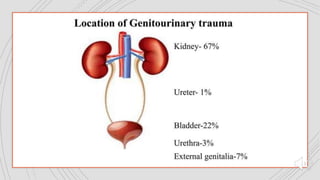
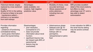
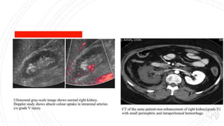
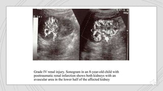
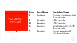
GENITOURINARY
TRAUMA
Dr. Niranjan Patil
Prof. Department of Radio-diagnosis
DY Patil Medical College and Hospital
Kolhapur
RENALTRAUMA
90% - Blunt , 10% -
Penetrating
~10% of patients
with significant
abdominal trauma.
When severe,
associated with
injuries to other
organs in upto 80%.
When the kidney is
the only organ
damaged the injury
is minor in around
98% of cases .
Mechansimof
renaltrauma
 Blunt trauma direct blow to the kidney.
 Penetrating trauma stab or gunshot injury.
 High velocity deceleration pedicle injury (avulsion of renal
vessel thrombosis of renal artery ).
 Major renal injury (grade 4 and grade5).
 Upto to 25% of blunt, and in upto 70% of penetrating renal
trauma.
Predisposing
factors
Pre-existing renal abnormalities
Vulnerable position (more anterior location and proximity to
rigid spine or iliac crest)
Tranplant kidneys
Horse shoe kidneys
Crossed fused ectopia
Increased bulk : tumours (angiomylipoma, RCC ), cysts
hydronephrosis
Pediatric kidneys larger size
Indications
forrenal
imaging
Penetrating
Gross hematuria
Microhematuria & systolic BP < 90mm Hg
Pediatric >50rbc /hpf
Mechanism of injury
-flank impact
- Deceleration injury (eg-fall from great height)
Roleof
radiology
 Most contemporary trends in trauma care, including renal
trauma,
 Call for less invasive procedures, trauma imaging by a
skilled radiologist is important.
 Accuretly distinguishing patients that can be managed
conservatively from those who need surgery, thus
improving the long – term outcome of patients.
 Radiologic interventions
- renal artery angiography and emboloisation/stenting.
- external drainage procedures (eg-Urinomas)
Classifictaionof
renaltrauma-
AAST
Imagingmodalities
CT scan IVU
Antegrade /
retrograde
pyelography
USG
Renal
angiography +/-
embolization
MRI
IVU USG CT scan MRI
Historical interest, largely
replaced by cross sectional
imaging.
Quality of IVU in the setting
of trauma is likely to be poor
: hypotensive patient –
minimal or no secretion
from both kidneys
Advantages :
easily available, non
invasive, high negative
predictive value
• Modality of choice, most
accurate technique
• Allows assessment of
entire abdomen,
including liver and
spleen.
MRI provides excellent
detail of the renal anatomy
but offers no clear
advantage over CT, and is
less able to detect
extravasation.
Provides information
regarding:
- presence of functioning
contralateral kidney.
- some gross information
about the injured kidney.
Disadvantages:
• poor resolution
(compared to CT)
• does not provide
information about renal
function.
• significant trauma may
be missed (upto 80% of
parenchymal
• lesions may be
overlooked)
• Arteriovenous phase
• Delayed scan @ 10-
20min ? Omit if normal
kidneys with no
perinephric,
retroperitoneal or pelvic
fluid
A rare indication for MRI in
the renal trauma setting
may be severe contrast
allergy.
FindingsinUSG
 Acute parenchymal, subcapsular and perinephric
hematomas – echo poor areas.
 More heterogenous and echogenic with time.
 Disruption of renal parenchyma with capsular tears and
urinomas.
CT ScanTechnique
The abdomen and pelvis should be scanned form the diaphragmatic dome to pubic
symphysis with contrast.
Protocol : >commence scanning 30 sec after the start of an injection of 50-100 ml of 300
strength contrast at 2-3 ml/sec, collimation 7mm, pitch 1.3
>addition of 400-600 ml of oral contrast (4% diatriazoate) immediately before scen helps in
dilenating associated bowel injuries (stomach duodenum and proximal jejunum)
To preform a complete evaluation, the entire scan must be scrutinized with 3 different
window/ level settings : soft tissue, lung and bone.
Grade I Grade II Grade III Grade IV Grade V
Contusions and
subcapsular hematoma
are key terms used In
the identification
Perinephric hematomas
& renal parenchymal
lacerations are key terms
Are deeper lacerations
(>1cm) that do not result in
urine leak. Any injury
occurring in the presence
of a vascular injury
Include injuries to
renal parenchyma
and collecting
system as well as
vascular injuries.
Shattered kidney,
avulsion or laceration of
the main renal artery ,
vein and devascularised
kidney with active
bleeding
Contusions appear on
portal venous phase
CT images as globular,
ovoid or round poorly
demarcated area of
relatively poor
enhancement
Hematomas contained
within the gerota fascia
has been postulated to
have a tamponade effect
on renal bleeding,
resulting in perinephric
clot formation
Vascular injuries include
pseudoaneurysm or
arteriovenous fistula.
Lacerations
extending deep to
involve the
collecting system
with urinary
extravasation are
included.
Loss of identifiable renal
parenchymal anatomy
Hilar vascular injuries
are not common.
Subcapsular
hematoma are
confined by renal
capsule. These are non
enhancing cresentric or
lentiform shaped fluid
collections
Lacerations are
superficial (<1cm) and
there is no urine leak
These have been
described as a focal
collection of vascular
contrast enhacement that
shows decreasing
attenuation with delayed
imaging
Delayed imaging
usually shows
urine leaking into
the perirenal
space
Complete arterial tears
result in retroperitoneal
hematomas with
possible active
bleeding.
Treatmentand
prognosis
 Grade I – conservative management
 Grade II – conservative managemnent under close
observation.
 Grade III – conservative management under close
observation may be managed surgically if undergoing
laparotomy for other abdominal injuries.
 Grade IV – surgical management, especially if undergoing
laparotomy for other abdominal injuries.
 Garde V – surgical management
Complications
 Urinoma
 Perinephric abscess
 Pseudoaneurysm
 Secondary bleeding
 Hypertension
 Renal insufficiency
 Urinary fistuala
 Pyleonephritis
Ureterictrauma
<1% of urinary tract trauma
Mechanism of injury :
External :
penetrating trauma
Deceleration injuries :
PUJ avulsion
Less often upper 1/3rd of ureter
More frequent in children (sufficient flexibility to produce hyperflexion injuries )
Iatrogenic : gynecologic surgery for malignancy
Hematuria may be absent in one third of cases
AASTUreteral
Injury Scale
ImagingFindings:
IVU Retrograde pyelogram CT
Mild to moderate fullness
of the PCS & extravasation
at the site of tear.
Complete tear – ureter
fails to opacify below the
tear.
Fistulations to other
structures.
Limited use in acute
setting
Delayed excretory phase
images are usually required to
make the diagnosis
 Contrast extravasation
 Formation of urinoma
 Occurance of ureteric
discontinuity
BLADDERTRAUMA
Trauma
Blunt Penetrating
Spontaneous
Pre-existing
bladder wall
abnormalities
Bladder tumor,
cystitis ,
perivesical
inflammation
Neurogenic
bladder ,
previous
radiotherapy
Iatrogenic
trauma
Classification
Imagingmodalities
Conventional
cystography
CT
Cystography
CONVENTIONALCYSTOGRAPHY
Advantages
• Nearly 100% sensitive for detecting rupture, provided that
adequate distension is accomplished and that post voiding
images are obtained.
Disadvantages
• Time consuming and not useful in evaluating trauma to other
viscera
• Require extra radiography in addition to necessary trauma
evaluation
CT
CYSTOGRAPHY
• performed with ~350-400ml of
contrast material administered in a
retrograde fashion before the study
Technique
Washout study
• Less time consuming
• Concomitant imjury to other viscera
(most importantly kidneys)
Advantages of CT
Cystography
TYPES
EXTRA PERITONEAL RUPTURE INTRA PERITONEAL RUPTURE
Commonest bladder injury associated with anterior
pelvic ring fracture
Contrast extravasates into peritoneal cavity and has a
more cloudy appearance
Extravasation of contrast into the perivesical space
Contrast may extend anterosuperiorly along the anterior
pelvic and abdominal wall upto umbilicus or posteriorly
around rectum in presacral space
Associated tear of urogenital diaphragm allowing the
contrast to appear within the perineum , thigh and
scrotum.
Usually tear is along the dome of bladder which is the
weakest part
The extravasated contrast stays close to the bladder
and has a sharp irregular margins
Associated pelvic fracture
Extraperitonealrupture
Intraperitonealrupture
URETHRALTRAUMA
 Almost entirely restricted to males unless there is major
pelvic trauma in females.
 Should be suspected
 Pelvic trauma associated with hematuria or retention.
 Especially blood at the urethral meatus.
ANTERIOR
URETHRAL
TRAUMA
 Mechanism
 Iatrogenic (attempted catheterization, instrumentation )
 Blunt perineal trauma (straddle injury) : bulbar urethra and
corpus spongiosum are compressed against the inferior aspect
of anterior pelvic ring
FINDINGS
Rupture
Partial : contrast extravasation with some filling of proximal urethra
Complete : failure of filling of proximal urethra
Stricture (usually <1cm or so in length).
Complications
Stricture
Impotence
(upto ~10%)
Incontinence
PENILEFRACTURE
Fracture of the penis occurs exclusively with an excretion, with
aggressive vaginal intercourse being the most common cause.
It is related to excessive bending of the erect penis and
thrusting against the pubic symphysis.
The patient usually reports a cracking sound, Immediate pain,
and rapid detumenscence.
SCROTALTRAUMA
 Causes :
Blunt
Penetrating
Degloving
Electric burn
 Ultrasound is the imaging technique of choice
in acute scrotal trauma except in degloving
injury.
genitourinary trauma.pptx

genitourinary trauma.pptx

  • 1.
    GENITOURINARY TRAUMA Dr. Niranjan Patil Prof.Department of Radio-diagnosis DY Patil Medical College and Hospital Kolhapur
  • 3.
    RENALTRAUMA 90% - Blunt, 10% - Penetrating ~10% of patients with significant abdominal trauma. When severe, associated with injuries to other organs in upto 80%. When the kidney is the only organ damaged the injury is minor in around 98% of cases .
  • 4.
    Mechansimof renaltrauma  Blunt traumadirect blow to the kidney.  Penetrating trauma stab or gunshot injury.  High velocity deceleration pedicle injury (avulsion of renal vessel thrombosis of renal artery ).  Major renal injury (grade 4 and grade5).  Upto to 25% of blunt, and in upto 70% of penetrating renal trauma.
  • 5.
    Predisposing factors Pre-existing renal abnormalities Vulnerableposition (more anterior location and proximity to rigid spine or iliac crest) Tranplant kidneys Horse shoe kidneys Crossed fused ectopia Increased bulk : tumours (angiomylipoma, RCC ), cysts hydronephrosis Pediatric kidneys larger size
  • 6.
    Indications forrenal imaging Penetrating Gross hematuria Microhematuria &systolic BP < 90mm Hg Pediatric >50rbc /hpf Mechanism of injury -flank impact - Deceleration injury (eg-fall from great height)
  • 7.
    Roleof radiology  Most contemporarytrends in trauma care, including renal trauma,  Call for less invasive procedures, trauma imaging by a skilled radiologist is important.  Accuretly distinguishing patients that can be managed conservatively from those who need surgery, thus improving the long – term outcome of patients.  Radiologic interventions - renal artery angiography and emboloisation/stenting. - external drainage procedures (eg-Urinomas)
  • 8.
  • 12.
    Imagingmodalities CT scan IVU Antegrade/ retrograde pyelography USG Renal angiography +/- embolization MRI
  • 13.
    IVU USG CTscan MRI Historical interest, largely replaced by cross sectional imaging. Quality of IVU in the setting of trauma is likely to be poor : hypotensive patient – minimal or no secretion from both kidneys Advantages : easily available, non invasive, high negative predictive value • Modality of choice, most accurate technique • Allows assessment of entire abdomen, including liver and spleen. MRI provides excellent detail of the renal anatomy but offers no clear advantage over CT, and is less able to detect extravasation. Provides information regarding: - presence of functioning contralateral kidney. - some gross information about the injured kidney. Disadvantages: • poor resolution (compared to CT) • does not provide information about renal function. • significant trauma may be missed (upto 80% of parenchymal • lesions may be overlooked) • Arteriovenous phase • Delayed scan @ 10- 20min ? Omit if normal kidneys with no perinephric, retroperitoneal or pelvic fluid A rare indication for MRI in the renal trauma setting may be severe contrast allergy.
  • 14.
    FindingsinUSG  Acute parenchymal,subcapsular and perinephric hematomas – echo poor areas.  More heterogenous and echogenic with time.  Disruption of renal parenchyma with capsular tears and urinomas.
  • 17.
    CT ScanTechnique The abdomenand pelvis should be scanned form the diaphragmatic dome to pubic symphysis with contrast. Protocol : >commence scanning 30 sec after the start of an injection of 50-100 ml of 300 strength contrast at 2-3 ml/sec, collimation 7mm, pitch 1.3 >addition of 400-600 ml of oral contrast (4% diatriazoate) immediately before scen helps in dilenating associated bowel injuries (stomach duodenum and proximal jejunum) To preform a complete evaluation, the entire scan must be scrutinized with 3 different window/ level settings : soft tissue, lung and bone.
  • 19.
    Grade I GradeII Grade III Grade IV Grade V Contusions and subcapsular hematoma are key terms used In the identification Perinephric hematomas & renal parenchymal lacerations are key terms Are deeper lacerations (>1cm) that do not result in urine leak. Any injury occurring in the presence of a vascular injury Include injuries to renal parenchyma and collecting system as well as vascular injuries. Shattered kidney, avulsion or laceration of the main renal artery , vein and devascularised kidney with active bleeding Contusions appear on portal venous phase CT images as globular, ovoid or round poorly demarcated area of relatively poor enhancement Hematomas contained within the gerota fascia has been postulated to have a tamponade effect on renal bleeding, resulting in perinephric clot formation Vascular injuries include pseudoaneurysm or arteriovenous fistula. Lacerations extending deep to involve the collecting system with urinary extravasation are included. Loss of identifiable renal parenchymal anatomy Hilar vascular injuries are not common. Subcapsular hematoma are confined by renal capsule. These are non enhancing cresentric or lentiform shaped fluid collections Lacerations are superficial (<1cm) and there is no urine leak These have been described as a focal collection of vascular contrast enhacement that shows decreasing attenuation with delayed imaging Delayed imaging usually shows urine leaking into the perirenal space Complete arterial tears result in retroperitoneal hematomas with possible active bleeding.
  • 27.
    Treatmentand prognosis  Grade I– conservative management  Grade II – conservative managemnent under close observation.  Grade III – conservative management under close observation may be managed surgically if undergoing laparotomy for other abdominal injuries.  Grade IV – surgical management, especially if undergoing laparotomy for other abdominal injuries.  Garde V – surgical management
  • 28.
    Complications  Urinoma  Perinephricabscess  Pseudoaneurysm  Secondary bleeding  Hypertension  Renal insufficiency  Urinary fistuala  Pyleonephritis
  • 29.
    Ureterictrauma <1% of urinarytract trauma Mechanism of injury : External : penetrating trauma Deceleration injuries : PUJ avulsion Less often upper 1/3rd of ureter More frequent in children (sufficient flexibility to produce hyperflexion injuries ) Iatrogenic : gynecologic surgery for malignancy Hematuria may be absent in one third of cases
  • 30.
  • 31.
    ImagingFindings: IVU Retrograde pyelogramCT Mild to moderate fullness of the PCS & extravasation at the site of tear. Complete tear – ureter fails to opacify below the tear. Fistulations to other structures. Limited use in acute setting Delayed excretory phase images are usually required to make the diagnosis  Contrast extravasation  Formation of urinoma  Occurance of ureteric discontinuity
  • 33.
    BLADDERTRAUMA Trauma Blunt Penetrating Spontaneous Pre-existing bladder wall abnormalities Bladdertumor, cystitis , perivesical inflammation Neurogenic bladder , previous radiotherapy Iatrogenic trauma
  • 34.
  • 35.
  • 36.
    CONVENTIONALCYSTOGRAPHY Advantages • Nearly 100%sensitive for detecting rupture, provided that adequate distension is accomplished and that post voiding images are obtained. Disadvantages • Time consuming and not useful in evaluating trauma to other viscera • Require extra radiography in addition to necessary trauma evaluation
  • 37.
    CT CYSTOGRAPHY • performed with~350-400ml of contrast material administered in a retrograde fashion before the study Technique Washout study • Less time consuming • Concomitant imjury to other viscera (most importantly kidneys) Advantages of CT Cystography
  • 38.
    TYPES EXTRA PERITONEAL RUPTUREINTRA PERITONEAL RUPTURE Commonest bladder injury associated with anterior pelvic ring fracture Contrast extravasates into peritoneal cavity and has a more cloudy appearance Extravasation of contrast into the perivesical space Contrast may extend anterosuperiorly along the anterior pelvic and abdominal wall upto umbilicus or posteriorly around rectum in presacral space Associated tear of urogenital diaphragm allowing the contrast to appear within the perineum , thigh and scrotum. Usually tear is along the dome of bladder which is the weakest part The extravasated contrast stays close to the bladder and has a sharp irregular margins Associated pelvic fracture
  • 39.
  • 40.
  • 41.
    URETHRALTRAUMA  Almost entirelyrestricted to males unless there is major pelvic trauma in females.  Should be suspected  Pelvic trauma associated with hematuria or retention.  Especially blood at the urethral meatus.
  • 46.
    ANTERIOR URETHRAL TRAUMA  Mechanism  Iatrogenic(attempted catheterization, instrumentation )  Blunt perineal trauma (straddle injury) : bulbar urethra and corpus spongiosum are compressed against the inferior aspect of anterior pelvic ring
  • 48.
    FINDINGS Rupture Partial : contrastextravasation with some filling of proximal urethra Complete : failure of filling of proximal urethra Stricture (usually <1cm or so in length).
  • 49.
  • 50.
    PENILEFRACTURE Fracture of thepenis occurs exclusively with an excretion, with aggressive vaginal intercourse being the most common cause. It is related to excessive bending of the erect penis and thrusting against the pubic symphysis. The patient usually reports a cracking sound, Immediate pain, and rapid detumenscence.
  • 52.
    SCROTALTRAUMA  Causes : Blunt Penetrating Degloving Electricburn  Ultrasound is the imaging technique of choice in acute scrotal trauma except in degloving injury.

Editor's Notes

  • #9 Grade I Subscapular hematoma, parenchymal contusion without laceration, or both Grade 2 Perirenal hematoma confined to gerota fascia Renal parenchymal laceration of <1-cm depth without urinary extravasation Grade 3 Renal parenchymal laceration of > 1 cm depth collecting system rupture or urinary etravasation Any injury in the presence of a kidney vascular injury or active bleeding contained within the gerota fascia Garde 4 Parenchymal laceration extending into urinary collecting system with urinary extravasation Renal pelvis laceration, complete urteropelvic disruption or both Active bleeding beyond the gerota fascia into retroperitoneum or peritoneum Segmental renal vein or artery injury or segmental or complete kidney infacrtion caused by vessel thrombosis without active bleeding Grade 5 Shattered kidney with loss of identifiable parenchymal renal anatomy Devascularised kidney with active bleeding Main renal artery or vein laceration or avulsion of hilum.
  • #14 IVU Single shot IVU (full length film 15min after contrast injection ) In hemodynamically unstable patients destined for ER laparotomy. Absence of unilateral excretion : major vascular injury (usually RA avulsion) Soft tissue swelling with loss of psoas outline : reropertioneal perinephric and subcapsular hematomas Disruption of
  • #19 Contusions and subcapsular hematoma are key terms used In the identification Contusions appear on portal venous phase CT images as globular, ovoid or round poorly demarcated area of relatively poor enhancement Subcapsular hematoma are confined by renal capsule. These are non enhancing cresentric or lentiform shaped fluid collections
  • #20 Grade III - active bleeding : it is defined by vascular contrast enhancement (focal or diffuse) that increases in size and attenuation in the delayed phase. If this is present, it must be contained within gerota fascia.
  • #21 Perinephric hematomas & renal parenchymal lacerations are ley terms Hematomas contained within the gerota fascia has been postulated to have a tamponade effect on renal bleeding, resulting in perinephric clot formation Lacerations are superficial (<1cm) and there is no urine leak
  • #22 Are deeper lacerations (>1cm) that do not result in urine leak. Any injury occurring in the presence of a vascular injury Vascular injuries include pseudoaneurysm or arteriovenous fistula. These have been described as a focal collection of vascular contrast enhacement that shows decreasing attenuation with delayed imaging
  • #23 Include injuries to renal parenchyma and collecting system as well as vascular injuries. Lacerations extending deep to involve the collecting system with urinary extravasation are included. Delayed imaging usually shows urine leaking into the perirenal space
  • #26 Shattered kidney, avulsion or laceration of the main renal artery , vein and devascularised kidney with active bleeding Loss of identifiable renal parenchymal anatomy Hilar vascular injuries are not common. Complete arterial tears result in retroperitoneal hematomas with possible active bleeding.
  • #34 Blunt trauma : most common In patients with multi injury trauma Predisposed by bladder at the time of trauma Pelvic fractures (especially anterior ring ) commomnly associated 7% symphysis pubis diastasis associated wit bladder trauma Iatrogenic surgery particulary caesarean section and transurethral bladder resection (usually for tumor)