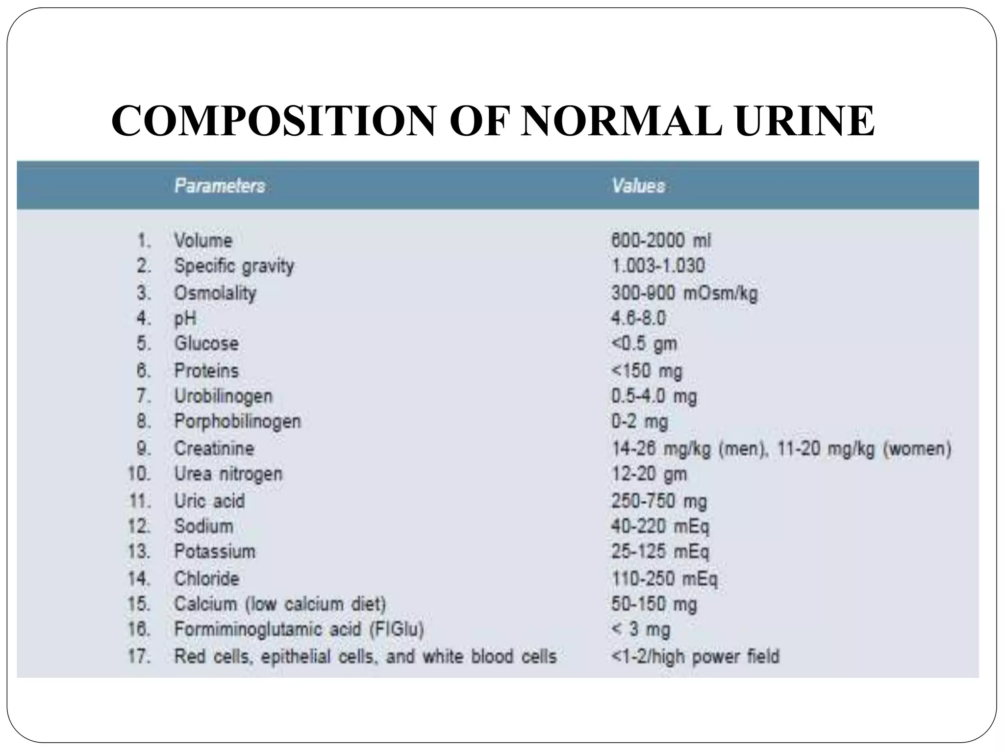
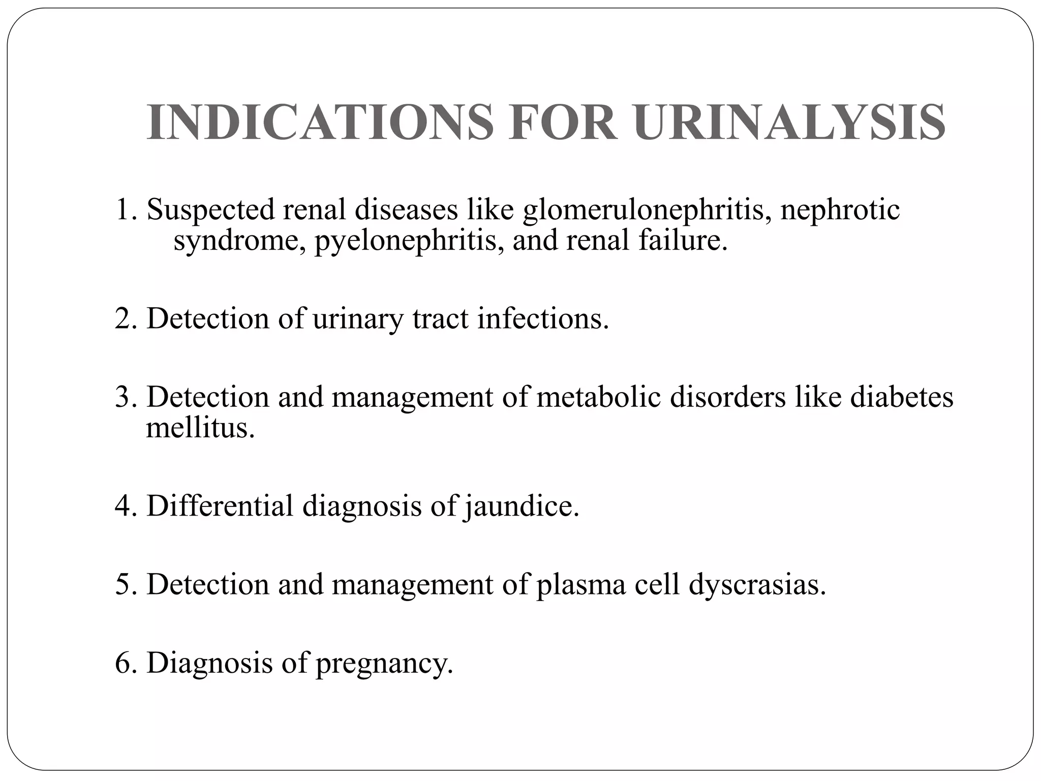
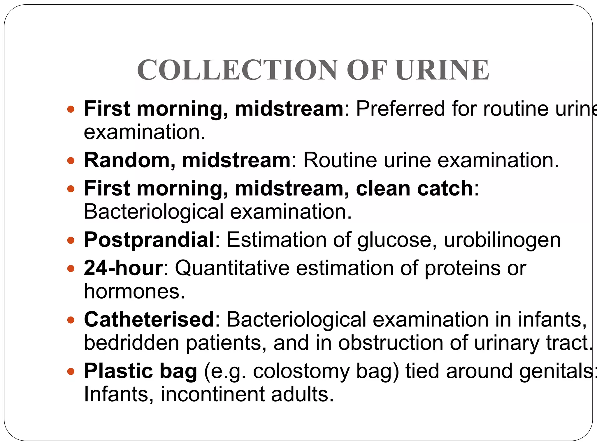
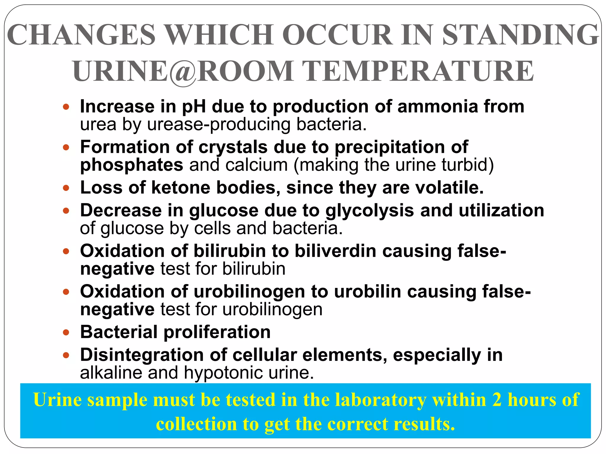
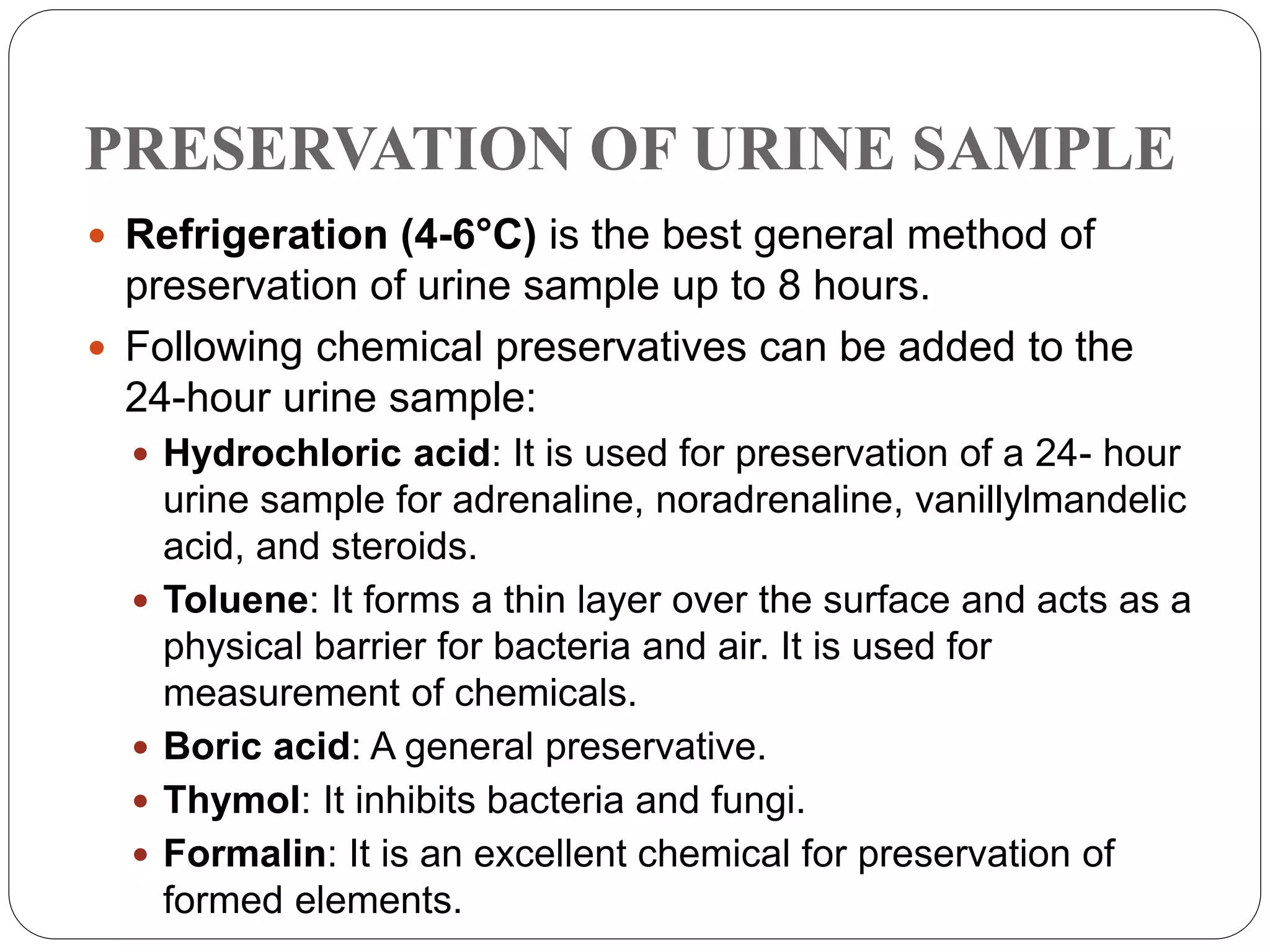
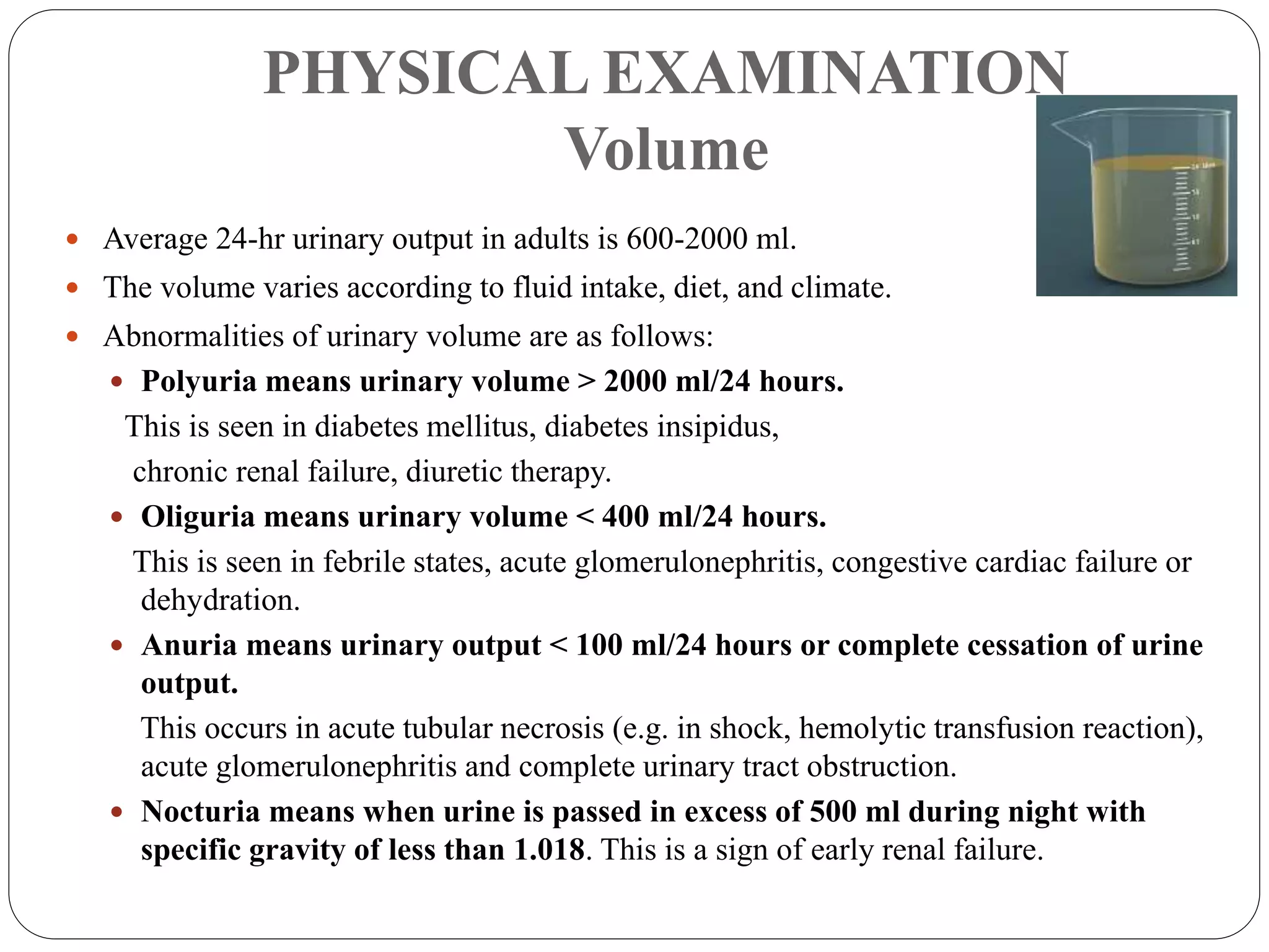
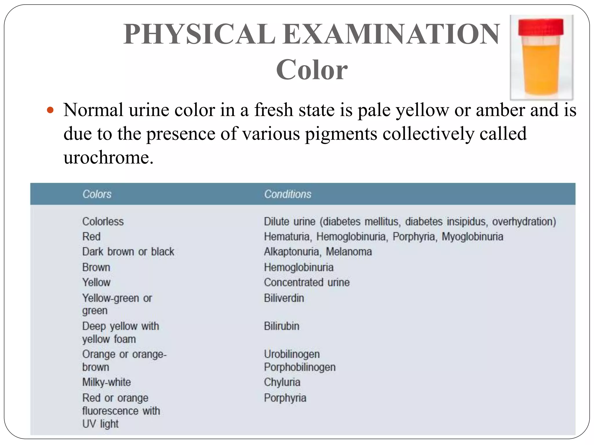
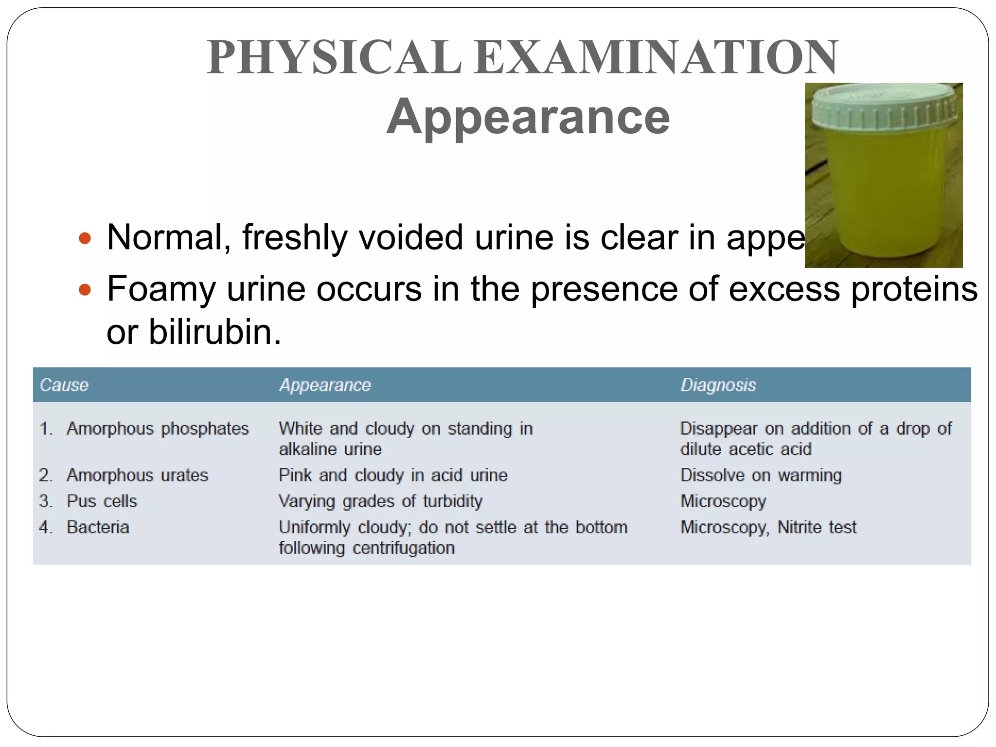
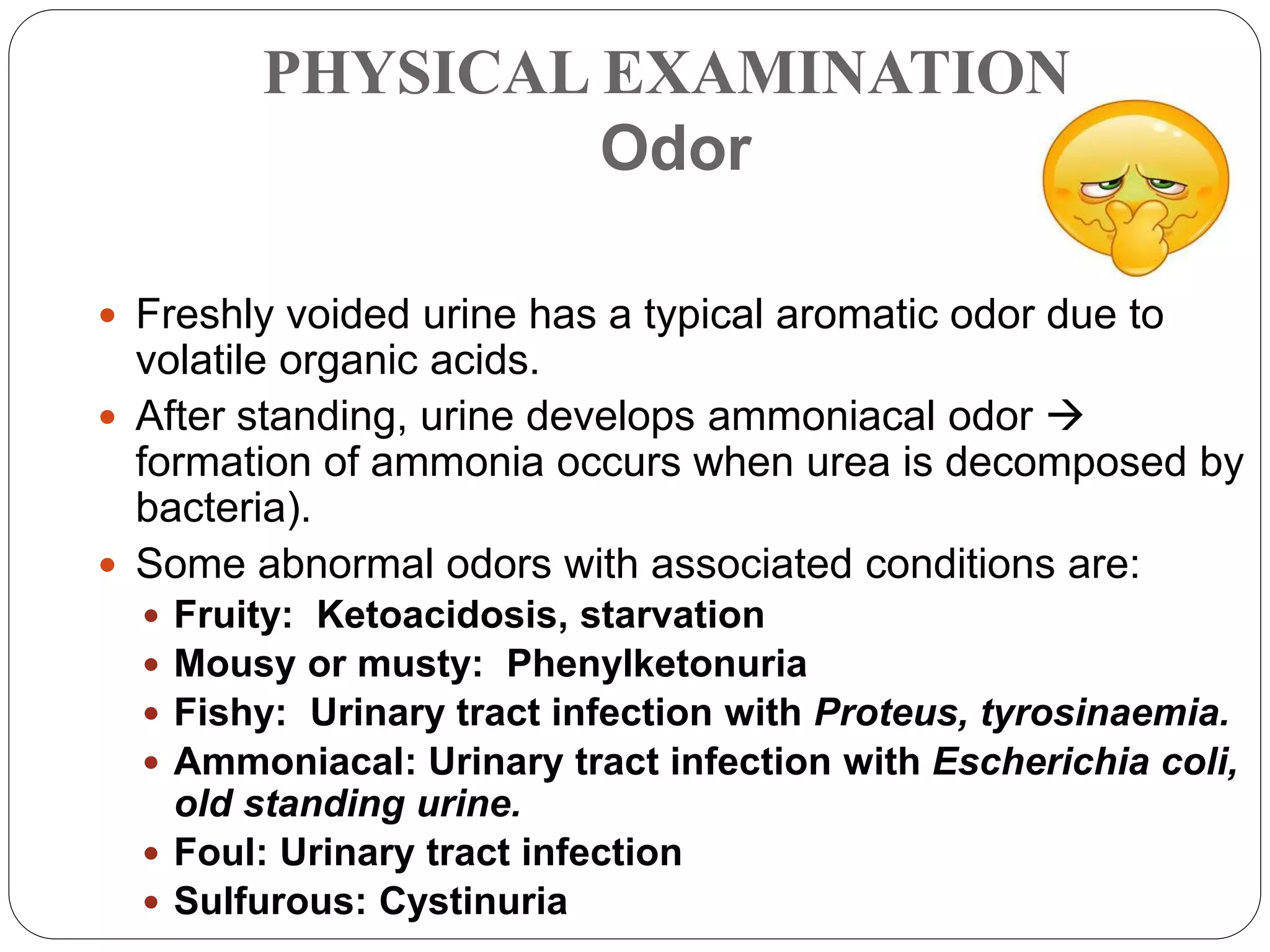
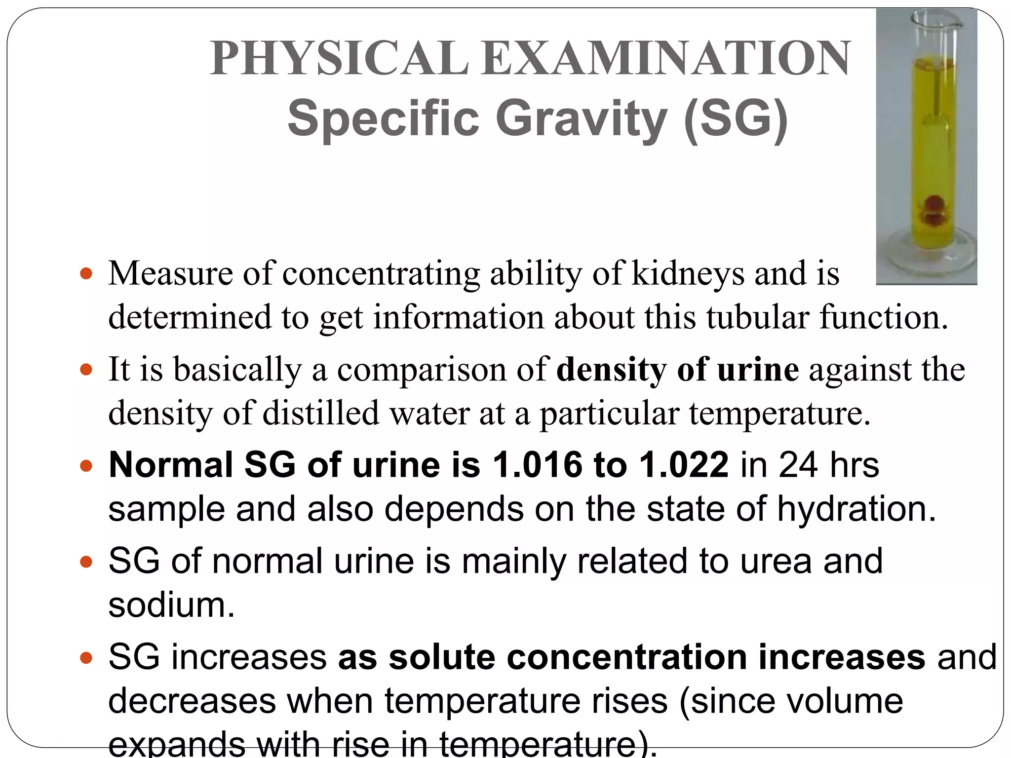
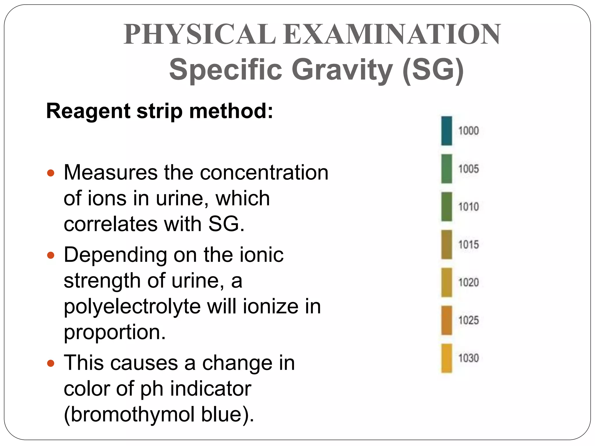
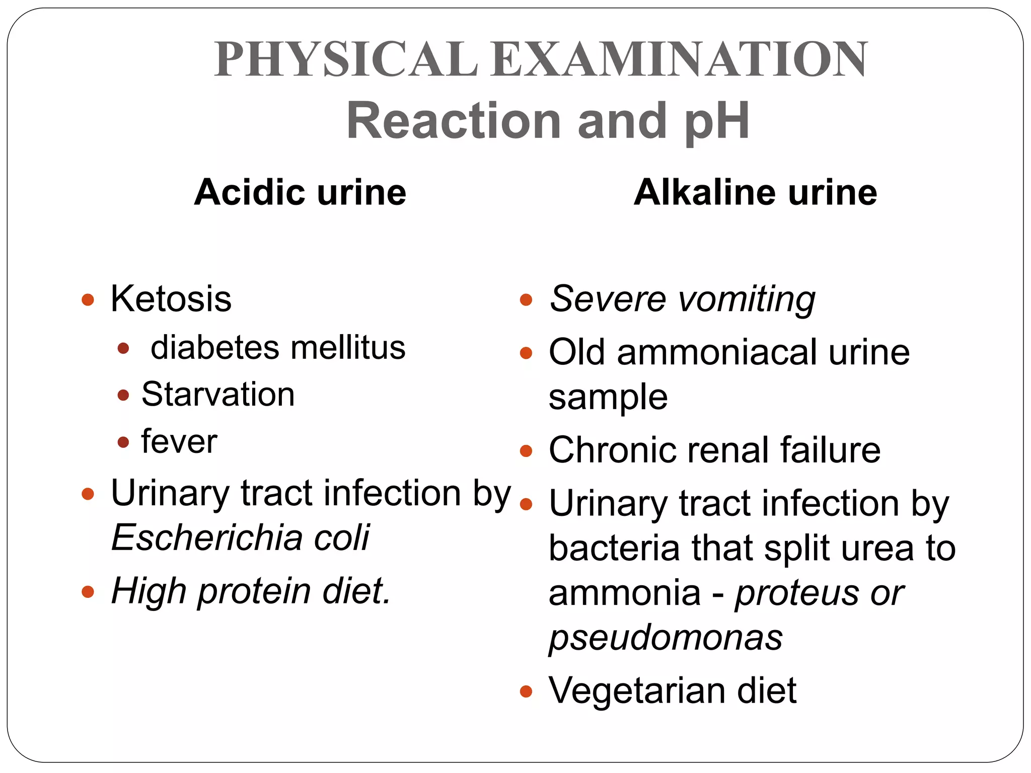
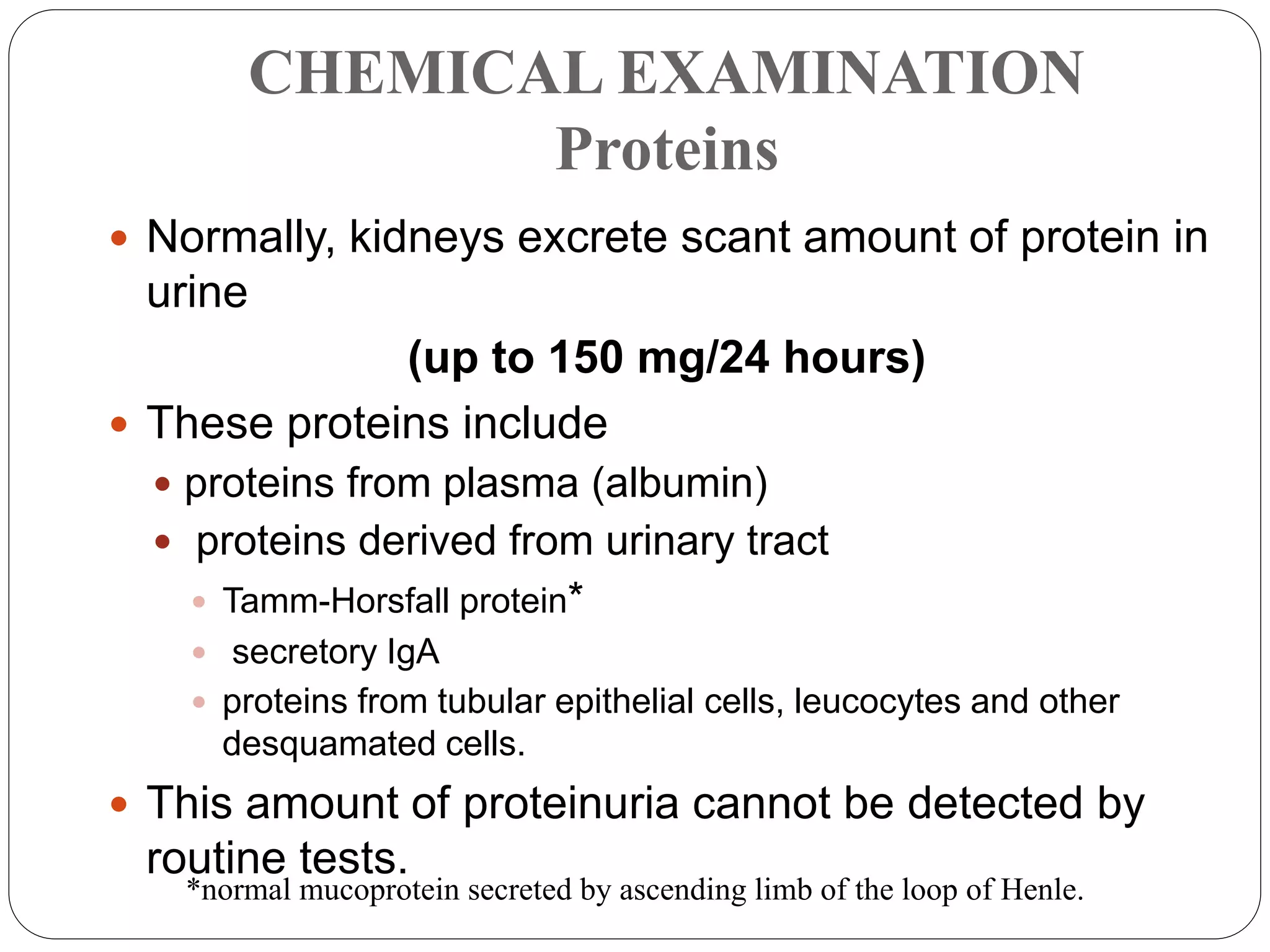
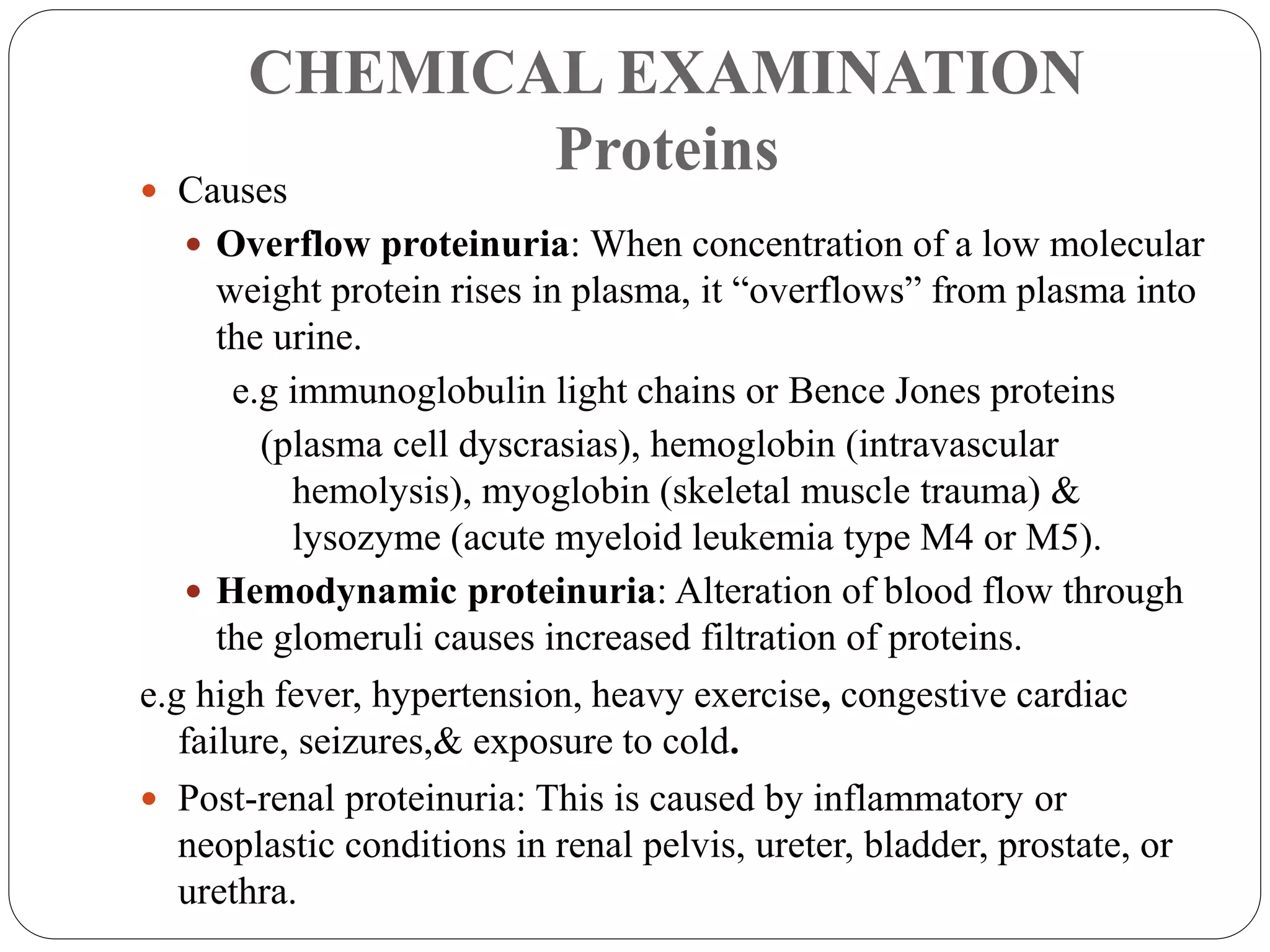
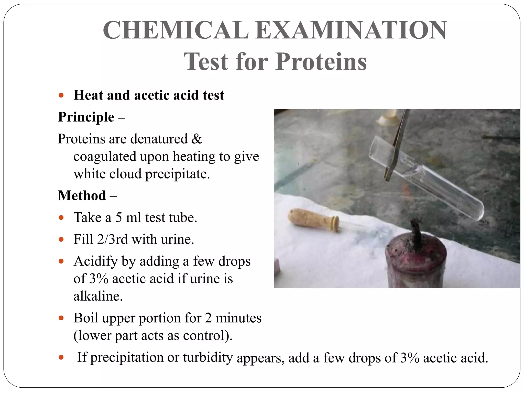
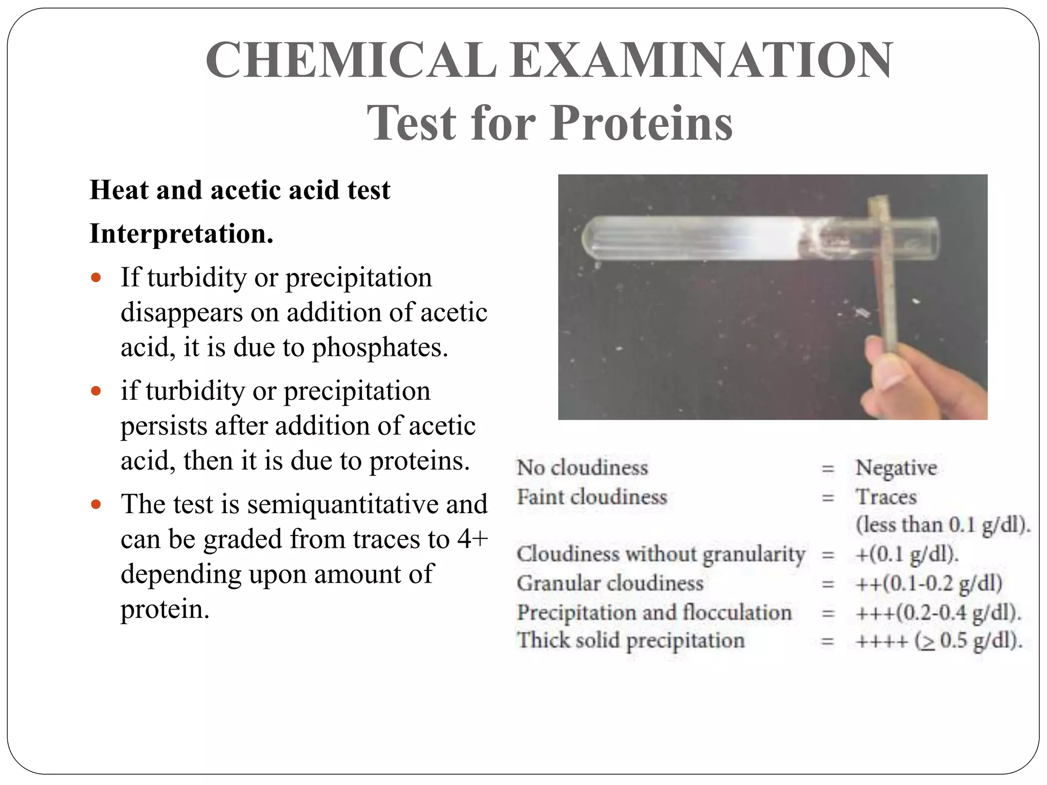
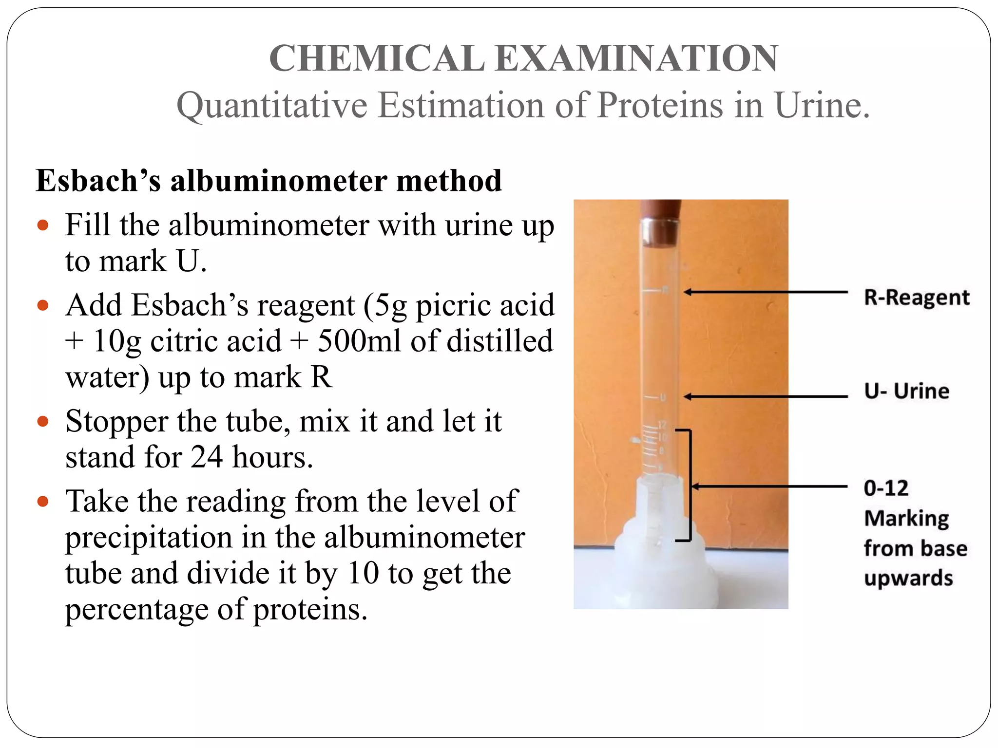
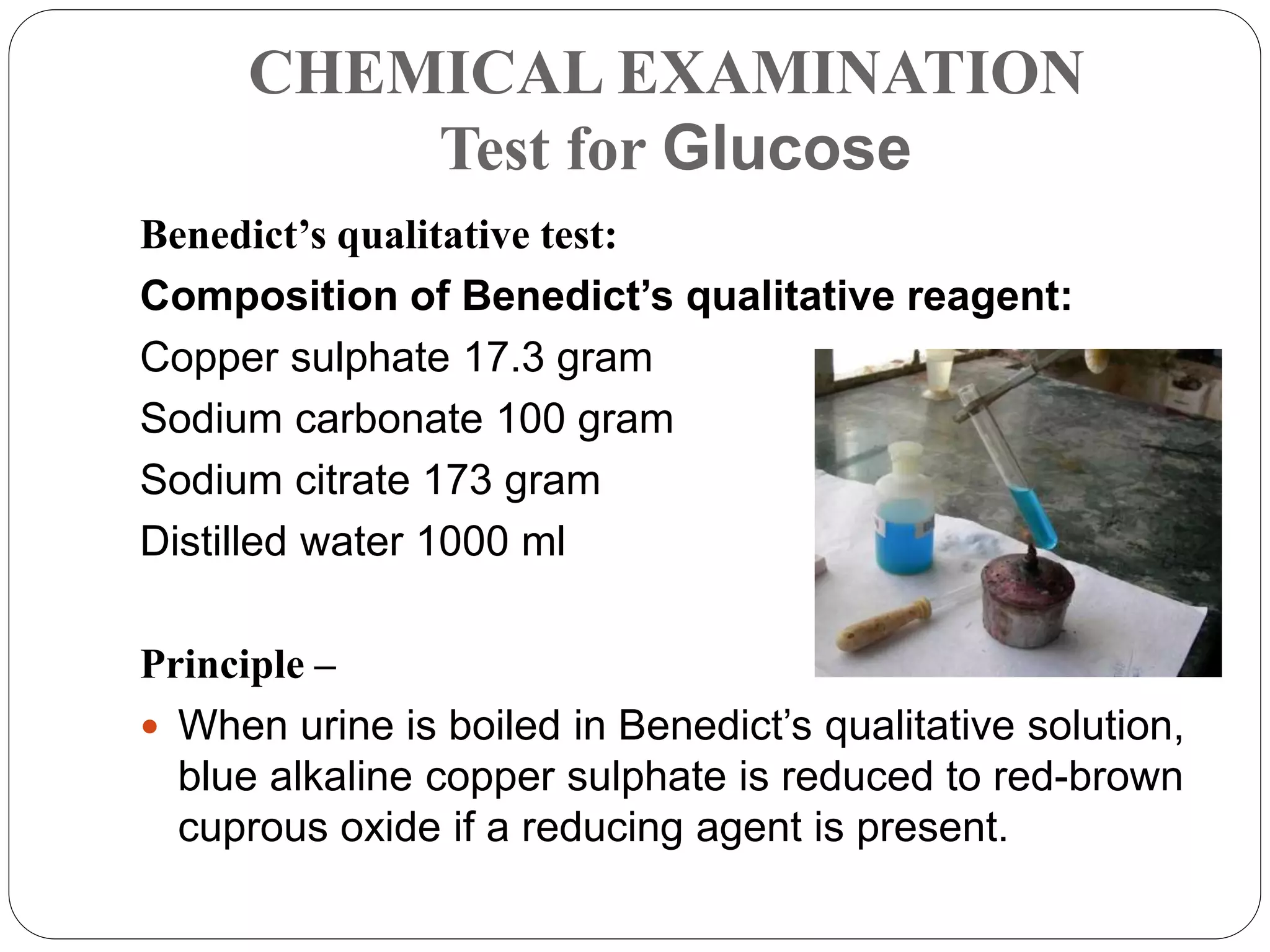
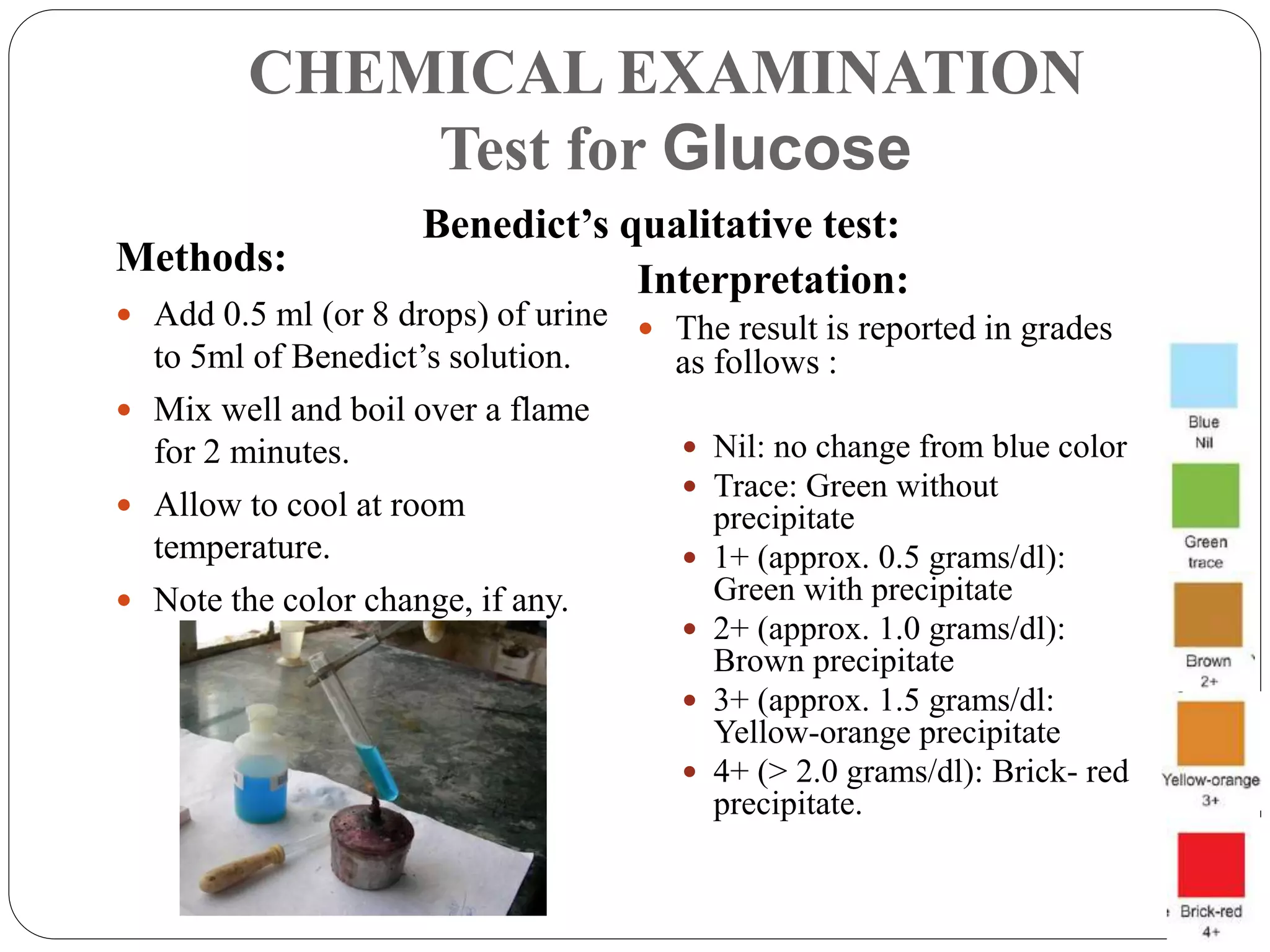
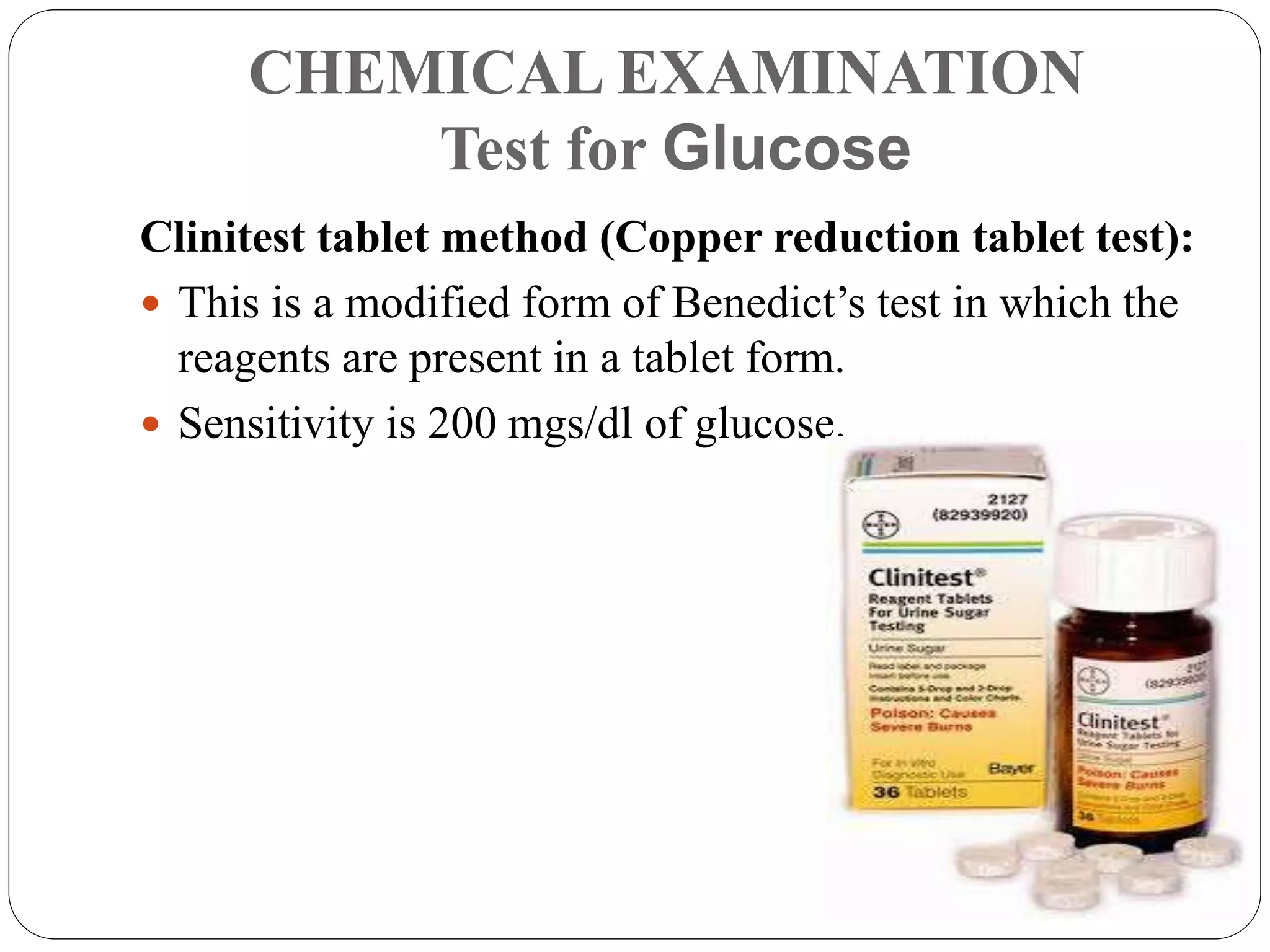
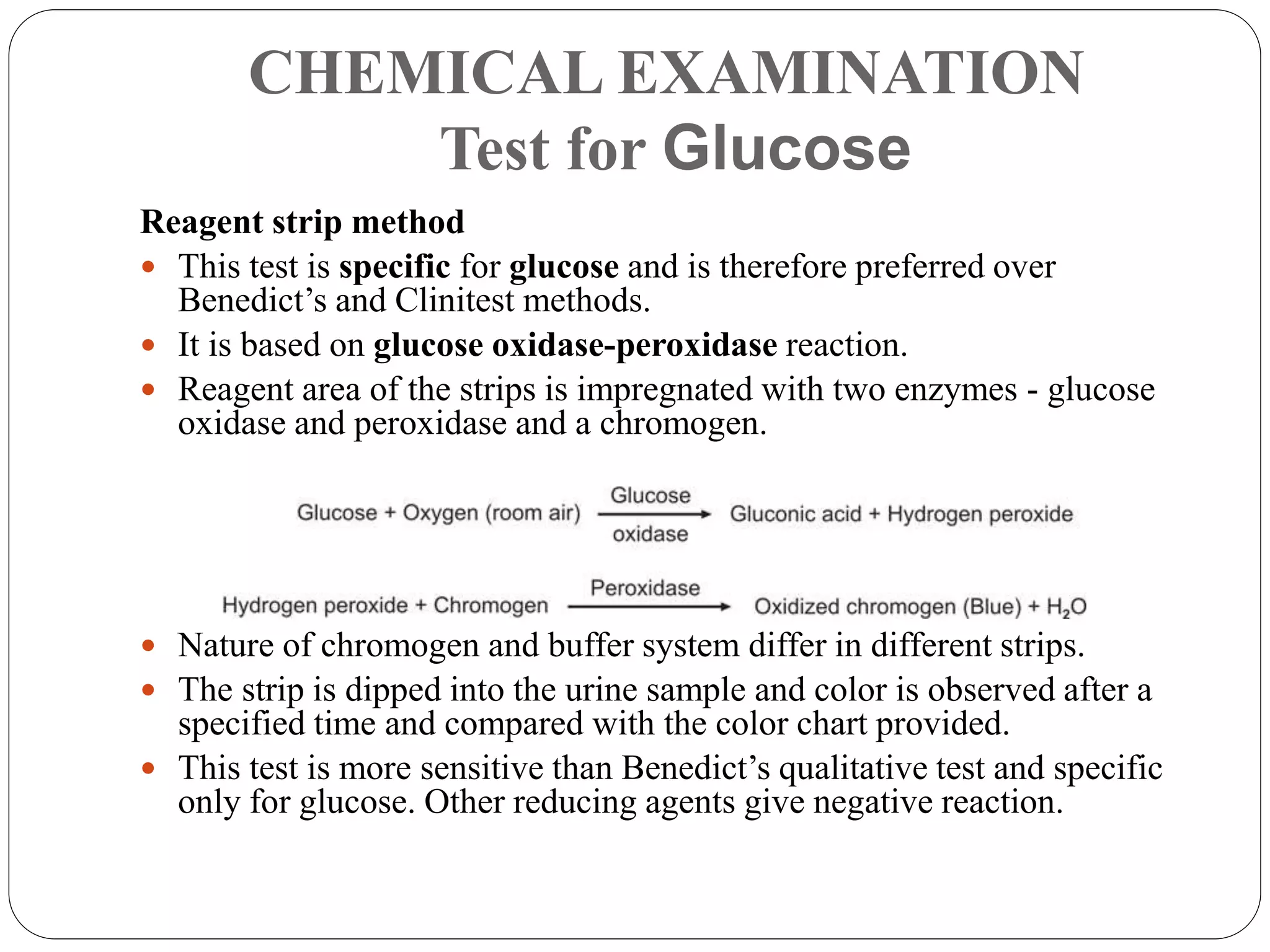
This document provides information about urinalysis, including indications for testing urine, sample collection methods, changes that occur in standing urine, preservation of samples, and the various examinations performed - physical, chemical, and microscopic. The physical examination assesses properties like volume, color, appearance, odor, specific gravity, and pH. The chemical examination tests for proteins, glucose, ketones, bilirubin, and other substances. Microscopic examination analyzes urine sediment. Proper collection and handling of urine samples is important for obtaining accurate test results.
This chapter is largely about the water and electrolytes ( salts )in your plasma and how the body manages to keep you from drying up and blowing away even if you are in the hot Texas sun and without liquid drink.
WHAT IS URINE ANALYSIS?
Urine analysis, also called Urinalysis – one of the oldest laboratory procedures in the practice of medicine.
Also knows as Urine- R&M (routine & microscopy)
Is an array of tests performed on urine
WHY URINALYSIS?
General evaluation of health
Diagnosis of disease or disorders of the kidneys or urinary tract
Diagnosis of other systemic disease that affect kidney function
Monitoring of patients with diabetes
Screening for drug abuse (eg. Sulfonamide or aminoglycosides)
COLLECTION OF URINE SPECIMENS
Improper collection---- may invalidate the results
Containers for collection of urine should be wide mouthed, clean and dry.
Analyzed within 2 hours of collection else requires refrigeration.
URINE CULTURE
Culture within 1 hour after collection or stored in a refrigerator at 4oC for no more than 18 hours.
Culture is performed when Polynephritis or Cystitis is suspected.
UTI is most frequent caused by E.Coli.
Other common agents are Enterobacter, Proteus, and Enterococcus faecalis.
URINALYSIS; WHAT TO LOOK FOR?
• Urinalysis consists of the following measurements:
Macroscopic or physical examination
Chemical examination
Microscopic examination of the sediment
Urine culture
PHYSICAL EXAMINATION OF URINE
Examination of physical characteristics:
Volume
Color
Odor
pH
Specific gravity
The refractometer or a reagent strip is used to measure specific gravity
PHYSICAL EXAMINATION
Normal- 1-2.5 L/day
Oliguria- Urine Output < 400ml/day
Dehydration
Shock
Acute glomerulonephritis
Renal Failure
Polyuria- Urine Output > 2.5 L/day
Increased water ingestion
Diabetes mellitus and insipidus.
Anuria- Urine output < 100ml/day
Seen in renal shut down Volume
PHYSICAL EXAMINATION
Normal
pale yellow in color due to pigments urochrome (different colour pigments in urine), urobilin (When urobilinogen- degraded product of bilirubin, is exposed to air, it is oxidized to urobilin, giving urine its yellow color) and uroerythrin (red pigment in urine).
Cloudiness
may be caused by excessive cellular material or protein, crystallization or precipitation of non pathological salts upon standing at room temperature or in the refrigerator.
Color
Colour of urine depending upon it’s constituents.
PHYSICAL EXAMINATION
Abnormal Colors:
Colorless – diabetes, diuretics.
Deep Yellow – concentrated urine, excess bile pigments, jaundice Color
Blue-Green – Methylene Blue, Pseudomonas (Bactrium), Riboflavin (Vitamin B2, in FAD give Yellow Orange Color)
Pink-Orange-Red – Hemoglobin, Myoglobin, Phenolphthalein, Porphyrins, Rifampicin (antibiotic against TB give orange color to urine)
Red-Brown-Black - Hemoglobin, Myoglobin, Red Blood Cells, Homogentisic acid (Homogentisic acid present in Blood and its oxidized form alkapton are excreted in the urine, giving it an unusually dark color), L-DOPA (Levodopa, is the most effective drug for Parkinson’s disease), Melanin (brown Pigment)
It gives basic things regarding urinalysis and will be very useful for medical students, house surgeons, laboratory technicians and postgraduates in medicine.
Significance of vascular endothelial growth factor and CD31 and morphometric analysis of microvessel density by CD31 receptor expression as an adjuvant tool in diagnosis of psoriatic lesions of skin
Tom Selleck Health: A Comprehensive Look at the Iconic Actor’s Wellness Journeygreendigital
Tom Selleck, an enduring figure in Hollywood. has captivated audiences for decades with his rugged charm, iconic moustache. and memorable roles in television and film. From his breakout role as Thomas Magnum in Magnum P.I. to his current portrayal of Frank Reagan in Blue Bloods. Selleck's career has spanned over 50 years. But beyond his professional achievements. fans have often been curious about Tom Selleck Health. especially as he has aged in the public eye.
Follow us on: Pinterest
Introduction
Many have been interested in Tom Selleck health. not only because of his enduring presence on screen but also because of the challenges. and lifestyle choices he has faced and made over the years. This article delves into the various aspects of Tom Selleck health. exploring his fitness regimen, diet, mental health. and the challenges he has encountered as he ages. We'll look at how he maintains his well-being. the health issues he has faced, and his approach to ageing .
Early Life and Career
Childhood and Athletic Beginnings
Tom Selleck was born on January 29, 1945, in Detroit, Michigan, and grew up in Sherman Oaks, California. From an early age, he was involved in sports, particularly basketball. which played a significant role in his physical development. His athletic pursuits continued into college. where he attended the University of Southern California (USC) on a basketball scholarship. This early involvement in sports laid a strong foundation for his physical health and disciplined lifestyle.
Transition to Acting
Selleck's transition from an athlete to an actor came with its physical demands. His first significant role in "Magnum P.I." required him to perform various stunts and maintain a fit appearance. This role, which he played from 1980 to 1988. necessitated a rigorous fitness routine to meet the show's demands. setting the stage for his long-term commitment to health and wellness.
Fitness Regimen
Workout Routine
Tom Selleck health and fitness regimen has evolved. adapting to his changing roles and age. During his "Magnum, P.I." days. Selleck's workouts were intense and focused on building and maintaining muscle mass. His routine included weightlifting, cardiovascular exercises. and specific training for the stunts he performed on the show.
Selleck adjusted his fitness routine as he aged to suit his body's needs. Today, his workouts focus on maintaining flexibility, strength, and cardiovascular health. He incorporates low-impact exercises such as swimming, walking, and light weightlifting. This balanced approach helps him stay fit without putting undue strain on his joints and muscles.
Importance of Flexibility and Mobility
In recent years, Selleck has emphasized the importance of flexibility and mobility in his fitness regimen. Understanding the natural decline in muscle mass and joint flexibility with age. he includes stretching and yoga in his routine. These practices help prevent injuries, improve posture, and maintain mobilit
New Directions in Targeted Therapeutic Approaches for Older Adults With Mantl...i3 Health
i3 Health is pleased to make the speaker slides from this activity available for use as a non-accredited self-study or teaching resource.
This slide deck presented by Dr. Kami Maddocks, Professor-Clinical in the Division of Hematology and
Associate Division Director for Ambulatory Operations
The Ohio State University Comprehensive Cancer Center, will provide insight into new directions in targeted therapeutic approaches for older adults with mantle cell lymphoma.
STATEMENT OF NEED
Mantle cell lymphoma (MCL) is a rare, aggressive B-cell non-Hodgkin lymphoma (NHL) accounting for 5% to 7% of all lymphomas. Its prognosis ranges from indolent disease that does not require treatment for years to very aggressive disease, which is associated with poor survival (Silkenstedt et al, 2021). Typically, MCL is diagnosed at advanced stage and in older patients who cannot tolerate intensive therapy (NCCN, 2022). Although recent advances have slightly increased remission rates, recurrence and relapse remain very common, leading to a median overall survival between 3 and 6 years (LLS, 2021). Though there are several effective options, progress is still needed towards establishing an accepted frontline approach for MCL (Castellino et al, 2022). Treatment selection and management of MCL are complicated by the heterogeneity of prognosis, advanced age and comorbidities of patients, and lack of an established standard approach for treatment, making it vital that clinicians be familiar with the latest research and advances in this area. In this activity chaired by Michael Wang, MD, Professor in the Department of Lymphoma & Myeloma at MD Anderson Cancer Center, expert faculty will discuss prognostic factors informing treatment, the promising results of recent trials in new therapeutic approaches, and the implications of treatment resistance in therapeutic selection for MCL.
Target Audience
Hematology/oncology fellows, attending faculty, and other health care professionals involved in the treatment of patients with mantle cell lymphoma (MCL).
Learning Objectives
1.) Identify clinical and biological prognostic factors that can guide treatment decision making for older adults with MCL
2.) Evaluate emerging data on targeted therapeutic approaches for treatment-naive and relapsed/refractory MCL and their applicability to older adults
3.) Assess mechanisms of resistance to targeted therapies for MCL and their implications for treatment selection
Title: Sense of Taste
Presenter: Dr. Faiza, Assistant Professor of Physiology
Qualifications:
MBBS (Best Graduate, AIMC Lahore)
FCPS Physiology
ICMT, CHPE, DHPE (STMU)
MPH (GC University, Faisalabad)
MBA (Virtual University of Pakistan)
Learning Objectives:
Describe the structure and function of taste buds.
Describe the relationship between the taste threshold and taste index of common substances.
Explain the chemical basis and signal transduction of taste perception for each type of primary taste sensation.
Recognize different abnormalities of taste perception and their causes.
Key Topics:
Significance of Taste Sensation:
Differentiation between pleasant and harmful food
Influence on behavior
Selection of food based on metabolic needs
Receptors of Taste:
Taste buds on the tongue
Influence of sense of smell, texture of food, and pain stimulation (e.g., by pepper)
Primary and Secondary Taste Sensations:
Primary taste sensations: Sweet, Sour, Salty, Bitter, Umami
Chemical basis and signal transduction mechanisms for each taste
Taste Threshold and Index:
Taste threshold values for Sweet (sucrose), Salty (NaCl), Sour (HCl), and Bitter (Quinine)
Taste index relationship: Inversely proportional to taste threshold
Taste Blindness:
Inability to taste certain substances, particularly thiourea compounds
Example: Phenylthiocarbamide
Structure and Function of Taste Buds:
Composition: Epithelial cells, Sustentacular/Supporting cells, Taste cells, Basal cells
Features: Taste pores, Taste hairs/microvilli, and Taste nerve fibers
Location of Taste Buds:
Found in papillae of the tongue (Fungiform, Circumvallate, Foliate)
Also present on the palate, tonsillar pillars, epiglottis, and proximal esophagus
Mechanism of Taste Stimulation:
Interaction of taste substances with receptors on microvilli
Signal transduction pathways for Umami, Sweet, Bitter, Sour, and Salty tastes
Taste Sensitivity and Adaptation:
Decrease in sensitivity with age
Rapid adaptation of taste sensation
Role of Saliva in Taste:
Dissolution of tastants to reach receptors
Washing away the stimulus
Taste Preferences and Aversions:
Mechanisms behind taste preference and aversion
Influence of receptors and neural pathways
Impact of Sensory Nerve Damage:
Degeneration of taste buds if the sensory nerve fiber is cut
Abnormalities of Taste Detection:
Conditions: Ageusia, Hypogeusia, Dysgeusia (parageusia)
Causes: Nerve damage, neurological disorders, infections, poor oral hygiene, adverse drug effects, deficiencies, aging, tobacco use, altered neurotransmitter levels
Neurotransmitters and Taste Threshold:
Effects of serotonin (5-HT) and norepinephrine (NE) on taste sensitivity
Supertasters:
25% of the population with heightened sensitivity to taste, especially bitterness
Increased number of fungiform papillae
Pulmonary Thromboembolism - etilogy, types, medical- Surgical and nursing man...VarunMahajani
Disruption of blood supply to lung alveoli due to blockage of one or more pulmonary blood vessels is called as Pulmonary thromboembolism. In this presentation we will discuss its causes, types and its management in depth.
Factory Supply Best Quality Pmk Oil CAS 28578–16–7 PMK Powder in Stockrebeccabio
Factory Supply Best Quality Pmk Oil CAS 28578–16–7 PMK Powder in Stock
Telegram: bmksupplier
signal: +85264872720
threema: TUD4A6YC
You can contact me on Telegram or Threema
Communicate promptly and reply
Free of customs clearance, Double Clearance 100% pass delivery to USA, Canada, Spain, Germany, Netherland, Poland, Italy, Sweden, UK, Czech Republic, Australia, Mexico, Russia, Ukraine, Kazakhstan.Door to door service
Hot Selling Organic intermediates
TEST BANK for Operations Management, 14th Edition by William J. Stevenson, Ve...kevinkariuki227
TEST BANK for Operations Management, 14th Edition by William J. Stevenson, Verified Chapters 1 - 19, Complete Newest Version.pdf
Title: Sense of Smell
Presenter: Dr. Faiza, Assistant Professor of Physiology
Qualifications:
MBBS (Best Graduate, AIMC Lahore)
FCPS Physiology
ICMT, CHPE, DHPE (STMU)
MPH (GC University, Faisalabad)
MBA (Virtual University of Pakistan)
Learning Objectives:
Describe the primary categories of smells and the concept of odor blindness.
Explain the structure and location of the olfactory membrane and mucosa, including the types and roles of cells involved in olfaction.
Describe the pathway and mechanisms of olfactory signal transmission from the olfactory receptors to the brain.
Illustrate the biochemical cascade triggered by odorant binding to olfactory receptors, including the role of G-proteins and second messengers in generating an action potential.
Identify different types of olfactory disorders such as anosmia, hyposmia, hyperosmia, and dysosmia, including their potential causes.
Key Topics:
Olfactory Genes:
3% of the human genome accounts for olfactory genes.
400 genes for odorant receptors.
Olfactory Membrane:
Located in the superior part of the nasal cavity.
Medially: Folds downward along the superior septum.
Laterally: Folds over the superior turbinate and upper surface of the middle turbinate.
Total surface area: 5-10 square centimeters.
Olfactory Mucosa:
Olfactory Cells: Bipolar nerve cells derived from the CNS (100 million), with 4-25 olfactory cilia per cell.
Sustentacular Cells: Produce mucus and maintain ionic and molecular environment.
Basal Cells: Replace worn-out olfactory cells with an average lifespan of 1-2 months.
Bowman’s Gland: Secretes mucus.
Stimulation of Olfactory Cells:
Odorant dissolves in mucus and attaches to receptors on olfactory cilia.
Involves a cascade effect through G-proteins and second messengers, leading to depolarization and action potential generation in the olfactory nerve.
Quality of a Good Odorant:
Small (3-20 Carbon atoms), volatile, water-soluble, and lipid-soluble.
Facilitated by odorant-binding proteins in mucus.
Membrane Potential and Action Potential:
Resting membrane potential: -55mV.
Action potential frequency in the olfactory nerve increases with odorant strength.
Adaptation Towards the Sense of Smell:
Rapid adaptation within the first second, with further slow adaptation.
Psychological adaptation greater than receptor adaptation, involving feedback inhibition from the central nervous system.
Primary Sensations of Smell:
Camphoraceous, Musky, Floral, Pepperminty, Ethereal, Pungent, Putrid.
Odor Detection Threshold:
Examples: Hydrogen sulfide (0.0005 ppm), Methyl-mercaptan (0.002 ppm).
Some toxic substances are odorless at lethal concentrations.
Characteristics of Smell:
Odor blindness for single substances due to lack of appropriate receptor protein.
Behavioral and emotional influences of smell.
Transmission of Olfactory Signals:
From olfactory cells to glomeruli in the olfactory bulb, involving lateral inhibition.
Primitive, less old, and new olfactory systems with different path
MANAGEMENT OF ATRIOVENTRICULAR CONDUCTION BLOCK.pdfJim Jacob Roy
Cardiac conduction defects can occur due to various causes.
Atrioventricular conduction blocks ( AV blocks ) are classified into 3 types.
This document describes the acute management of AV block.
HOT NEW PRODUCT! BIG SALES FAST SHIPPING NOW FROM CHINA!! EU KU DB BK substit...GL Anaacs
Contact us if you are interested:
Email / Skype : kefaya1771@gmail.com
Threema: PXHY5PDH
New BATCH Ku !!! MUCH IN DEMAND FAST SALE EVERY BATCH HAPPY GOOD EFFECT BIG BATCH !
Contact me on Threema or skype to start big business!!
Hot-sale products:
NEW HOT EUTYLONE WHITE CRYSTAL!!
5cl-adba precursor (semi finished )
5cl-adba raw materials
ADBB precursor (semi finished )
ADBB raw materials
APVP powder
5fadb/4f-adb
Jwh018 / Jwh210
Eutylone crystal
Protonitazene (hydrochloride) CAS: 119276-01-6
Flubrotizolam CAS: 57801-95-3
Metonitazene CAS: 14680-51-4
Payment terms: Western Union,MoneyGram,Bitcoin or USDT.
Deliver Time: Usually 7-15days
Shipping method: FedEx, TNT, DHL,UPS etc.Our deliveries are 100% safe, fast, reliable and discreet.
Samples will be sent for your evaluation!If you are interested in, please contact me, let's talk details.
We specializes in exporting high quality Research chemical, medical intermediate, Pharmaceutical chemicals and so on. Products are exported to USA, Canada, France, Korea, Japan,Russia, Southeast Asia and other countries.
3. INDICATIONS FOR URINALYSIS
1. Suspected renal diseases like glomerulonephritis, nephrotic
syndrome, pyelonephritis, and renal failure.
2. Detection of urinary tract infections.
3. Detection and management of metabolic disorders like diabetes
mellitus.
4. Differential diagnosis of jaundice.
5. Detection and management of plasma cell dyscrasias.
6. Diagnosis of pregnancy.
4. COLLECTION OF URINE
First morning, midstream: Preferred for routine urine
examination.
Random, midstream: Routine urine examination.
First morning, midstream, clean catch:
Bacteriological examination.
Postprandial: Estimation of glucose, urobilinogen
24-hour: Quantitative estimation of proteins or
hormones.
Catheterised: Bacteriological examination in infants,
bedridden patients, and in obstruction of urinary tract.
Plastic bag (e.g. colostomy bag) tied around genitals:
Infants, incontinent adults.
5. CHANGES WHICH OCCUR IN STANDING
URINE@ROOM TEMPERATURE
Increase in pH due to production of ammonia from
urea by urease-producing bacteria.
Formation of crystals due to precipitation of
phosphates and calcium (making the urine turbid)
Loss of ketone bodies, since they are volatile.
Decrease in glucose due to glycolysis and utilization
of glucose by cells and bacteria.
Oxidation of bilirubin to biliverdin causing false-
negative test for bilirubin
Oxidation of urobilinogen to urobilin causing false-
negative test for urobilinogen
Bacterial proliferation
Disintegration of cellular elements, especially in
alkaline and hypotonic urine.
Urine sample must be tested in the laboratory within 2 hours of
collection to get the correct results.
6. PRESERVATION OF URINE SAMPLE
Refrigeration (4-6°C) is the best general method of
preservation of urine sample up to 8 hours.
Following chemical preservatives can be added to the
24-hour urine sample:
Hydrochloric acid: It is used for preservation of a 24- hour
urine sample for adrenaline, noradrenaline, vanillylmandelic
acid, and steroids.
Toluene: It forms a thin layer over the surface and acts as a
physical barrier for bacteria and air. It is used for
measurement of chemicals.
Boric acid: A general preservative.
Thymol: It inhibits bacteria and fungi.
Formalin: It is an excellent chemical for preservation of
formed elements.
8. PHYSICAL EXAMINATION
Volume
Average 24-hr urinary output in adults is 600-2000 ml.
The volume varies according to fluid intake, diet, and climate.
Abnormalities of urinary volume are as follows:
Polyuria means urinary volume > 2000 ml/24 hours.
This is seen in diabetes mellitus, diabetes insipidus,
chronic renal failure, diuretic therapy.
Oliguria means urinary volume < 400 ml/24 hours.
This is seen in febrile states, acute glomerulonephritis, congestive cardiac failure or
dehydration.
Anuria means urinary output < 100 ml/24 hours or complete cessation of urine
output.
This occurs in acute tubular necrosis (e.g. in shock, hemolytic transfusion reaction),
acute glomerulonephritis and complete urinary tract obstruction.
Nocturia means when urine is passed in excess of 500 ml during night with
specific gravity of less than 1.018. This is a sign of early renal failure.
9. PHYSICAL EXAMINATION
Color
Normal urine color in a fresh state is pale yellow or amber and is
due to the presence of various pigments collectively called
urochrome.
10. PHYSICAL EXAMINATION
Appearance
Normal, freshly voided urine is clear in appearance.
Foamy urine occurs in the presence of excess proteins
or bilirubin.
Causes of cloudy or turbid urine
11. PHYSICAL EXAMINATION
Odor
Freshly voided urine has a typical aromatic odor due to
volatile organic acids.
After standing, urine develops ammoniacal odor
formation of ammonia occurs when urea is decomposed by
bacteria).
Some abnormal odors with associated conditions are:
Fruity: Ketoacidosis, starvation
Mousy or musty: Phenylketonuria
Fishy: Urinary tract infection with Proteus, tyrosinaemia.
Ammoniacal: Urinary tract infection with Escherichia coli,
old standing urine.
Foul: Urinary tract infection
Sulfurous: Cystinuria
12. PHYSICAL EXAMINATION
Specific Gravity (SG)
Measure of concentrating ability of kidneys and is
determined to get information about this tubular function.
It is basically a comparison of density of urine against the
density of distilled water at a particular temperature.
Normal SG of urine is 1.016 to 1.022 in 24 hrs
sample and also depends on the state of hydration.
SG of normal urine is mainly related to urea and
sodium.
SG increases as solute concentration increases and
decreases when temperature rises (since volume
expands with rise in temperature).
13. PHYSICAL EXAMINATION
Specific Gravity (SG)
Hypersthenuria - Causes of increase in SG of urine
are diabetes mellitus, glycosuria, albuminuria, fever
and dehydration.
Hyposthenuria - Causes of decrease in SG of urine
are diabetes insipidus, pyelonephritis, diuretics and
alcohol
Isosthenuria - Low and fixed SG at 1.010 due to loss
of concentrating ability of tubules seen in end stage
renal failure.
14. PHYSICAL EXAMINATION
Specific Gravity (SG)
Urinometer
1. Fill a measuring cylinder with 50 ml
of urine.
2. Lower urinometer gently into the
urine and let it float freely.
3. Let urinometer settle; it should not
touch the sides or bottom of the
cylinder.
4. Take the reading of SG on the scale
(lowest point of meniscus) at the
surface of the urine.
5. Take out the urinometer and
immediately note the temperature of
urine with a thermometer.
6. For every 3°C increase / decrease
add / subtract 0.001
16. PHYSICAL EXAMINATION
Specific Gravity (SG)
Refractometer method:
SG can be precisely determined
by a refractometer, which
measures the refractive index of
the total soluble solids.
Higher the concentration of
total dissolved solids, higher is
the refractive index.
Extent of refraction of a beam
of light passed through urine is
a measure of solute
concentration, and thus of SG.
• The method is simple and requires
only 1-2 drops of urine.
• Result is read from a scale or from
digital display.
17. PHYSICAL EXAMINATION
Specific Gravity (SG)
Reagent strip method:
Measures the concentration
of ions in urine, which
correlates with SG.
Depending on the ionic
strength of urine, a
polyelectrolyte will ionize in
proportion.
This causes a change in
color of ph indicator
(bromothymol blue).
18. PHYSICAL EXAMINATION
Reaction and pH
pH is the scale for measuring acidity or alkalinity.
Fresh urine is required for correct estimation of pH upon
standing, urine becomes alkaline owing to loss of CO2 &
production of ammonia from urea.
Normal value ranges from 4.6-8
Urine pH depends on diet, acid base balance, water balance, and
renal tubular function
Various methods for determination of reaction of urine:
Litmus paper
pH indicator paper
pH meter
Reagent strip tests
19. PHYSICAL EXAMINATION
Reaction and pH
1. Litmus paper test:
A small strip of litmus paper is dipped in urine and any color
change is noted.
2. pH indicator paper:
Reagent area (impregnated with bromothymol blue and methyl
red) of indicator paper strip is dipped in urine sample and the
color change is compared with the color guide provided.
Approximate pH is obtained.
20. PHYSICAL EXAMINATION
Reaction and pH
3. pH meter:
An electrode of pH meter is dipped in urine sample and pH
is read off directly from the digital display.
It is used if exact pH is required.
4. Reagent strip test:
The test area contains polyionic polymer bound to H+
On reaction with cations in urine, H+ is released causing
change in color of the pH-sensitive dye.
21. PHYSICAL EXAMINATION
Reaction and pH
Acidic urine
Ketosis
diabetes mellitus
Starvation
fever
Urinary tract infection by
Escherichia coli
High protein diet.
Alkaline urine
Severe vomiting
Old ammoniacal urine
sample
Chronic renal failure
Urinary tract infection by
bacteria that split urea to
ammonia - proteus or
pseudomonas
Vegetarian diet
23. CHEMICAL EXAMINATION
The chemical examination is carried out for the following
substances :-
Proteins
Glucose
Ketones
Bilirubin
Bile salts
Urobilinogen
Blood
Hemoglobin
Myoglobin
Nitrite or leukocyte esterase
24. CHEMICAL EXAMINATION
Proteins
Normally, kidneys excrete scant amount of protein in
urine
(up to 150 mg/24 hours)
These proteins include
proteins from plasma (albumin)
proteins derived from urinary tract
Tamm-Horsfall protein*
secretory IgA
proteins from tubular epithelial cells, leucocytes and other
desquamated cells.
This amount of proteinuria cannot be detected by
routine tests.
*normal mucoprotein secreted by ascending limb of the loop of Henle.
25. CHEMICAL EXAMINATION
Proteins
Proteinuria refers to protein excretion in urine >150
mg/24 hours in adults.
Causes
Glomerular proteinuria due to increased permeability
of glomerular capillary wall.
e.g. nephrotic syndrome.
Tubular proteinuria due to excretion of low molecular
weight proteins which are actively reabsorbed by proximal
renal tubules in diseased conditions of tubules.
e.g. acute and chronic pyelonephritis, heavy metal poisoning,
tuberculosis of kidney, interstitial nephritis, cystinosis, Fanconi
syndrome and rejection of kidney transplant.
26. CHEMICAL EXAMINATION
Proteins
Causes
Overflow proteinuria: When concentration of a low molecular
weight protein rises in plasma, it “overflows” from plasma into
the urine.
e.g immunoglobulin light chains or Bence Jones proteins
(plasma cell dyscrasias), hemoglobin (intravascular
hemolysis), myoglobin (skeletal muscle trauma) &
lysozyme (acute myeloid leukemia type M4 or M5).
Hemodynamic proteinuria: Alteration of blood flow through
the glomeruli causes increased filtration of proteins.
e.g high fever, hypertension, heavy exercise, congestive cardiac
failure, seizures,& exposure to cold.
Post-renal proteinuria: This is caused by inflammatory or
neoplastic conditions in renal pelvis, ureter, bladder, prostate, or
urethra.
27. CHEMICAL EXAMINATION
Test for Proteins
Heat and acetic acid test
Principle –
Proteins are denatured &
coagulated upon heating to give
white cloud precipitate.
Method –
Take a 5 ml test tube.
Fill 2/3rd with urine.
Acidify by adding a few drops
of 3% acetic acid if urine is
alkaline.
Boil upper portion for 2 minutes
(lower part acts as control).
If precipitation or turbidity appears, add a few drops of 3% acetic acid.
28. CHEMICAL EXAMINATION
Test for Proteins
Heat and acetic acid test
Interpretation.
If turbidity or precipitation
disappears on addition of acetic
acid, it is due to phosphates.
if turbidity or precipitation
persists after addition of acetic
acid, then it is due to proteins.
The test is semiquantitative and
can be graded from traces to 4+
depending upon amount of
protein.
29. CHEMICAL EXAMINATION
Test for Proteins
Sulphosalicylic Acid Test
Principle –
Addition of sulphosalicylic acid
to the urine causes formation of a
white precipitate if proteins are
present.
Method –
Take 2 ml of clear urine in a test
tube.
If reaction of urine is neutral or
alkaline, a drop of glacial acetic
acid is added.
Add 2-3 drops of sulphosalicylic
acid (3 to 5%) and examine for
turbidity against a dark
background
Interpretation.
Appearance of turbidity
which persists after
heating indicates
presence of proteins.
30. CHEMICAL EXAMINATION
Test for Proteins
Heller’s Test
Method –
Take 2 ml of concentrated nitric acid in a test
tube.
Add urine drop by drop by the side of test tube.
Interpretation-
Appearance of white ring at the junction indicates
presence of protein.
31. CHEMICAL EXAMINATION
Test for Proteins
Reagent Strip Method
Principle –
The reagent area of the strip
is coated with bromophenol
blue indicator and buffered to
an acid pH which changes
color in the presence of
proteins.
Method –
Bromophenol coated strip is
dipped in urine.
Change in colour of strip
indicates presence of proteins
in urine and is compared with
the colour chart provided for
semiquantitative grading.
Interpretation -
Reagent strip test is mainly reactive to albumin.
It is false-negative in the presence of Bence Jones proteins, myoglobin,
32. CHEMICAL EXAMINATION
Microalbuminuria
Urinary excretion of 30 to 300 mg/24 hours (or 2-20 mg/dl) of
albumin in urine.
Significance –
earliest sign of renal damage in diabetes mellitus (diabetic
nephropathy).
independent risk factor for cardiovascular disease in diabetes
mellitus.
Detection –
Measurement of albumin-creatinine ratio in a random urine sample.
Measurement of albumin in an early morning or random urine
sample.
Measurement of albumin in a 24 hr sample .
Test strips that screen for microalbuminuria are available
commercially.
33. CHEMICAL EXAMINATION
Quantitative Estimation of Proteins in Urine.
Esbach’s albuminometer method
Fill the albuminometer with urine up
to mark U.
Add Esbach’s reagent (5g picric acid
+ 10g citric acid + 500ml of distilled
water) up to mark R
Stopper the tube, mix it and let it
stand for 24 hours.
Take the reading from the level of
precipitation in the albuminometer
tube and divide it by 10 to get the
percentage of proteins.
34. CHEMICAL EXAMINATION
Quantitative Estimation of Proteins in Urine.
Turbidimetric method
Take 1 ml of urine and 1 ml standard in two separate
tubes.
Add 4 ml of trichloroacetic acid to each tube.
After 5 minutes take the reading with red filter (680
nm).
35. CHEMICAL EXAMINATION
Bence Jones Proteinuria
Bence Jones proteins are monoclonal immunoglobulin
light chains (either κ or λ) that are synthesized by
neoplastic plasma cells.
Excess production of these light chains occurs in plasma
cell dyscrasias like multiple myeloma and primary
amyloidosis.
Because of their low molecular weight and high
concentration they are excreted in urine (overflow
proteinuria).
36. CHEMICAL EXAMINATION
Test for Bence Jones Proteinuria
Upon Δ, Bence Jones proteins precipitate at temperatures
between 40-60°C.
(other proteins precipitate between 60-70°C)
The precipitate disappears on further heating at 85-100°C.
(while precipitate of other proteins does not)
When cooled (60-85°C),
•This test is not specific for Bence Jones proteins and both false-positive
and negative results can occur.
• This test has been replaced by protein electrophoresis of concentrated
urine sample.
• Protein electrophoresis – Movement of charged particles through an
electrolyte subjected to an electric field. e.g. M band in multiple myeloma
there is reappearance of
precipitate of Bence Jones proteins.
37. CHEMICAL EXAMINATION
Glucose
Main indication for testing glucose in urine is detection
of unsuspected diabetes mellitus or follow-up of
known diabetic patients.
All of the glucose filtered by the glomeruli is
reabsorbed by the proximal renal tubules and
returned to circulation.
Normally a very small amount of glucose is excreted
in urine that cannot be detected by the routine tests.
(< 500 mg/24 hours or <15 mg/dl)
Presence of detectable amounts of glucose in urine is
glycosuria.
Glycosuria results if the filtered glucose load exceeds
the capacity of renal tubular reabsorption.
38. CHEMICAL EXAMINATION
Glucose
Causes of Glycosuria
Glycosuria with hyperglycemia:
Endocrine diseases: diabetes mellitus, acromegaly, Cushing’s
syndrome, hyperthyroidism, pancreatic disease.
Non-endocrine diseases: central nervous system diseases, liver
disorders.
Drugs: adrenocorticotrophic hormone, corticosteroids, thiazides.
Alimentary glycosuria (Lag-storage glycosuria):
After a meal, there is rapid intestinal absorption of glucose leading to
transient elevation of blood glucose above renal threshold.
This can occur in persons with gastrectomy or gastrojejunostomy
and in hyperthyroidism.
Glucose tolerance test reveals a peak at 1 hour above renal threshold
(which causes glycosuria); the fasting and 2-hour glucose values are
normal.
39. CHEMICAL EXAMINATION
Glucose
Causes of Glycosuria
Glycosuria without hyperglycemia: Renal glycosuria
This accounts for 5% of cases of glycosuria in general
population.
Renal threshold is the highest glucose level in blood at which
glucose appears in urine and which is detectable by routine
laboratory tests.
The normal renal threshold for glucose is 180 mg/dl.
The renal threshold is set below 180 mgs/dl but glucose
tolerance is normal
The disorder is transmitted as autosomal dominant.
40. CHEMICAL EXAMINATION
Test for Glucose
Benedict’s qualitative test:
Composition of Benedict’s qualitative reagent:
Copper sulphate 17.3 gram
Sodium carbonate 100 gram
Sodium citrate 173 gram
Distilled water 1000 ml
Principle –
When urine is boiled in Benedict’s qualitative solution,
blue alkaline copper sulphate is reduced to red-brown
cuprous oxide if a reducing agent is present.
41. CHEMICAL EXAMINATION
Test for Glucose
Methods:
Add 0.5 ml (or 8 drops) of urine
to 5ml of Benedict’s solution.
Mix well and boil over a flame
for 2 minutes.
Allow to cool at room
temperature.
Note the color change, if any.
Interpretation:
The result is reported in grades
as follows :
Nil: no change from blue color
Trace: Green without
precipitate
1+ (approx. 0.5 grams/dl):
Green with precipitate
2+ (approx. 1.0 grams/dl):
Brown precipitate
3+ (approx. 1.5 grams/dl:
Yellow-orange precipitate
4+ (> 2.0 grams/dl): Brick- red
precipitate.
Benedict’s qualitative test:
42. CHEMICAL EXAMINATION
Test for Glucose
Clinitest tablet method (Copper reduction tablet test):
This is a modified form of Benedict’s test in which the
reagents are present in a tablet form.
Sensitivity is 200 mgs/dl of glucose.
43. CHEMICAL EXAMINATION
Test for Glucose
Reagent strip method
This test is specific for glucose and is therefore preferred over
Benedict’s and Clinitest methods.
It is based on glucose oxidase-peroxidase reaction.
Reagent area of the strips is impregnated with two enzymes - glucose
oxidase and peroxidase and a chromogen.
Nature of chromogen and buffer system differ in different strips.
The strip is dipped into the urine sample and color is observed after a
specified time and compared with the color chart provided.
This test is more sensitive than Benedict’s qualitative test and specific
only for glucose. Other reducing agents give negative reaction.