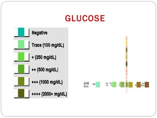
“Urine may be a waste material for man but is an important
guide for a physician”
Dr. Mohammed Kamal
Nephrology Specialist
New Mansoura General Hospital (international)
URINE ANALYSIS
Urine should be analysed as rapidly as possible.
 ideally within 30 minutes.
If not possible:
it should be refrigerated immediately and stored for preferably no more than 6–12
hours after collection.
Refrigerated urine should be brought to room temperature and thoroughly mixed
before analysis
Urine should not be frozen if sediment analysis is to be performed.
Casts are particularly vulnerable to disintegration and will only be detected if fresh urine is
examined very soon after collection.
URINALYSIS
.
A. Physical Examination
Includes:
1. Volume.
2. Colour.
3. Odour.
4. Reaction (pH).
5. Specific gravity.
B. Biochemical Examination
Includes:
1. Proteins.
2. Sugar.
3. Ketone bodies.
4. Bile salts.
5. Bile Pigments.
6. Blood.
C. Microscopic Tests
Include:
1. Cells.
2. Crystals.
3. Casts.
4. Microorganism
5. Parasites.
6.Contamination
COLLECTION OF URINE SPECIMENS
First morning sample –concentrated urine ---
biochemical analysis, casts and crystals.
Random specimen - chemical screening, microscopic
examinations.
24 sample urine sample - quantitative estimation of
proteins, sugars, electrolytes, and hormones
Mid stream urine specimen
Clean catch urine
PHYSICAL EXAMINATION
Volume –
Normal -- 1.2-2 L /day.
The day is 3-4 times > night.
Night is < 400 ml.
Polyuria >3000ml / day.
Oliguria <400ml / day.
Anuria <100 ml per day.
APPEARANCE
COLOUR
Normal - amber yellow (due to the presence
of urobilin, uroerythrin and urochromes ).
Colorless -Very dilute urine (Diabetes,
Polyuria).
Yellow orange (high colored) - Concentrated
urine, Excess urobilin, Bile pigments, Intake of
carrots.
Red/ smoky - RBC, Myoglobin, Beetroot ,Aniline dyes, Menstrual contamination.
Cloudy - Phosphates & Carbonates, Urates & Uric acid, Pus cells, Bacteria,
Spermatozoabacteria,Yeast, Spermatozoa.
Milky - Pyuria, Fat, Chyluria.
Brown black – Methemoglobin, Homogenestic acid
( alkaptonuria ),Melanin.
Orange- Bile pigments, Drugs like
Rifampicin- orange red
Nitrofurantoin-dark yellow to brown
Levodopa -brown to black;
Amitryptyline- green or blue-green.
Imipenem–cilastatin -brown urine
TURBIDITY
Freshly voided: Clear and transparent
Pus cells : form white precipitate
Bacteria growth : gives uniform cloudiness.
Mucus : it forms bulky deposits
Red cells : gives turbid smoky urine
Chyluria : gives turbid milky urine
ODOUR OF URINE
Normal odour
Fresh urine has aromatic odour
Abnormal odours
a. Ammonia smell: after prolonged standing
b. Fecal smell: due to urinary infection.
c. Fruity smell: ketosis
d. Mousy order : phenylketonuria.
e. Rancid :Tyrosinaemia.
f. Maple syrup odour : MSUD
pH
Normal pH for urine ranges from 4.5 – 8.0.
A pH < 7 indicates acid urine and a pH > 7 alkaline urine.
Some foods (such as citrus fruits and dairy products) and
medications (such as antacids) can affect urine pH.
In a diet high in protein the urine is more acidic, while a diet
high in vegetable material yields a urine that is more alkaline.
CAUSES OF ACIDIC URINE
Acidosis
Uncontrolled diabetes
Diarrhea
Starvation and dehydration
Respiratory Acidosis
CAUSES OF ALKALINE URINE
UTI with urease producing org.
Feeding
Salicylate intoxication
Urinary retention due to obstruction
Chronic renal failure
Respiratory alkalosis
Renal tubular acidosis
SPECIFIC GRAVITY
The specific gravity (SG) of urine is a useful
indicator of renal concentrating ability.
 This can be readily obtained by measuring
the refractive index (RI) in specially
calibrated refractometer.
The specific gravity of a solution refers to the ratio of
its weight to that of an equal volume of water at the
same temperature.
 For urine, the specific gravity is a function of the
number and weight of the dissolved solute particles.
Specific gravity measures the concentrating and
diluting abilities of the kidney.
Urine always has SG greater than that of distilled water, which has an
SG of 1.000.
Normal adults with adequate fluid intake: 1.016 and 1.022 (in a 24
hours specimen).
The SG of urine is increased by large amounts of glucose, protein,
lipid and contrast material.
LOW SPECIFIC GRAVITY
 HYPOSTHENURIA :indicates dilute urine, which
may be caused by:
- Diabetes insipidus ( can be as low as 1.001).
- Drinking excessive amounts of liquid.
- Pyelonephritis, glomerulonephritis.
- Use of diuretics.
HIGH SPECIFIC GRAVITY
 HYPERSTHENURIA : indicates very concentrated
urine, which may be caused by:
- Dehydration
- Diabetes mellitus.
- Adrenal insufficiency.
- Toxemia of pregnancy (protein in the urine).
ISOSTHENURIC
There is little or no variability between several specimens
from a patient , and SG is fixed at about 1.010.
It indicates : severe renal damage in which there is
disruption of both concentrating and diluting abilities.
BIOCHEMICAL EXAMINATION
The Dipstick method
1.Tests for proteins
Test – HEAT &ACETICACIDTEST
Principle-proteins are denatured & coagulated on
heating to give white cloud precipitate.
Method-take 2/3 of test tube with urine, heat only
the upper part keeping lower part as control.
Presence of phosphates, carbonates, proteins gives a
white cloud formation.Add acetic acid 1-2 drops, if
the cloud persists it indicates it is protein(acetic acid
dissolves the carbonates/phosphates)
proteinuria
Other tests
Sulphosalicylic acid test
Dipsticks
Esbach’s albuminometer- for quantitative
estimation of proteins
Causes of proteinuria
Prerenal causes-Heavy
exercise,Fever,hypertension, multiple myeloma,
eclampsia
Renal –acute & chronic glomerulonephritis,Renal
tubular dysfunction,Polycystic kidney, nephrotic
syndrome
Post renal- acute & chronic cystitis, tuberculosis
cystitis
Selective proteinuria
Nonselective proteinuria
Microalbuminuria
The level of albumin protein produced by microalbuminuria cannot be
detected by urine dipstick methods. In a properly functioning body,
albumin is not normally present in urine because it is retained in the
bloodstream by the kidneys. Microalbuminuria is diagnosed from a 24-hour
urine collection
Significance of microalbuminuria
an indicator of subclinical cardiovascular disease
an important prognostic marker for kidney disease
in diabetes mellitus
in hypertension
increasing microalbuminuria during the first 48 hours after
admission to an intensive care unit predicts elevated risk for
acute respiratory failure , multiple organ failure , and overall
mortality
Bence Jones proteins
These are light chain globulins seen in multiple
myeloma, macroglobulimias, lymphoma.
Test-Thermal method(waterbath):
Proteins has unusual property of precipitating at
400 -600c & then dissolving when the urine is
brought to boiling(1000c) & reappears when the
urine is cooled.
GLUCOSURIA
 Under normal circumstances glucose in not excreted in urine.
 Glucose is freely filtered then reabsorbed in the proximal tubule, but resorptive
capacity is limited.
 Glucosuria occurs when : blood glucose exceeds this renal threshold, for example
Diabetes mellitus { bl.glucose >160-180 mg /dl }
 Glucosuria in the absence of hyperglycaemia reflects:
- a tubular resorption defect eg: Fanconi syndrome
GLUCOSE
KETONURIA
Ketonuria is usually associated with diabetic ketoacidosis
and starvation.
Dipsticks detect acetoacetate and to a lesser extent
acetone but do not detect betahydroxybutyrate (BHB).
TESTS
1.Dipstick test
2.Rothera’s test
KETONES
Ketones are excreted when the body metabolizes fats incompletely (ketonuria).
BILE IN URINE
The constituents are :
1. Bilirubin (bile pigments),
2. Bile salts,
3. Urobilin and Urobilinogen.
• Bilirubin appears IN JAUNDICE.
• Increased bilirubinuria may be caused by liver diseses,
cholestasis or haemolytic anaemia.
• Bilirubin in urine is in the form of conjugated bilirubin
BLOOD IN URINE
Red blood cells or Haemoglobin in urine.
When hemolysis occurs in circulation or urine.
Normally an occasional red cell may be found on microscopic
examination of the urine sediment.
In women during menstruation, the urine may get contaminated with
menstrual blood
HAEMATURIA: Denotes the presence of red
blood cells in urine.
- Renal disorders, Infections
or Neoplasm orTrauma related to any part of
urinary tract.
Types:
-Microscopic
-Macroscopic {gross}haematuria
HEMOGLOBINURIA: is the presence of blood pigments in the
urine without the presence of red blood cells.
- Hemolytic anemia, Transfusion reactions, Malaria, Paroxysmal
Nocturnal Hemoglobinuria.
MICROSCOPY
In this test, urine is spun in a centrifuge so the solid materials (sediment)
settle out.The sediment is spread on a slide and examined under a
microscope.
Types of materials that may be found include:
Red blood cells
White blood cells
Casts
Epithelial cells
Bacteria
Crystals
CELLS
Several types of cells can be found in the urine, some of
which come from the blood and others from the different
types of epithelium that line the urinary tract.
TYPE
1.Erythrocytes
2. Leucocytes
3.Tubular cells
4.Uroepithelial cells
5.Squamous cells
ERYTHROCYTES (RBC’S)
Hematuria is the presence of abnormal numbers of red blood
cells in urine
Due to glomerular damage, kidney trauma, urinary tract stones,
urinary tract infections, blood toxins, and physical stress.
Contaminate from the vagina in menstruating women.
Some RBC may be present even in healthy individuals.
HAEMATURIA CAN BE
Non-glomerular : 80 % of the erythrocytes show a regular
(or isomorphic) appearance.
Glomerular : when a similar proportion of erythrocytes
are changed (or dysmorphic).
Mixed : when the two types of cells are approximately in
the same proportion.
(a) Isomorphic erythrocytes (dark cells have lost their
hemoglobin content)
(b) Dysmorphic erythrocytes.
LEUCOCYTES (WBC)
Pyuria refers to the presence
of abnormal numbers ofWBC
that may appear with infection
in the urinary tract.
WBC from the vagina, especially in the presence of vaginal
and cervical infections, or the urethra in men and women
may contaminate the urine.
NEUTROPHILS
 Appear as cells with an average
diameter of about 10 mμ
and a granular cytoplasm
surrounding a lobulated
nucleus.
Found in UTI , active proliferative glomerulonephritis,
acute or chronic interstitial nephritis, and urological
disorders.
In women, consequence of urine contamination from
genital secretions.
 In such cases, they are associated with large
amounts of squamous epithelial cells and bacteria.
LYMPHOCYTES
Indicates Chronic inflmmatory conditions , viral
diseases, renal transplant rejection
The gradual or abrupt appearance of
lymphocyturia in renal graft recipients is an
early and sensitive marker of acute cellular
rejection.
EOSINOPHILS
 Marker of Acute interstitial nephritis caused by:
- Drugs such as methycillin.
- Extracapillary glomerulonephritis.
- Atheroembolic renal disease.
- Urinary tract infection,
- Prostatitis.
- Urinary schistosomiasis.
RENAL TUBULAR CELLS
Round to ovoid mononucleated cells,
13um. Few tubular cells are rectangular,
polygonal or even columnar.
Tubular cells are a found in:
- acute tubular necrosis
- acute interstitial nephritis
- acute cellular allograft rejection and
- acute nephritic or nephrotic syndrome.
UROTHELIAL CELLS
These come from the urothelium, a multilayered
epithelium lining the urinary excretory tract from
the calyces to the bladder in the female and to the
proximal urethra in the male.
Two main types of urothelial cells are found.
1. Deriving from the deep layers :
.have club-like or ovoid
appearance, a thin cytoplasm, and
a mean diameter of about 18 m.μ
.found in urolithiasis, bladder
cancer, hydronephrosis, ureteric
stents or prolonged bladder
catheterization.
2. Deriving from the superficial
layers :
.are round to oval and are
much larger having a mean
diameter of about 30 m.μ
.found in UTI.
SQUAMOUS CELLS
Have abundant cytoplasm with few granules and a small, central
nucleus.
They are the largest cells found in the urine, with a mean diameter
of about 55 m.μ
They are found routinely in small numbers, being exfoliated from
the urethra.
When found in large numbers, they indicate a contamination of
urine from vaginal discharge.
LIPIDS
Lipids are present in urine mainly as droplets.
These can be either isolated or in aggregates —or within casts and cells.
In casts or cells, they can form 'oval fat bodies', which are tubular cells or macrophages
gorged with lipids.
Under polarized light: when containing free cholesterol and cholesterol esters,
they appear as 'Maltese crosses', which are bright particles cut by symmetrical crosses.
CAUSES:
Nephrotic syndrome or heavy proteinuria.
PCKD or with non-glomerular diseases.
In primary abnormalities of lipid metabolism, such as Fabry's disease.
(a) A large aggregate of lipid droplets.
(b) A macrophage partly gorged with lipid droplets (a so-called 'oval fat body').
(c) Maltese crosses
CASTS
Casts are elongated elements with a basic cylindrical
shape that has some possible variation due to
bending, wrinkling, and irregular edges.
Kidney is the sole site of origin.
TAMM-HORSFALL PROTEIN
A glycoprotein secreted by thick part of ascending loop of henle and early
distal convoluted tubules.
Constitutes 1/3 of total urinary protein.
Forms the matrix of all casts.
The protein forms a meshwork of fibrils that can trap any elements present in
the tubular filtrate including cells, cell fragments or granular material.
CLASSIFICATION OF CASTS
1.MATRIX
- Hyaline.
-Waxy.
2.INCLUSIONS
- Granules- proteins, cell debris.
- Fat globules- triglycerides, cholestrol esters.
- Hemosiderin granules.
- Crystals- uncommon.
- Melanin granules- rare.
3.PIGMENTS
- Haemoglobin.
- Myoglobin.
- Bilirubin .
- Drugs.
4.CELLS
- Erythocyte casts.
- Leucocytes : neutrophils, lymphocytes, monocytes.
- Renal tubular epithelial cells.
- Mixed cells : erythrocytes, neutrophils and renal
tubular cells.
- Bacteria.
HYALINE CASTS
Most frequently observed casts.
Consists almost entirely ofTamm-Horsfall protein.
Low refractive index so not easily visualized with
brightfield microscopy.
Easily visualized with phase contrast microscopy.
Can be found normally and also seen in:
1.Exercise
2.Diuretics
3.Heat exposure
4.Dehydration
5.Fever
WAXY CASTS
In chronic renal diseases some casts become
denser in appearance and known as waxy casts.
High refractive index so easily visualized with
brightfield microscopy.
Commonly associated with tubular
inflammation and degeneration.
SEEN IN:
Chronic renal failure.
Acute and chronic renal allogratft rejection.
When unusually broad waxy casts are found known as
renal failure casts.
-They imply advanced tubular atrophy and/or dilatation ,
in turn reflecting ESRD and extreme stasis of urine flow.
HYALINE–GRANULAR CASTS
These are hyaline casts
containing variable amounts of
fine granules.
They are the most frequent
casts seen in patients with
glomerulonephritis.
GRANULAR CASTS
These casts can contain either fine or
coarse granules.
Originate from plasma protein
aggregates that pass into tubules from
damaged glomeruli.
Also from cellular remnants ofWBC,
RBC, damaged renal tubular cells.
APPEAR IN :
Glomerular and tubular diseases.
Tubulointerstitial disease.
Renal allogratft rejection.
Pyelonephritis, viral infection, chronic lead poisoning.
Coarse granular casts occur with Haematuria in renal
papillary necrosis.
INCLUSION CASTS
FATTY CASTS
Fatty material is incorporated
into the cast matrix from lipid-
laden renal tubular cells.
Commonly seen with heavy
proteinuria, so feature of
nephrotic syndrome
INCLUSION CASTS
CRYSTAL CAST
Casts containing urates, calcium oxalates and sulphonamides.
Occasionally seen.
Indicate deposition of crystals in the tubule or collecting duct.
Hematuria related to tubular damage accompanies crystal casts.
PIGMENT CASTS
HAEMOGLOBIN CASTS.
MYOGLOBIN CASTS- red brown in colour and occur with myoglobinuria
following acute muscle damage. May be associated with acute renal
failure.
BILIRUBIN CASTS- seen in obstructive jaundice as deep yellow brown
colored.
DRUGS- phenazopyridine cause a bright yellow to orange colour in acid
urine and will color casts and cells.
HAEMOGLOBIN
CASTS
An erythrocyte
cast. Inset: a
haemoglobin cast
Appear yellow to red or even pale.
Usually seen with erythrocyte casts
and glomerular disease.
Rarely seen with tubular bleeding and
hemoglobinuria.
When the erythrocytes embedded in
the matrix of cast undergo
degenerative processes haemoglobin
casts are formed.
CELLULAR CASTS
1.ERYTHROCYTE (RBC) CASTS
contain variable amounts of erythrocytes embedded in the
matrix of the cast.
Indicator of bleeding with in nephron.
considered as a highly specific marker of
glomerular bleeding.
Glomerular damage allows rbc to escape into tubule and if
there is concomittant proteinuria and optimal conditions for
cast formation , rbc casts form in distal nephron.
ERYTHROCYTE CASTSAPPEAR IN:
Acute glomerulonephritis
IgA Nephropathy
Lupus nephritis
Renal infarction
Severe pyelonephritis
Renal relapse in patients with SLE
LEUCOCYTE (WBC) CASTS
Contain variable amounts of neutrophils and
indicate the renal origin of leucocytes.
Value in patients with urinary tract infection,
since their presence suggests the involvement of
the renal parenchyma.
May also be found in acute interstitial nephritis
and proliferative active glomerulonephritis.
EPITHELIAL CASTS
These contain tubular epithelial cells.
found in all conditions associated with tubular damage such as:
 acute tubular necrosis.
 acute interstitial nephritis.
acute renal allograft cellular rejection.
acute nephritic syndrome.
nephrotic syndrome.
Viral diseases (CMV).
Heavy metal poisoning, ethylene glycol, salicylate intoxication.
CRYSTALS
Formed by precipitation of urinary salts when alteration in multiple
factors affect their solubility like pH, temperature, concentration.
Urine can contain several types of crystals.
 They are found in both acidic urine and alkaline urine.
Some are birefringent under polarized light.
URIC ACID
These crystals precipitate at a pH <5.4.
 Wide range of shapes.
 Appear mostly as lozenges which have a typical amber colour.
Under polarized light show polychromatic birefringence.
1.Reflect increased nucleoprotein turnover eg
chemotherapy for leukemia
2.Evidence of uric acid stones lodged in ureter.
3.Urate nephropathy of gout.
CALCIUM OXALATE
Two main types of calcium oxalate crystals:
1. Monohydrated: dumb-bell or as biconcave/biconvex discs , birefringent.
2. Bihydrated: bipyramidal shape.
can cause extensive tubular injury
.Causes:
.Chronic renal disease.
.Ethylene glycol toxicity.
.Increased absorption of oxalates from food following small bowel resection, crohns disease.
CYSTINE
Are thin, hexagonal, birefringent plates with irregular sides.
Can be isolated, heaped upon one another, or in clumps.
Found mostly in acidic urine.
Marker of cystinuria.
 Also found in cystine calculi.
CRYSTALS DUE TO DRUGS
Sulfadiazine
Acyclovir and indinavir
Triamterene
Coronary dilator piridoxylate
Barbiturate primidone,
Vasodilator naftidrofuryl oxalate,
Vitamin C
Amoxycillin
CHOLESTEROL CRYSTALS
Appear as brownish,
transparent thin plates, with
sharp edges and corners.
Found with other lipid
particles, in the urine of
patients with nephrotic
syndrome or heavy proteinuria.
A plate of cholesterol crystal (on its
lowest corner, a few small lipid
droplets; on the background, a hyaline
cast)
MICROORGANISMS
BACTERIA
Seen as rods or cocci.
May be found due to contamination rather
than infection.
The presence of leucocytes increases the
probability of a real infection, especially in
women, but leucocytes and bacteria may
contaminate urine from genitalia.
In patients with acute pyelonephritis,
bacterial casts can be seen.
FUNGI
• Candida - most frequently found yeast
• Elongated, ovoid, or spherical.
• Presence of buds.
• Most frequent cause is contamination from the
genitalia.
• Grows in the urinary tract, mostly in patients with
diabetes, structural abnormalities, indwelling
catheters, prolonged antibiotic treatment or
immunosuppression.
• Candidal casts are found in urine of patients with
renal candidiasis.
PARASITES
SCHISTOSOMA HAEMATOBIUM
The adult form lives and lays the eggs in the vesical plexus
and veins draining the ureters.
Endemic in Nile valley,West Africa,Arabia.
Causes haematuria, chronic renal failure due to
obstructive uropathy, glomerulonephritis, or bladder
cancer.
Eggs : spindle shaped, a rounded anterior and a conical
posterior end tapering into a delicate terminal spine.
Take home message
Urine analysis screening is effective in identifying patients with a symptomatic renal
disease for further evaluation.
Urine microscopy should be performed in any patient who has persistent haematuria
or proteinuria & may be useful if the urine dipstick is suggestive of UTI.
Quantitative urine analysis test may be performed for diagnosis of many specific
disorders such as endocrine diseases, bladder cancer &osteoporosis.
Kamal

Kamal

  • 1.
    “Urine may bea waste material for man but is an important guide for a physician” Dr. Mohammed Kamal Nephrology Specialist New Mansoura General Hospital (international) URINE ANALYSIS
  • 2.
    Urine should beanalysed as rapidly as possible.  ideally within 30 minutes. If not possible: it should be refrigerated immediately and stored for preferably no more than 6–12 hours after collection. Refrigerated urine should be brought to room temperature and thoroughly mixed before analysis Urine should not be frozen if sediment analysis is to be performed. Casts are particularly vulnerable to disintegration and will only be detected if fresh urine is examined very soon after collection.
  • 3.
    URINALYSIS . A. Physical Examination Includes: 1.Volume. 2. Colour. 3. Odour. 4. Reaction (pH). 5. Specific gravity. B. Biochemical Examination Includes: 1. Proteins. 2. Sugar. 3. Ketone bodies. 4. Bile salts. 5. Bile Pigments. 6. Blood. C. Microscopic Tests Include: 1. Cells. 2. Crystals. 3. Casts. 4. Microorganism 5. Parasites. 6.Contamination
  • 4.
    COLLECTION OF URINESPECIMENS First morning sample –concentrated urine --- biochemical analysis, casts and crystals. Random specimen - chemical screening, microscopic examinations. 24 sample urine sample - quantitative estimation of proteins, sugars, electrolytes, and hormones Mid stream urine specimen Clean catch urine
  • 5.
    PHYSICAL EXAMINATION Volume – Normal-- 1.2-2 L /day. The day is 3-4 times > night. Night is < 400 ml. Polyuria >3000ml / day. Oliguria <400ml / day. Anuria <100 ml per day.
  • 6.
    APPEARANCE COLOUR Normal - amberyellow (due to the presence of urobilin, uroerythrin and urochromes ). Colorless -Very dilute urine (Diabetes, Polyuria). Yellow orange (high colored) - Concentrated urine, Excess urobilin, Bile pigments, Intake of carrots.
  • 7.
    Red/ smoky -RBC, Myoglobin, Beetroot ,Aniline dyes, Menstrual contamination. Cloudy - Phosphates & Carbonates, Urates & Uric acid, Pus cells, Bacteria, Spermatozoabacteria,Yeast, Spermatozoa. Milky - Pyuria, Fat, Chyluria. Brown black – Methemoglobin, Homogenestic acid ( alkaptonuria ),Melanin. Orange- Bile pigments, Drugs like Rifampicin- orange red Nitrofurantoin-dark yellow to brown Levodopa -brown to black; Amitryptyline- green or blue-green. Imipenem–cilastatin -brown urine
  • 8.
    TURBIDITY Freshly voided: Clearand transparent Pus cells : form white precipitate Bacteria growth : gives uniform cloudiness. Mucus : it forms bulky deposits Red cells : gives turbid smoky urine Chyluria : gives turbid milky urine
  • 9.
    ODOUR OF URINE Normalodour Fresh urine has aromatic odour Abnormal odours a. Ammonia smell: after prolonged standing b. Fecal smell: due to urinary infection. c. Fruity smell: ketosis d. Mousy order : phenylketonuria. e. Rancid :Tyrosinaemia. f. Maple syrup odour : MSUD
  • 10.
    pH Normal pH forurine ranges from 4.5 – 8.0. A pH < 7 indicates acid urine and a pH > 7 alkaline urine. Some foods (such as citrus fruits and dairy products) and medications (such as antacids) can affect urine pH. In a diet high in protein the urine is more acidic, while a diet high in vegetable material yields a urine that is more alkaline.
  • 11.
    CAUSES OF ACIDICURINE Acidosis Uncontrolled diabetes Diarrhea Starvation and dehydration Respiratory Acidosis
  • 12.
    CAUSES OF ALKALINEURINE UTI with urease producing org. Feeding Salicylate intoxication Urinary retention due to obstruction Chronic renal failure Respiratory alkalosis Renal tubular acidosis
  • 13.
    SPECIFIC GRAVITY The specificgravity (SG) of urine is a useful indicator of renal concentrating ability.  This can be readily obtained by measuring the refractive index (RI) in specially calibrated refractometer.
  • 14.
    The specific gravityof a solution refers to the ratio of its weight to that of an equal volume of water at the same temperature.  For urine, the specific gravity is a function of the number and weight of the dissolved solute particles. Specific gravity measures the concentrating and diluting abilities of the kidney.
  • 15.
    Urine always hasSG greater than that of distilled water, which has an SG of 1.000. Normal adults with adequate fluid intake: 1.016 and 1.022 (in a 24 hours specimen). The SG of urine is increased by large amounts of glucose, protein, lipid and contrast material.
  • 16.
    LOW SPECIFIC GRAVITY HYPOSTHENURIA :indicates dilute urine, which may be caused by: - Diabetes insipidus ( can be as low as 1.001). - Drinking excessive amounts of liquid. - Pyelonephritis, glomerulonephritis. - Use of diuretics.
  • 17.
    HIGH SPECIFIC GRAVITY HYPERSTHENURIA : indicates very concentrated urine, which may be caused by: - Dehydration - Diabetes mellitus. - Adrenal insufficiency. - Toxemia of pregnancy (protein in the urine).
  • 18.
    ISOSTHENURIC There is littleor no variability between several specimens from a patient , and SG is fixed at about 1.010. It indicates : severe renal damage in which there is disruption of both concentrating and diluting abilities.
  • 19.
  • 20.
  • 21.
    1.Tests for proteins Test– HEAT &ACETICACIDTEST Principle-proteins are denatured & coagulated on heating to give white cloud precipitate. Method-take 2/3 of test tube with urine, heat only the upper part keeping lower part as control. Presence of phosphates, carbonates, proteins gives a white cloud formation.Add acetic acid 1-2 drops, if the cloud persists it indicates it is protein(acetic acid dissolves the carbonates/phosphates) proteinuria
  • 22.
    Other tests Sulphosalicylic acidtest Dipsticks Esbach’s albuminometer- for quantitative estimation of proteins
  • 23.
    Causes of proteinuria Prerenalcauses-Heavy exercise,Fever,hypertension, multiple myeloma, eclampsia Renal –acute & chronic glomerulonephritis,Renal tubular dysfunction,Polycystic kidney, nephrotic syndrome Post renal- acute & chronic cystitis, tuberculosis cystitis
  • 24.
  • 25.
    Microalbuminuria The level ofalbumin protein produced by microalbuminuria cannot be detected by urine dipstick methods. In a properly functioning body, albumin is not normally present in urine because it is retained in the bloodstream by the kidneys. Microalbuminuria is diagnosed from a 24-hour urine collection
  • 26.
    Significance of microalbuminuria anindicator of subclinical cardiovascular disease an important prognostic marker for kidney disease in diabetes mellitus in hypertension increasing microalbuminuria during the first 48 hours after admission to an intensive care unit predicts elevated risk for acute respiratory failure , multiple organ failure , and overall mortality
  • 27.
    Bence Jones proteins Theseare light chain globulins seen in multiple myeloma, macroglobulimias, lymphoma. Test-Thermal method(waterbath): Proteins has unusual property of precipitating at 400 -600c & then dissolving when the urine is brought to boiling(1000c) & reappears when the urine is cooled.
  • 28.
    GLUCOSURIA  Under normalcircumstances glucose in not excreted in urine.  Glucose is freely filtered then reabsorbed in the proximal tubule, but resorptive capacity is limited.  Glucosuria occurs when : blood glucose exceeds this renal threshold, for example Diabetes mellitus { bl.glucose >160-180 mg /dl }  Glucosuria in the absence of hyperglycaemia reflects: - a tubular resorption defect eg: Fanconi syndrome
  • 29.
  • 30.
    KETONURIA Ketonuria is usuallyassociated with diabetic ketoacidosis and starvation. Dipsticks detect acetoacetate and to a lesser extent acetone but do not detect betahydroxybutyrate (BHB). TESTS 1.Dipstick test 2.Rothera’s test
  • 31.
    KETONES Ketones are excretedwhen the body metabolizes fats incompletely (ketonuria).
  • 32.
    BILE IN URINE Theconstituents are : 1. Bilirubin (bile pigments), 2. Bile salts, 3. Urobilin and Urobilinogen. • Bilirubin appears IN JAUNDICE. • Increased bilirubinuria may be caused by liver diseses, cholestasis or haemolytic anaemia. • Bilirubin in urine is in the form of conjugated bilirubin
  • 33.
    BLOOD IN URINE Redblood cells or Haemoglobin in urine. When hemolysis occurs in circulation or urine. Normally an occasional red cell may be found on microscopic examination of the urine sediment. In women during menstruation, the urine may get contaminated with menstrual blood
  • 34.
    HAEMATURIA: Denotes thepresence of red blood cells in urine. - Renal disorders, Infections or Neoplasm orTrauma related to any part of urinary tract. Types: -Microscopic -Macroscopic {gross}haematuria HEMOGLOBINURIA: is the presence of blood pigments in the urine without the presence of red blood cells. - Hemolytic anemia, Transfusion reactions, Malaria, Paroxysmal Nocturnal Hemoglobinuria.
  • 35.
    MICROSCOPY In this test,urine is spun in a centrifuge so the solid materials (sediment) settle out.The sediment is spread on a slide and examined under a microscope. Types of materials that may be found include: Red blood cells White blood cells Casts Epithelial cells Bacteria Crystals
  • 36.
    CELLS Several types ofcells can be found in the urine, some of which come from the blood and others from the different types of epithelium that line the urinary tract. TYPE 1.Erythrocytes 2. Leucocytes 3.Tubular cells 4.Uroepithelial cells 5.Squamous cells
  • 37.
    ERYTHROCYTES (RBC’S) Hematuria isthe presence of abnormal numbers of red blood cells in urine Due to glomerular damage, kidney trauma, urinary tract stones, urinary tract infections, blood toxins, and physical stress. Contaminate from the vagina in menstruating women. Some RBC may be present even in healthy individuals.
  • 38.
    HAEMATURIA CAN BE Non-glomerular: 80 % of the erythrocytes show a regular (or isomorphic) appearance. Glomerular : when a similar proportion of erythrocytes are changed (or dysmorphic). Mixed : when the two types of cells are approximately in the same proportion.
  • 39.
    (a) Isomorphic erythrocytes(dark cells have lost their hemoglobin content) (b) Dysmorphic erythrocytes.
  • 40.
    LEUCOCYTES (WBC) Pyuria refersto the presence of abnormal numbers ofWBC that may appear with infection in the urinary tract. WBC from the vagina, especially in the presence of vaginal and cervical infections, or the urethra in men and women may contaminate the urine.
  • 41.
    NEUTROPHILS  Appear ascells with an average diameter of about 10 mμ and a granular cytoplasm surrounding a lobulated nucleus.
  • 42.
    Found in UTI, active proliferative glomerulonephritis, acute or chronic interstitial nephritis, and urological disorders. In women, consequence of urine contamination from genital secretions.  In such cases, they are associated with large amounts of squamous epithelial cells and bacteria.
  • 43.
    LYMPHOCYTES Indicates Chronic inflmmatoryconditions , viral diseases, renal transplant rejection The gradual or abrupt appearance of lymphocyturia in renal graft recipients is an early and sensitive marker of acute cellular rejection.
  • 44.
    EOSINOPHILS  Marker ofAcute interstitial nephritis caused by: - Drugs such as methycillin. - Extracapillary glomerulonephritis. - Atheroembolic renal disease. - Urinary tract infection, - Prostatitis. - Urinary schistosomiasis.
  • 45.
    RENAL TUBULAR CELLS Roundto ovoid mononucleated cells, 13um. Few tubular cells are rectangular, polygonal or even columnar. Tubular cells are a found in: - acute tubular necrosis - acute interstitial nephritis - acute cellular allograft rejection and - acute nephritic or nephrotic syndrome.
  • 46.
    UROTHELIAL CELLS These comefrom the urothelium, a multilayered epithelium lining the urinary excretory tract from the calyces to the bladder in the female and to the proximal urethra in the male. Two main types of urothelial cells are found.
  • 47.
    1. Deriving fromthe deep layers : .have club-like or ovoid appearance, a thin cytoplasm, and a mean diameter of about 18 m.μ .found in urolithiasis, bladder cancer, hydronephrosis, ureteric stents or prolonged bladder catheterization.
  • 48.
    2. Deriving fromthe superficial layers : .are round to oval and are much larger having a mean diameter of about 30 m.μ .found in UTI.
  • 49.
    SQUAMOUS CELLS Have abundantcytoplasm with few granules and a small, central nucleus. They are the largest cells found in the urine, with a mean diameter of about 55 m.μ They are found routinely in small numbers, being exfoliated from the urethra. When found in large numbers, they indicate a contamination of urine from vaginal discharge.
  • 50.
    LIPIDS Lipids are presentin urine mainly as droplets. These can be either isolated or in aggregates —or within casts and cells. In casts or cells, they can form 'oval fat bodies', which are tubular cells or macrophages gorged with lipids. Under polarized light: when containing free cholesterol and cholesterol esters, they appear as 'Maltese crosses', which are bright particles cut by symmetrical crosses. CAUSES: Nephrotic syndrome or heavy proteinuria. PCKD or with non-glomerular diseases. In primary abnormalities of lipid metabolism, such as Fabry's disease.
  • 51.
    (a) A largeaggregate of lipid droplets. (b) A macrophage partly gorged with lipid droplets (a so-called 'oval fat body'). (c) Maltese crosses
  • 52.
    CASTS Casts are elongatedelements with a basic cylindrical shape that has some possible variation due to bending, wrinkling, and irregular edges. Kidney is the sole site of origin.
  • 53.
    TAMM-HORSFALL PROTEIN A glycoproteinsecreted by thick part of ascending loop of henle and early distal convoluted tubules. Constitutes 1/3 of total urinary protein. Forms the matrix of all casts. The protein forms a meshwork of fibrils that can trap any elements present in the tubular filtrate including cells, cell fragments or granular material.
  • 54.
    CLASSIFICATION OF CASTS 1.MATRIX -Hyaline. -Waxy. 2.INCLUSIONS - Granules- proteins, cell debris. - Fat globules- triglycerides, cholestrol esters. - Hemosiderin granules. - Crystals- uncommon. - Melanin granules- rare.
  • 55.
    3.PIGMENTS - Haemoglobin. - Myoglobin. -Bilirubin . - Drugs. 4.CELLS - Erythocyte casts. - Leucocytes : neutrophils, lymphocytes, monocytes. - Renal tubular epithelial cells. - Mixed cells : erythrocytes, neutrophils and renal tubular cells. - Bacteria.
  • 56.
    HYALINE CASTS Most frequentlyobserved casts. Consists almost entirely ofTamm-Horsfall protein. Low refractive index so not easily visualized with brightfield microscopy. Easily visualized with phase contrast microscopy.
  • 57.
    Can be foundnormally and also seen in: 1.Exercise 2.Diuretics 3.Heat exposure 4.Dehydration 5.Fever
  • 58.
    WAXY CASTS In chronicrenal diseases some casts become denser in appearance and known as waxy casts. High refractive index so easily visualized with brightfield microscopy. Commonly associated with tubular inflammation and degeneration.
  • 59.
    SEEN IN: Chronic renalfailure. Acute and chronic renal allogratft rejection. When unusually broad waxy casts are found known as renal failure casts. -They imply advanced tubular atrophy and/or dilatation , in turn reflecting ESRD and extreme stasis of urine flow.
  • 60.
    HYALINE–GRANULAR CASTS These arehyaline casts containing variable amounts of fine granules. They are the most frequent casts seen in patients with glomerulonephritis.
  • 61.
    GRANULAR CASTS These castscan contain either fine or coarse granules. Originate from plasma protein aggregates that pass into tubules from damaged glomeruli. Also from cellular remnants ofWBC, RBC, damaged renal tubular cells.
  • 62.
    APPEAR IN : Glomerularand tubular diseases. Tubulointerstitial disease. Renal allogratft rejection. Pyelonephritis, viral infection, chronic lead poisoning. Coarse granular casts occur with Haematuria in renal papillary necrosis.
  • 63.
    INCLUSION CASTS FATTY CASTS Fattymaterial is incorporated into the cast matrix from lipid- laden renal tubular cells. Commonly seen with heavy proteinuria, so feature of nephrotic syndrome
  • 64.
    INCLUSION CASTS CRYSTAL CAST Castscontaining urates, calcium oxalates and sulphonamides. Occasionally seen. Indicate deposition of crystals in the tubule or collecting duct. Hematuria related to tubular damage accompanies crystal casts.
  • 65.
    PIGMENT CASTS HAEMOGLOBIN CASTS. MYOGLOBINCASTS- red brown in colour and occur with myoglobinuria following acute muscle damage. May be associated with acute renal failure. BILIRUBIN CASTS- seen in obstructive jaundice as deep yellow brown colored. DRUGS- phenazopyridine cause a bright yellow to orange colour in acid urine and will color casts and cells.
  • 66.
    HAEMOGLOBIN CASTS An erythrocyte cast. Inset:a haemoglobin cast Appear yellow to red or even pale. Usually seen with erythrocyte casts and glomerular disease. Rarely seen with tubular bleeding and hemoglobinuria. When the erythrocytes embedded in the matrix of cast undergo degenerative processes haemoglobin casts are formed.
  • 67.
    CELLULAR CASTS 1.ERYTHROCYTE (RBC)CASTS contain variable amounts of erythrocytes embedded in the matrix of the cast. Indicator of bleeding with in nephron. considered as a highly specific marker of glomerular bleeding. Glomerular damage allows rbc to escape into tubule and if there is concomittant proteinuria and optimal conditions for cast formation , rbc casts form in distal nephron.
  • 68.
    ERYTHROCYTE CASTSAPPEAR IN: Acuteglomerulonephritis IgA Nephropathy Lupus nephritis Renal infarction Severe pyelonephritis Renal relapse in patients with SLE
  • 69.
    LEUCOCYTE (WBC) CASTS Containvariable amounts of neutrophils and indicate the renal origin of leucocytes. Value in patients with urinary tract infection, since their presence suggests the involvement of the renal parenchyma. May also be found in acute interstitial nephritis and proliferative active glomerulonephritis.
  • 70.
    EPITHELIAL CASTS These containtubular epithelial cells. found in all conditions associated with tubular damage such as:  acute tubular necrosis.  acute interstitial nephritis. acute renal allograft cellular rejection. acute nephritic syndrome. nephrotic syndrome. Viral diseases (CMV). Heavy metal poisoning, ethylene glycol, salicylate intoxication.
  • 71.
    CRYSTALS Formed by precipitationof urinary salts when alteration in multiple factors affect their solubility like pH, temperature, concentration. Urine can contain several types of crystals.  They are found in both acidic urine and alkaline urine. Some are birefringent under polarized light.
  • 72.
    URIC ACID These crystalsprecipitate at a pH <5.4.  Wide range of shapes.  Appear mostly as lozenges which have a typical amber colour. Under polarized light show polychromatic birefringence. 1.Reflect increased nucleoprotein turnover eg chemotherapy for leukemia 2.Evidence of uric acid stones lodged in ureter. 3.Urate nephropathy of gout.
  • 73.
    CALCIUM OXALATE Two maintypes of calcium oxalate crystals: 1. Monohydrated: dumb-bell or as biconcave/biconvex discs , birefringent. 2. Bihydrated: bipyramidal shape. can cause extensive tubular injury .Causes: .Chronic renal disease. .Ethylene glycol toxicity. .Increased absorption of oxalates from food following small bowel resection, crohns disease.
  • 74.
    CYSTINE Are thin, hexagonal,birefringent plates with irregular sides. Can be isolated, heaped upon one another, or in clumps. Found mostly in acidic urine. Marker of cystinuria.  Also found in cystine calculi.
  • 75.
    CRYSTALS DUE TODRUGS Sulfadiazine Acyclovir and indinavir Triamterene Coronary dilator piridoxylate Barbiturate primidone, Vasodilator naftidrofuryl oxalate, Vitamin C Amoxycillin
  • 76.
    CHOLESTEROL CRYSTALS Appear asbrownish, transparent thin plates, with sharp edges and corners. Found with other lipid particles, in the urine of patients with nephrotic syndrome or heavy proteinuria. A plate of cholesterol crystal (on its lowest corner, a few small lipid droplets; on the background, a hyaline cast)
  • 77.
    MICROORGANISMS BACTERIA Seen as rodsor cocci. May be found due to contamination rather than infection. The presence of leucocytes increases the probability of a real infection, especially in women, but leucocytes and bacteria may contaminate urine from genitalia. In patients with acute pyelonephritis, bacterial casts can be seen.
  • 78.
    FUNGI • Candida -most frequently found yeast • Elongated, ovoid, or spherical. • Presence of buds. • Most frequent cause is contamination from the genitalia. • Grows in the urinary tract, mostly in patients with diabetes, structural abnormalities, indwelling catheters, prolonged antibiotic treatment or immunosuppression. • Candidal casts are found in urine of patients with renal candidiasis.
  • 79.
    PARASITES SCHISTOSOMA HAEMATOBIUM The adultform lives and lays the eggs in the vesical plexus and veins draining the ureters. Endemic in Nile valley,West Africa,Arabia. Causes haematuria, chronic renal failure due to obstructive uropathy, glomerulonephritis, or bladder cancer. Eggs : spindle shaped, a rounded anterior and a conical posterior end tapering into a delicate terminal spine.
  • 80.
    Take home message Urineanalysis screening is effective in identifying patients with a symptomatic renal disease for further evaluation. Urine microscopy should be performed in any patient who has persistent haematuria or proteinuria & may be useful if the urine dipstick is suggestive of UTI. Quantitative urine analysis test may be performed for diagnosis of many specific disorders such as endocrine diseases, bladder cancer &osteoporosis.

Editor's Notes

  • #4 &amp;lt;number&amp;gt;
  • #10 &amp;lt;number&amp;gt;