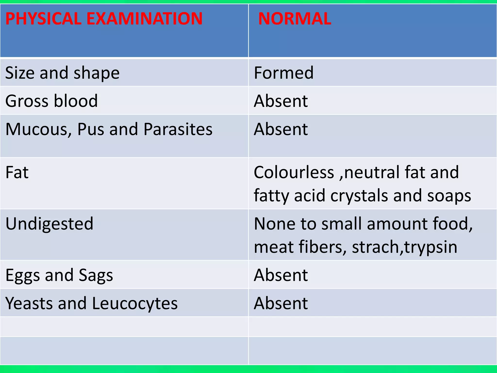
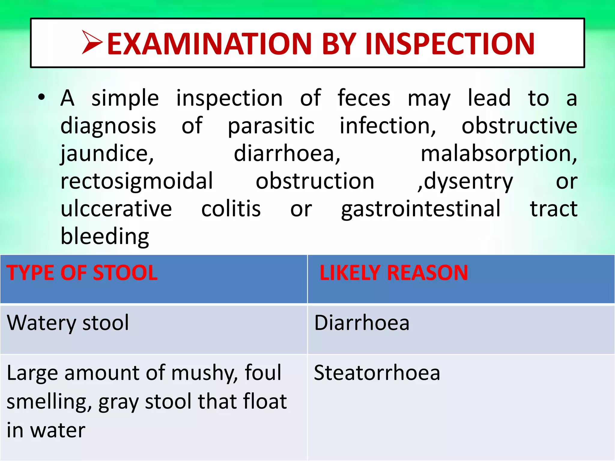
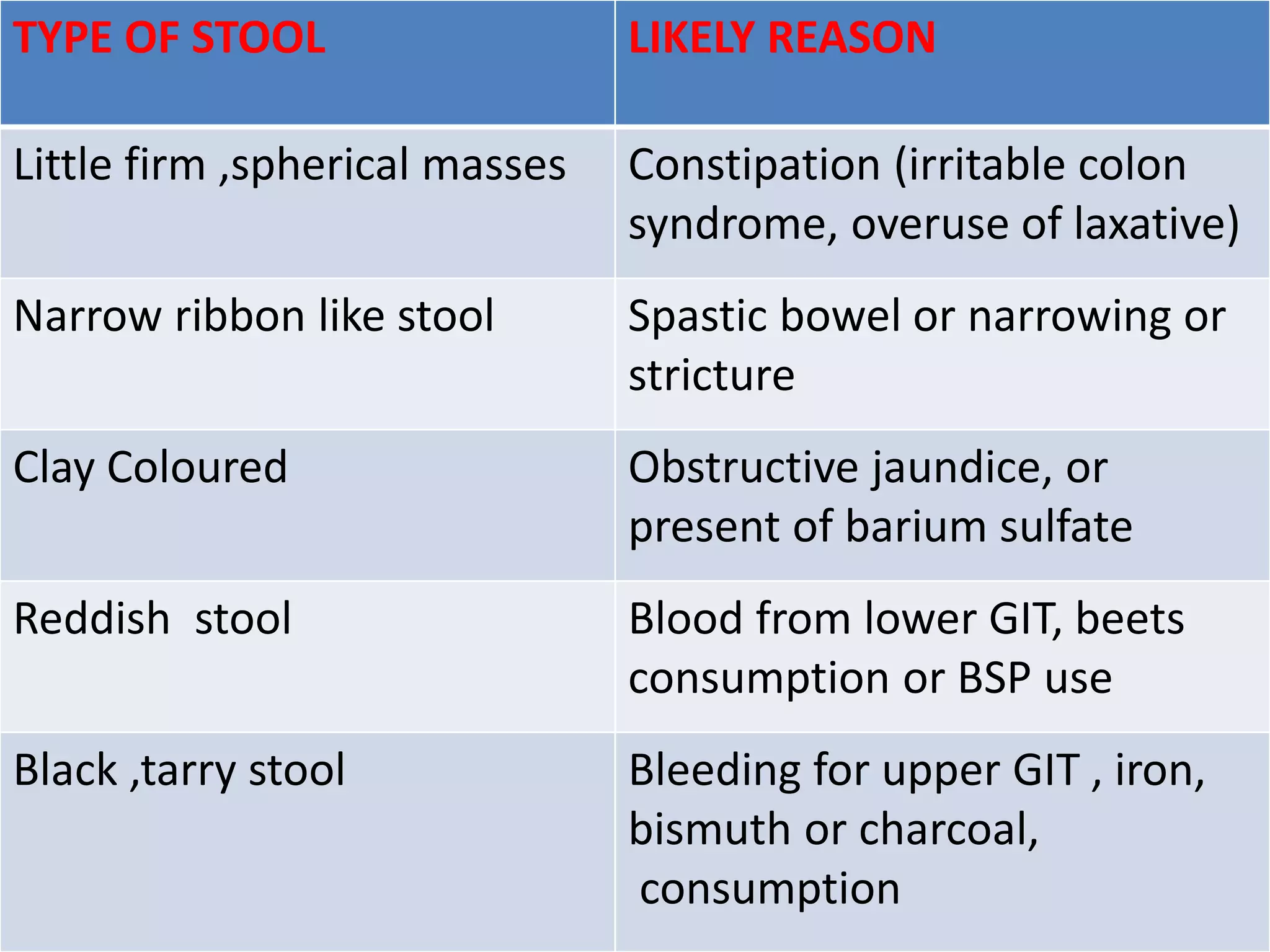
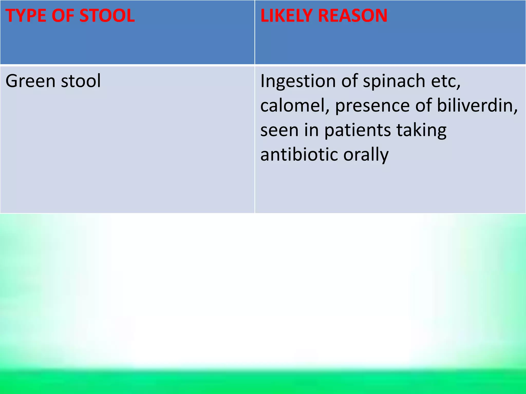
STOOL EXAMINATION
PRESENTED BY:-
DR.RAJESH UIKEY
Assist. Prof
DEPT. OF ROG NIDAN EVAM VIKRITI VIGYAN
GENERAL INTRODUCTION
• Average healthy adults defecate from three times
a day to three time a week.
• Common pattern is once a day .
• The stool tends to be soft and bulky on a diet
high in vegetables and small and dry on a diet
high in meat.
• Two thirds of the stool weight is attributable to its
water content ,The normal brown color is of still
undetermined origin.
• The odour results from indole and skatole
produced by bacteria from tryptophan.
FECES ARE COMPOSED OF:-
1. Waste residue of indigestible material in food
2. Bile (pigments and salts).
3. Intestinal secretion ,including mucous.
4. Leucocytes that migrate from the blood stream
5. Shed epithelial cells
6. Large numbers of bacteria that make up to one
third of total solids
7. Inorganic material (10-20%) that is chiefly
calcium and phosphate
8. Digested food (present in very small quantities)
TYPE OF FECES
Bristol Stool Scale is a medical aid designed to classify
the form of human feces into seven categories.
STOOL COLLECTION
Universal precaution :-
• Collect stool in a dry ,clean container
• Faces should be urine free when collected.
and transfer to another container by a
the laboratory immediatetly after collection.
• Worm stool are best for detecting ova and
refrigerate for ova and parasites.
Continue:-
• Some coliform bacilli produce antibiotics
substances that destroy enteric pathogens.
• A diarrhoeal stool will usually give good
results
• A freshly passed stool is the specimen of
choice
• Preferably stool specimens should be collect
before antibiotics therapy is initiated and as
early in the course of disease as possible
Continue:-
• Only a small amount of stool is a needed the
size of walnut . If mucous and blood are
present ,they should be indicated in part of
the specimen to be examined.
• Do not use a stool that has been passed into
the toilet bowl or that has been contaminated
with barium or other x-ray
• Label all stool specimen with patients name,
date and reason for examination testing.
INTERFERING FACTORS
• Meat interferes with some tests and should
usually be omitted from the diet for 3 day
before a test for blood
• Stool specimen from patients receiving
barium, bismuth, oil, or antibiotics are not
satisfactory
• Bismuth from paper towels and toilet tissues
interferes with tests.
NORMAL VALUES IN STOOL ANALYSIS
PHYSICAL EXAMINATION NORMAL
Colour brown
odour Varies with pH stool and
depend upon bacterial
fermentation and
putrefaction
Consistency Plastic ,not unusual to see
seeds and veg. skin ,soft and
bulky in a high veg. diet ,
small and dry in a high meat
diet
PHYSICAL EXAMINATION
PHYSICAL EXAMINATION NORMAL
Size and shape Formed
Gross blood Absent
Mucous, Pus and Parasites Absent
Fat Colourless ,neutral fat and
fatty acid crystals and soaps
Undigested None to small amount food,
meat fibers, strach,trypsin
Eggs and Sags Absent
Yeasts and Leucocytes Absent
CHEMICAL EXAMINATION
CHEMICAL EXAMINATION NORMAL
pH Neutral to weakly alkaline
Adult= 7.0-7.5
New born= 5.0-7.0
Bottle fed infant= neutral to
slightly alkaline pH of=7.0-8.0
Breast fed infant=Slightly
acidic
CHEMICAL EXAMINATION NORMAL
Occult blood Negative
Urobilinogen 50-300mg/24hrs
Porphyrins Coproporphyrins<200µg/24hrs
Protoporphyrins,1500µg/24hrs
Uroporphyrin<100µg/24hrs
Nitrogen 1-2 gm/24 hrs
Bile Negative in adult,positive in
childeren
Trypsin Positive in small amount in
adult, in greater amount in
normal children
EXAMINATION BY INSPECTION
• A simple inspection of feces may lead to a
diagnosis of parasitic infection, obstructive
jaundice, diarrhoea, malabsorption,
rectosigmoidal obstruction ,dysentry or
ulccerative colitis or gastrointestinal tract
bleeding
TYPE OF STOOL LIKELY REASON
Watery stool Diarrhoea
Large amount of mushy, foul
smelling, gray stool that float
in water
Steatorrhoea
TYPE OF STOOL LIKELY REASON
Little firm ,spherical masses Constipation (irritable colon
syndrome, overuse of laxative)
Narrow ribbon like stool Spastic bowel or narrowing or
stricture
Clay Coloured Obstructive jaundice, or
present of barium sulfate
Reddish stool Blood from lower GIT, beets
consumption or BSP use
Black ,tarry stool Bleeding for upper GIT , iron,
bismuth or charcoal,
consumption
TYPE OF STOOL LIKELY REASON
Green stool Ingestion of spinach etc,
calomel, presence of biliverdin,
seen in patients taking
antibiotic orally
MICROSCOPIC EXAMINATION
COVERSLIP PREPARATION FOR MICROSCOPIC EXAMINATION:-
Normal saline
one drop
Iodine
solution one
drop
1.On a clean and dry slide add one drop each of normal
saline and iodine solution
2. By using an applicator stick take a small portion
•From formed sample:-portion from well inside and surface
•From liquid stool containing mucus. Take blood stained
mucus and other small portion
3. Mix the sample with the drop of sodium chloride on the
Slide .Mix the sample with iodine solution drop
4.Place a coverslip over each drop (apply coverslips as
Shown to avoid the formation of air bubbles)
5. Label the slide with identification number
6. Examine the preparation under the microscopic
•First under – low power objective and afterwards
With high power objective
CONCENTRATION METHOD OF MICROSCOPIC EXAMINATION
Method:- When stools are mixed with a saturated solution of
sodium chloride the ova float to surface.
This method is recommended for ova of round worm
hookworm, whipworm, hymnolopis nana, Tenia etc
The method is not suittable for ova of schistosomes
and Flukes and protozoa cysts
1. Place small portion of stool in a penicillin bottle .
Add saturated sodium chloride (half bottle)
2. Mix by using applicator stick and add a saturated sodium
chloride and fill the bottle completely
3. Place a coverslip on the bottle mouth
4. Remove the coverslip with care , a drop of liquid should
On it. Place it on the glass slide
5.Examine under microscope(first under low power objective
And afterwards under high power objective)
MICROSCOPIC ANALYSIS
NO. DETECTION
OF
NORMAL
FINDING
ABNORMAL
FINDING
PATHOLOG.CONDI
TION
DIAGRAM
1 CELLS:-
Pus cells
Present , few Present ,
many
Bacillary dysentry ,
ulcerative colitis
Epithelial cells -do- -do- Inflammation of
the bowel
macrophages Occasional many Bacillary dysentry,
ulcerative colitis
Erythrocytes Absent Present Lesion is in the
colon , rectum or
anus, the clump in
amoebiasis
NO. DETECTION OF NORMAL
FINDING
ABNORM
AL
FINDING
PATHOLOG.CON
DITION
DIAGRAM
2 Crystals:-
Triple phosphate
and calcium oxalate
Present due to
indigestion of
certain food I,e.
spinach, berries,
tomato etyc
- -
Charcot-leyden
crystals
Absent Present ulcerative
colitis
amoebiasis
Haematoidin
crystals
Absent Present Intestinal
haemorrhage
3 Vegetable matter:-
Vegetable cells,
spirals, fibers, hairs
etc
Present in
residuel
constituent
- -
NO. DETECTION OF NORMAL FINDING ABNORMAL
FINDING
PATHOLOG.
CONDITIO
N
DIAGRAM
4 Animal matter:-
Connective tissue,
musle fibers and
elastic tissue
Present in residuel
constituent or
undigested fibers -
-
-
5 Undigested
ingredients:-
starch
Absent Present in
high
proportion
Indigestion
Fat Absent Present in
high
proportion
Indigestion
6 Other Findings:-
Yests
Present especialy
blastocystis
hominis
- -
Bacteria Constitute about
one third of the
weight of dried
faces
- -
INTESTINAL PROTOZOA
ORGANIUM TYPE DIAGRAM
Entamoeba histolytica cyst Pathogenic
Entamoeba coli cyst Non pathogenic
Giardia lamblia cyst Pathogenic
ORGANIUM TYPE DIAGRAM
Balantidium coli cyst Pathogenic
Enterobius vermicularis (pin
worm)
pathogenic
Trichiura (wipworm) Pathogenic
ORGANIUM TYPE DIAGRAM
Ascaris lumbricoides (round
worm)
Pathogenic
Ancylostoma duodenale (hook
worm)
pathogenic
Necator americanus Pathogenic
ORGANIUM TYPE DIAGRAM
Strongyloides stercoralis Pathogenic
Taenia solium (tape worm) pathogenic
Schistosoma haematobium Pathogenic
References
1. Godkar Praful B., Godar Darshan P, Text book
of medical laboratory technology, Parasitology
and faeces Examination 42, Second ed,
Mumbai: Balwani publishing house, 2003.
 Stool Examination for Routine Practice

Stool Examination for Routine Practice

  • 2.
    STOOL EXAMINATION PRESENTED BY:- DR.RAJESHUIKEY Assist. Prof DEPT. OF ROG NIDAN EVAM VIKRITI VIGYAN
  • 3.
    GENERAL INTRODUCTION • Averagehealthy adults defecate from three times a day to three time a week. • Common pattern is once a day . • The stool tends to be soft and bulky on a diet high in vegetables and small and dry on a diet high in meat. • Two thirds of the stool weight is attributable to its water content ,The normal brown color is of still undetermined origin. • The odour results from indole and skatole produced by bacteria from tryptophan.
  • 4.
    FECES ARE COMPOSEDOF:- 1. Waste residue of indigestible material in food 2. Bile (pigments and salts). 3. Intestinal secretion ,including mucous. 4. Leucocytes that migrate from the blood stream 5. Shed epithelial cells 6. Large numbers of bacteria that make up to one third of total solids 7. Inorganic material (10-20%) that is chiefly calcium and phosphate 8. Digested food (present in very small quantities)
  • 5.
    TYPE OF FECES BristolStool Scale is a medical aid designed to classify the form of human feces into seven categories.
  • 6.
    STOOL COLLECTION Universal precaution:- • Collect stool in a dry ,clean container • Faces should be urine free when collected. and transfer to another container by a the laboratory immediatetly after collection. • Worm stool are best for detecting ova and refrigerate for ova and parasites.
  • 7.
    Continue:- • Some coliformbacilli produce antibiotics substances that destroy enteric pathogens. • A diarrhoeal stool will usually give good results • A freshly passed stool is the specimen of choice • Preferably stool specimens should be collect before antibiotics therapy is initiated and as early in the course of disease as possible
  • 8.
    Continue:- • Only asmall amount of stool is a needed the size of walnut . If mucous and blood are present ,they should be indicated in part of the specimen to be examined. • Do not use a stool that has been passed into the toilet bowl or that has been contaminated with barium or other x-ray • Label all stool specimen with patients name, date and reason for examination testing.
  • 9.
    INTERFERING FACTORS • Meatinterferes with some tests and should usually be omitted from the diet for 3 day before a test for blood • Stool specimen from patients receiving barium, bismuth, oil, or antibiotics are not satisfactory • Bismuth from paper towels and toilet tissues interferes with tests.
  • 10.
    NORMAL VALUES INSTOOL ANALYSIS PHYSICAL EXAMINATION NORMAL Colour brown odour Varies with pH stool and depend upon bacterial fermentation and putrefaction Consistency Plastic ,not unusual to see seeds and veg. skin ,soft and bulky in a high veg. diet , small and dry in a high meat diet PHYSICAL EXAMINATION
  • 11.
    PHYSICAL EXAMINATION NORMAL Sizeand shape Formed Gross blood Absent Mucous, Pus and Parasites Absent Fat Colourless ,neutral fat and fatty acid crystals and soaps Undigested None to small amount food, meat fibers, strach,trypsin Eggs and Sags Absent Yeasts and Leucocytes Absent
  • 12.
    CHEMICAL EXAMINATION CHEMICAL EXAMINATIONNORMAL pH Neutral to weakly alkaline Adult= 7.0-7.5 New born= 5.0-7.0 Bottle fed infant= neutral to slightly alkaline pH of=7.0-8.0 Breast fed infant=Slightly acidic
  • 13.
    CHEMICAL EXAMINATION NORMAL Occultblood Negative Urobilinogen 50-300mg/24hrs Porphyrins Coproporphyrins<200µg/24hrs Protoporphyrins,1500µg/24hrs Uroporphyrin<100µg/24hrs Nitrogen 1-2 gm/24 hrs Bile Negative in adult,positive in childeren Trypsin Positive in small amount in adult, in greater amount in normal children
  • 14.
    EXAMINATION BY INSPECTION •A simple inspection of feces may lead to a diagnosis of parasitic infection, obstructive jaundice, diarrhoea, malabsorption, rectosigmoidal obstruction ,dysentry or ulccerative colitis or gastrointestinal tract bleeding TYPE OF STOOL LIKELY REASON Watery stool Diarrhoea Large amount of mushy, foul smelling, gray stool that float in water Steatorrhoea
  • 15.
    TYPE OF STOOLLIKELY REASON Little firm ,spherical masses Constipation (irritable colon syndrome, overuse of laxative) Narrow ribbon like stool Spastic bowel or narrowing or stricture Clay Coloured Obstructive jaundice, or present of barium sulfate Reddish stool Blood from lower GIT, beets consumption or BSP use Black ,tarry stool Bleeding for upper GIT , iron, bismuth or charcoal, consumption
  • 16.
    TYPE OF STOOLLIKELY REASON Green stool Ingestion of spinach etc, calomel, presence of biliverdin, seen in patients taking antibiotic orally
  • 17.
    MICROSCOPIC EXAMINATION COVERSLIP PREPARATIONFOR MICROSCOPIC EXAMINATION:- Normal saline one drop Iodine solution one drop 1.On a clean and dry slide add one drop each of normal saline and iodine solution
  • 18.
    2. By usingan applicator stick take a small portion •From formed sample:-portion from well inside and surface •From liquid stool containing mucus. Take blood stained mucus and other small portion
  • 19.
    3. Mix thesample with the drop of sodium chloride on the Slide .Mix the sample with iodine solution drop
  • 20.
    4.Place a coverslipover each drop (apply coverslips as Shown to avoid the formation of air bubbles)
  • 21.
    5. Label theslide with identification number
  • 22.
    6. Examine thepreparation under the microscopic •First under – low power objective and afterwards With high power objective
  • 23.
    CONCENTRATION METHOD OFMICROSCOPIC EXAMINATION Method:- When stools are mixed with a saturated solution of sodium chloride the ova float to surface. This method is recommended for ova of round worm hookworm, whipworm, hymnolopis nana, Tenia etc The method is not suittable for ova of schistosomes and Flukes and protozoa cysts
  • 24.
    1. Place smallportion of stool in a penicillin bottle . Add saturated sodium chloride (half bottle) 2. Mix by using applicator stick and add a saturated sodium chloride and fill the bottle completely
  • 25.
    3. Place acoverslip on the bottle mouth 4. Remove the coverslip with care , a drop of liquid should On it. Place it on the glass slide
  • 26.
    5.Examine under microscope(firstunder low power objective And afterwards under high power objective)
  • 27.
    MICROSCOPIC ANALYSIS NO. DETECTION OF NORMAL FINDING ABNORMAL FINDING PATHOLOG.CONDI TION DIAGRAM 1CELLS:- Pus cells Present , few Present , many Bacillary dysentry , ulcerative colitis Epithelial cells -do- -do- Inflammation of the bowel macrophages Occasional many Bacillary dysentry, ulcerative colitis Erythrocytes Absent Present Lesion is in the colon , rectum or anus, the clump in amoebiasis
  • 28.
    NO. DETECTION OFNORMAL FINDING ABNORM AL FINDING PATHOLOG.CON DITION DIAGRAM 2 Crystals:- Triple phosphate and calcium oxalate Present due to indigestion of certain food I,e. spinach, berries, tomato etyc - - Charcot-leyden crystals Absent Present ulcerative colitis amoebiasis Haematoidin crystals Absent Present Intestinal haemorrhage 3 Vegetable matter:- Vegetable cells, spirals, fibers, hairs etc Present in residuel constituent - -
  • 29.
    NO. DETECTION OFNORMAL FINDING ABNORMAL FINDING PATHOLOG. CONDITIO N DIAGRAM 4 Animal matter:- Connective tissue, musle fibers and elastic tissue Present in residuel constituent or undigested fibers - - - 5 Undigested ingredients:- starch Absent Present in high proportion Indigestion Fat Absent Present in high proportion Indigestion 6 Other Findings:- Yests Present especialy blastocystis hominis - - Bacteria Constitute about one third of the weight of dried faces - -
  • 30.
    INTESTINAL PROTOZOA ORGANIUM TYPEDIAGRAM Entamoeba histolytica cyst Pathogenic Entamoeba coli cyst Non pathogenic Giardia lamblia cyst Pathogenic
  • 31.
    ORGANIUM TYPE DIAGRAM Balantidiumcoli cyst Pathogenic Enterobius vermicularis (pin worm) pathogenic Trichiura (wipworm) Pathogenic
  • 32.
    ORGANIUM TYPE DIAGRAM Ascarislumbricoides (round worm) Pathogenic Ancylostoma duodenale (hook worm) pathogenic Necator americanus Pathogenic
  • 33.
    ORGANIUM TYPE DIAGRAM Strongyloidesstercoralis Pathogenic Taenia solium (tape worm) pathogenic Schistosoma haematobium Pathogenic
  • 34.
    References 1. Godkar PrafulB., Godar Darshan P, Text book of medical laboratory technology, Parasitology and faeces Examination 42, Second ed, Mumbai: Balwani publishing house, 2003.

Editor's Notes