Downloaded 149 times

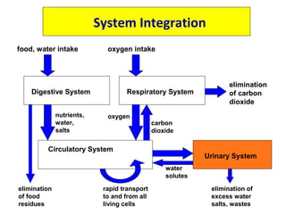
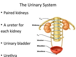
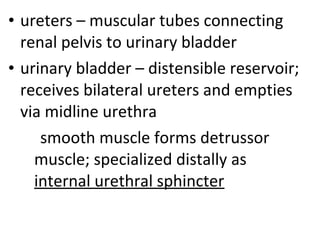
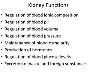
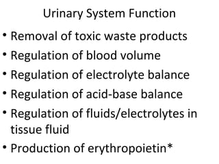
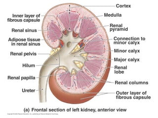

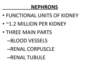
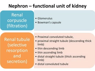
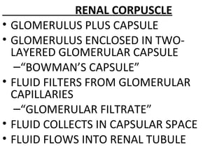
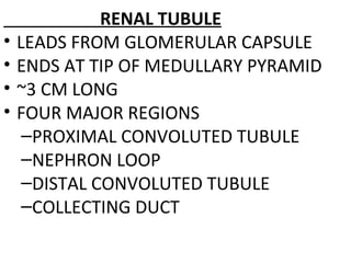
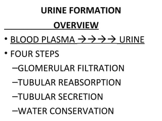
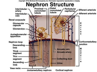
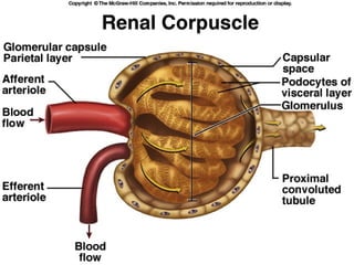
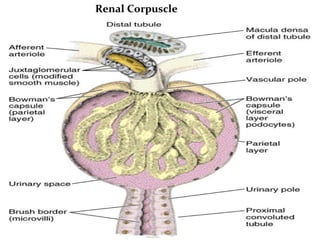
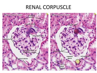

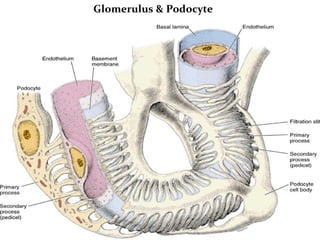
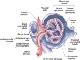
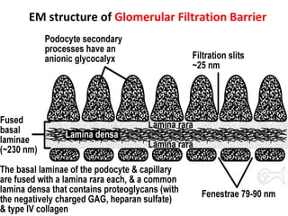
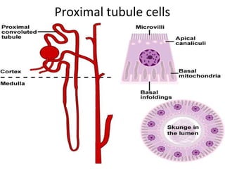


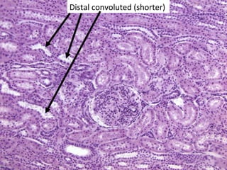
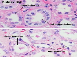
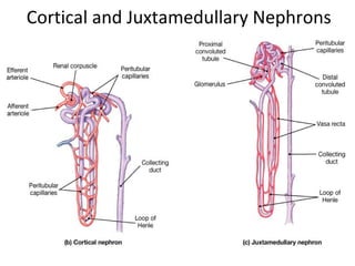
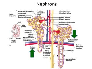
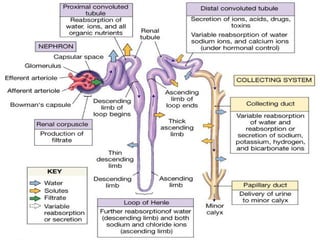
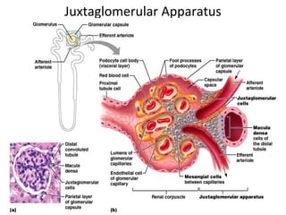


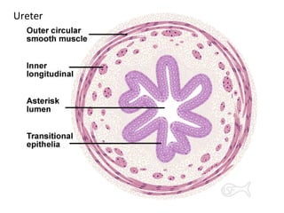
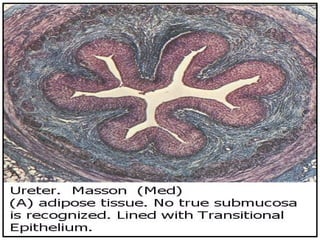
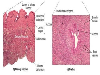


The urinary system consists of the kidneys, ureters, urinary bladder, and urethra. The kidneys contain millions of nephrons, which are the functional filtering units. Each nephron contains a renal corpuscle with glomerulus and Bowman's capsule, where blood is filtered, and a renal tubule with various segments that modify the filtrate and produce urine. The ureters carry urine from the kidneys to the bladder. The bladder stores urine until urination, when urine exits the body through the urethra. The urinary system removes wastes and regulates fluid and electrolyte balance.