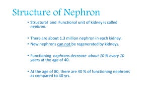
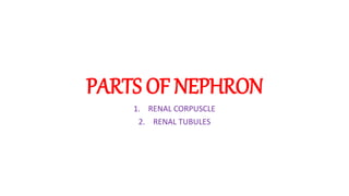
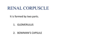
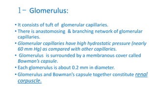
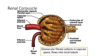
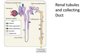
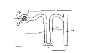
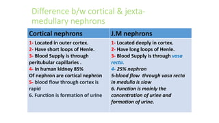
NEPHRON
Source: The Guyton and Hall physiology
Maryam Fida(o-1827)
• Structural and Functional unit of kidney is called
nephron.
• There are about 1.3 million nephron in each kidney.
• New nephrons can not be regenerated by kidneys.
• Functioning nephrons decrease about 10 % every 10
years at the age of 40.
• At the age of 80, there are 40 % of functioning nephrons
as compared to 40 yrs.
Structure of Nephron
PARTS OF NEPHRON
1. RENAL CORPUSCLE
2. RENAL TUBULES
It is formed by two parts.
1. GLOMERULUS
2. BOWMAN’S CAPSULE
RENAL CORPUSCLE
1- Glomerulus:
• It consists of tuft of glomerular capillaries.
• There is anastomosing & branching network of glomerular
capillaries.
• Glomerular capillaries have high hydrostatic pressure (nearly
60 mm Hg) as compared with other capillaries.
• Glomerulus is surrounded by a membranous cover called
Bowman’s capsule.
• Each glomerulus is about 0.2 mm in diameter.
• Glomerulus and Bowman’s capsule together constitute renal
corpuscle.
Glomerular filtrate collects in capsular
space, flows into renal tubule
Renal Corpuscle
• Fluid which is filter
Renal tubules
and collecting
Duct
2- Renal Tubules:
Each renal tubule is divided into
various part as they have different
functions.
i- Proximal convulated tubule.
It is continuation of Bowman’s
capsule.
ii- Loop of Henle. It is continuation of
prox. conv. tubule.
• * Loop of Henle has three parts.
a- descending limb,
b- u turn or bend in medulla and
c- ascending limb.
• Ascending limb has initial thin
segment followed by thick segment.
• At the end of thick ascending limb,
there is short segment called
macula densa, which plays
important role in controlling
functions of nephron.
• iii- Distal convulated tubule. It is
continuation of loop of Henle,
starting in renal cortex.
• Last portion of it is called cortical
collecting tubule.
• iv- Collecting duct.
It is formed by fusion of many
collecting tubules.
*It carries urine into pelvi-calyceal
spaces
i.e renal pelvis through tips of renal
papillae.
• Vasa recta extend downward
into medulla lying side by side
with loops of Henle.
• This specialized network of
capillaries in medulla plays a
vital role in formation of
concentrated urine.
Cortical nephrons J.M nephrons
1- Located in outer cortex.
2- Have short loops of Henle.
3- Blood Supply is through
peritubular capillaries .
4- In human kidney 85%
Of nephron are cortical nephron
5- blood flow through cortex is
rapid
6. Function is formation of urine
1- Located deeply in cortex.
2- Have long loops of Henle.
3- Blood Supply is through vasa
recta.
4- 25% nephron
5-blood flow through vasa recta
in medulla is slow
6. Function is mainly the
concentration of urine and
formation of urine.
Difference b/w cortical & jexta-
medullary nephrons
Nephron (The Guyton and Hall physiology)

Nephron (The Guyton and Hall physiology)

  • 1.
    NEPHRON Source: The Guytonand Hall physiology Maryam Fida(o-1827)
  • 2.
    • Structural andFunctional unit of kidney is called nephron. • There are about 1.3 million nephron in each kidney. • New nephrons can not be regenerated by kidneys. • Functioning nephrons decrease about 10 % every 10 years at the age of 40. • At the age of 80, there are 40 % of functioning nephrons as compared to 40 yrs. Structure of Nephron
  • 3.
    PARTS OF NEPHRON 1.RENAL CORPUSCLE 2. RENAL TUBULES
  • 5.
    It is formedby two parts. 1. GLOMERULUS 2. BOWMAN’S CAPSULE RENAL CORPUSCLE
  • 6.
    1- Glomerulus: • Itconsists of tuft of glomerular capillaries. • There is anastomosing & branching network of glomerular capillaries. • Glomerular capillaries have high hydrostatic pressure (nearly 60 mm Hg) as compared with other capillaries. • Glomerulus is surrounded by a membranous cover called Bowman’s capsule. • Each glomerulus is about 0.2 mm in diameter. • Glomerulus and Bowman’s capsule together constitute renal corpuscle.
  • 7.
    Glomerular filtrate collectsin capsular space, flows into renal tubule Renal Corpuscle
  • 8.
    • Fluid whichis filter Renal tubules and collecting Duct
  • 9.
    2- Renal Tubules: Eachrenal tubule is divided into various part as they have different functions. i- Proximal convulated tubule. It is continuation of Bowman’s capsule. ii- Loop of Henle. It is continuation of prox. conv. tubule. • * Loop of Henle has three parts. a- descending limb, b- u turn or bend in medulla and c- ascending limb. • Ascending limb has initial thin segment followed by thick segment. • At the end of thick ascending limb, there is short segment called macula densa, which plays important role in controlling functions of nephron.
  • 10.
    • iii- Distalconvulated tubule. It is continuation of loop of Henle, starting in renal cortex. • Last portion of it is called cortical collecting tubule. • iv- Collecting duct. It is formed by fusion of many collecting tubules. *It carries urine into pelvi-calyceal spaces i.e renal pelvis through tips of renal papillae. • Vasa recta extend downward into medulla lying side by side with loops of Henle. • This specialized network of capillaries in medulla plays a vital role in formation of concentrated urine.
  • 12.
    Cortical nephrons J.Mnephrons 1- Located in outer cortex. 2- Have short loops of Henle. 3- Blood Supply is through peritubular capillaries . 4- In human kidney 85% Of nephron are cortical nephron 5- blood flow through cortex is rapid 6. Function is formation of urine 1- Located deeply in cortex. 2- Have long loops of Henle. 3- Blood Supply is through vasa recta. 4- 25% nephron 5-blood flow through vasa recta in medulla is slow 6. Function is mainly the concentration of urine and formation of urine. Difference b/w cortical & jexta- medullary nephrons