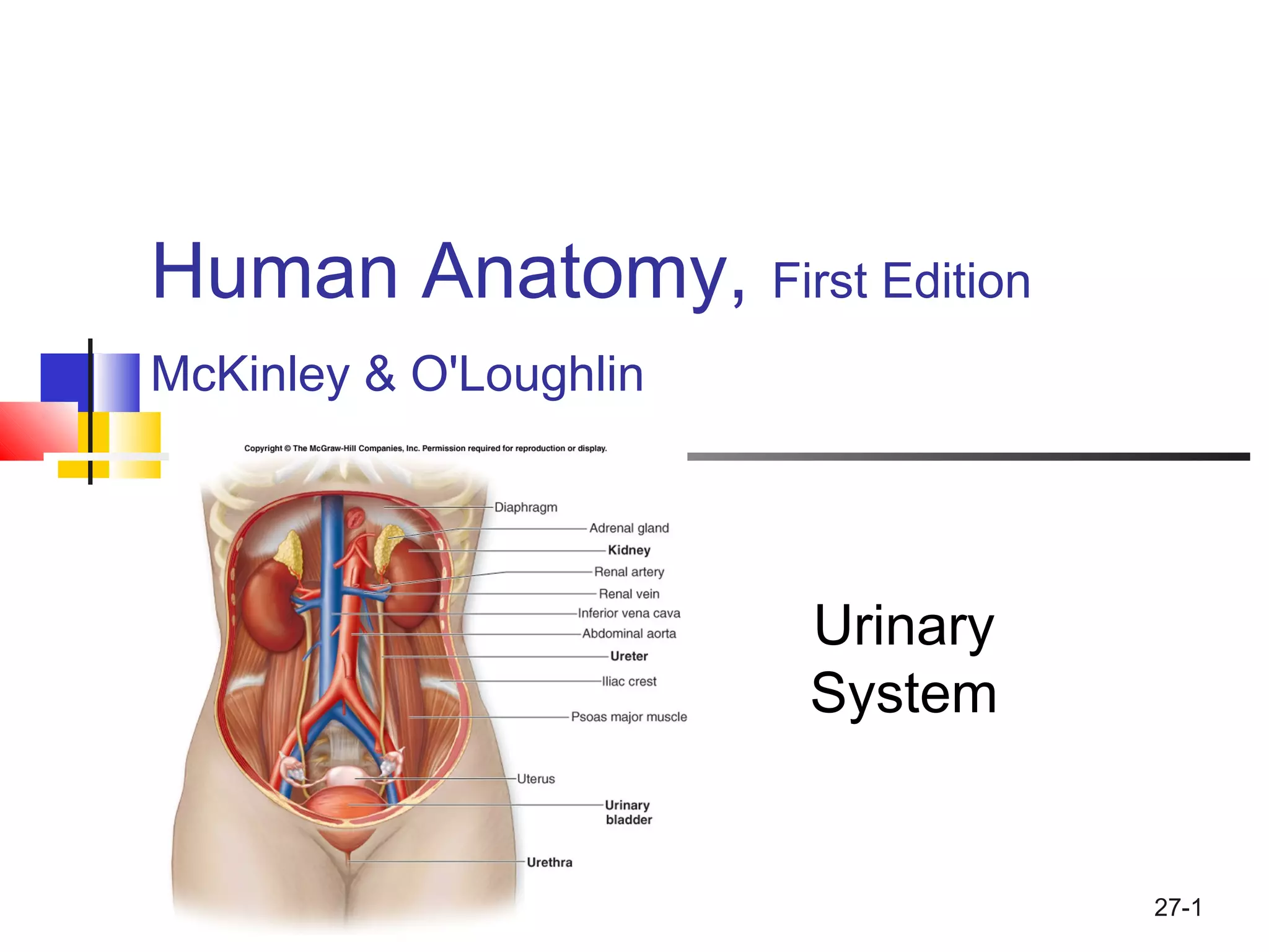
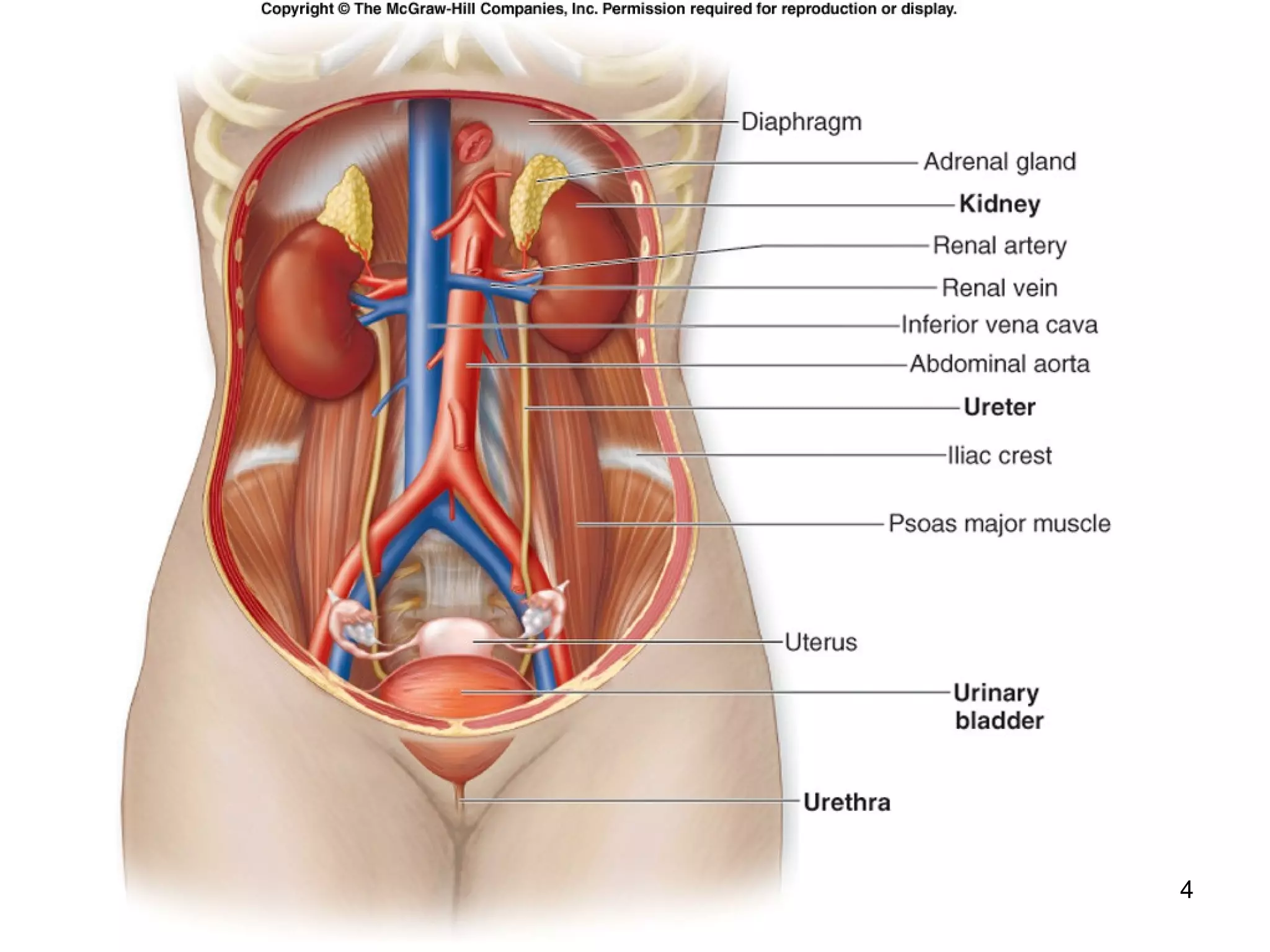
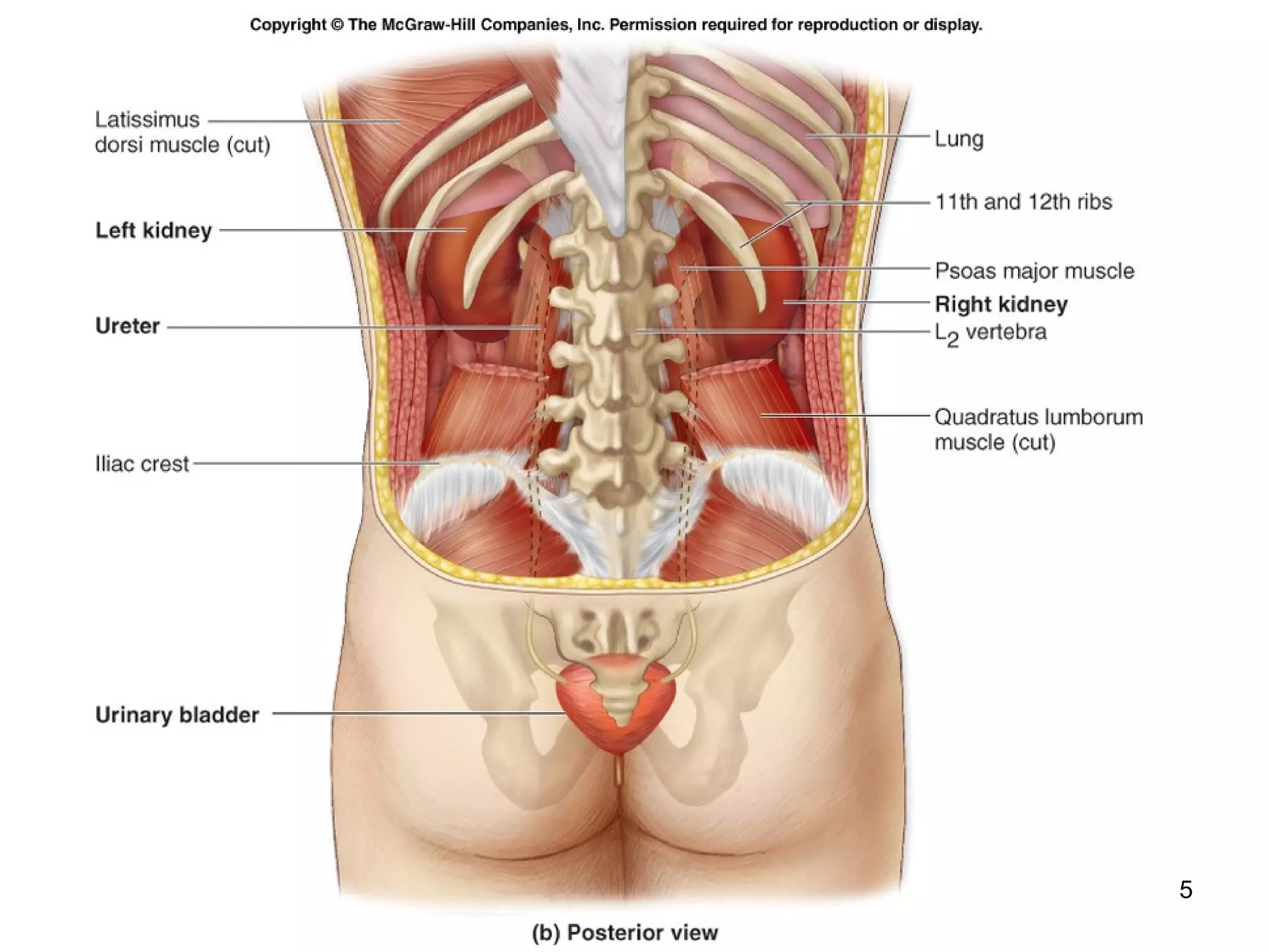
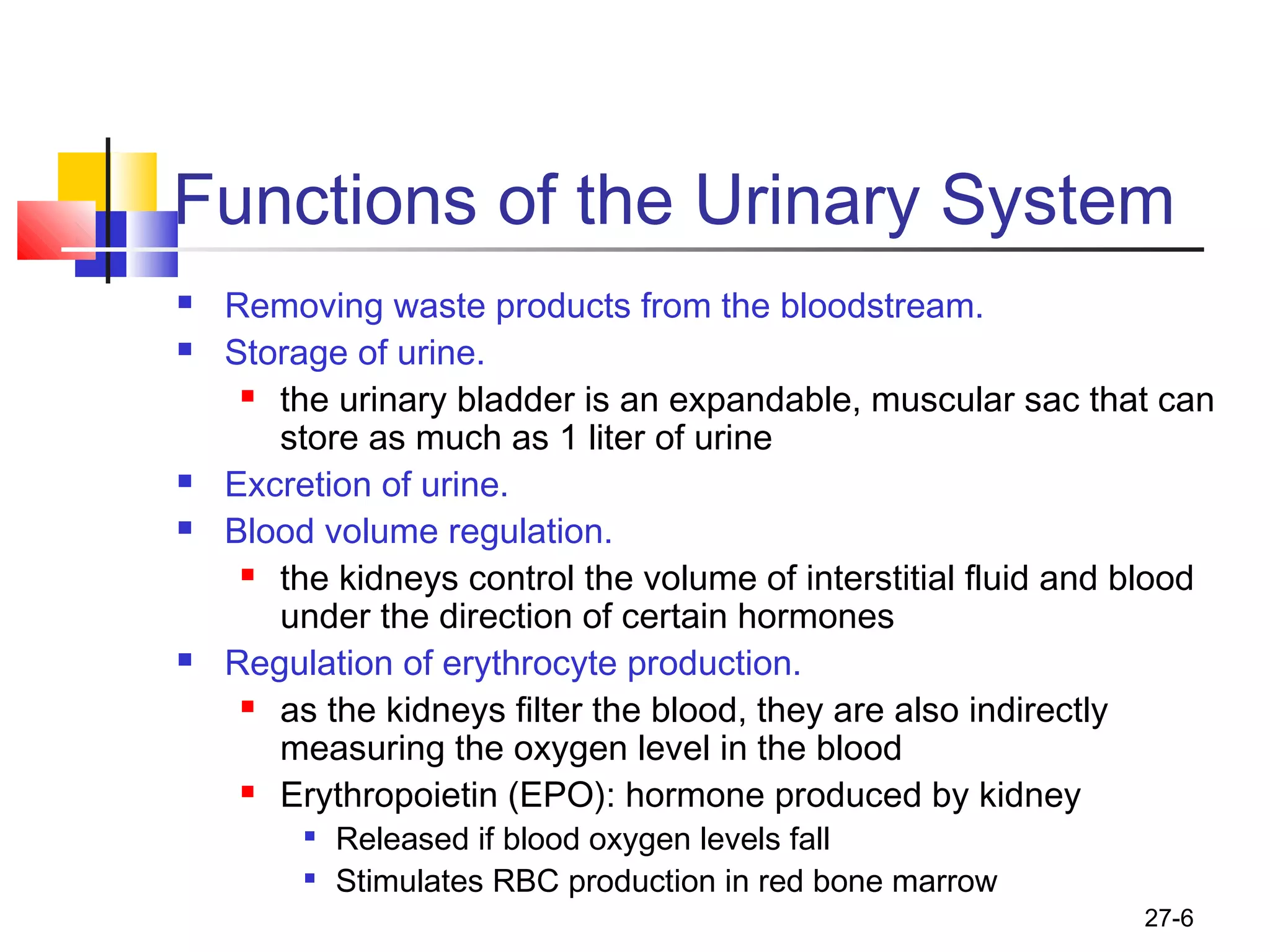
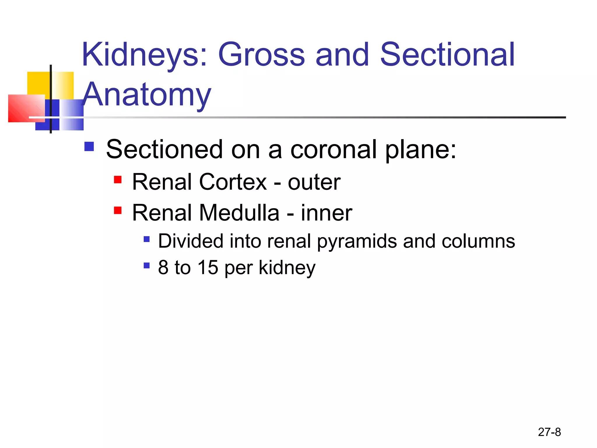
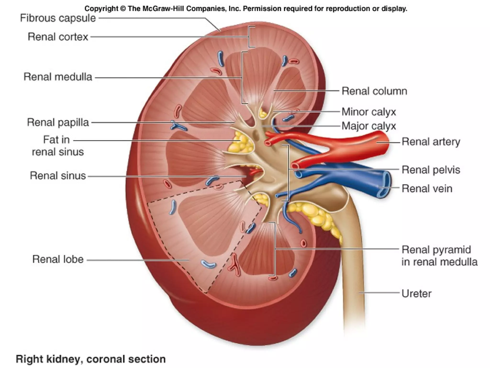
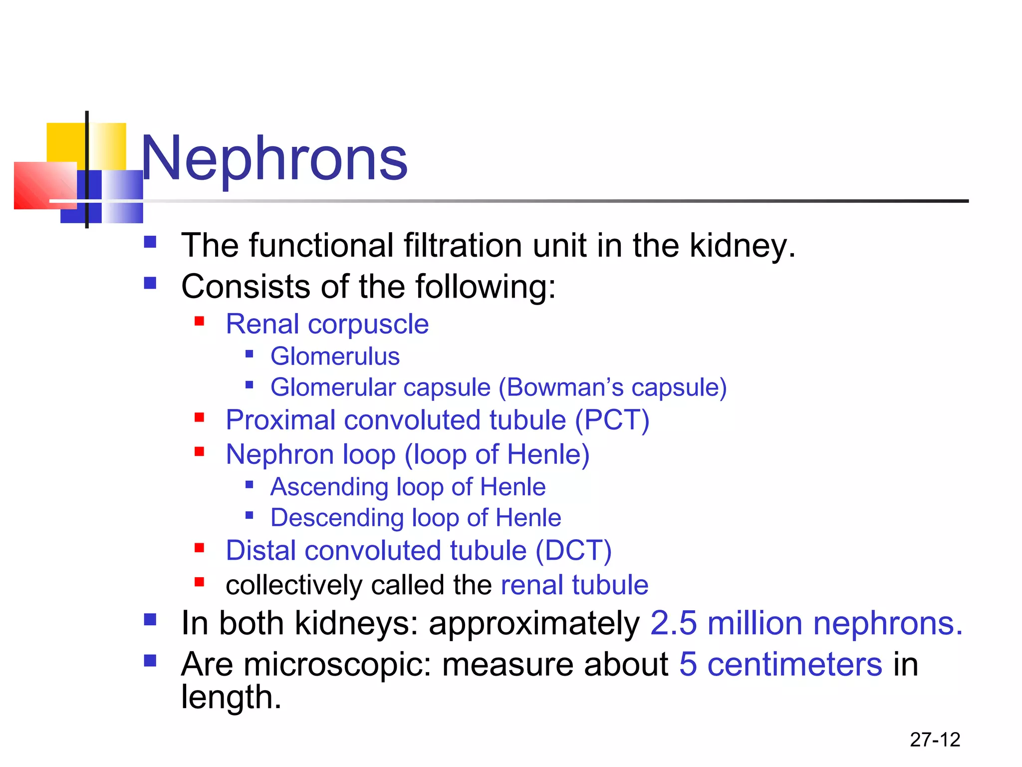
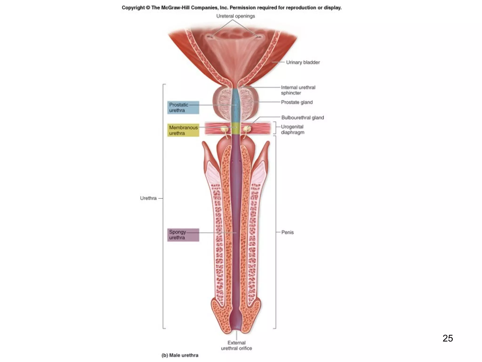
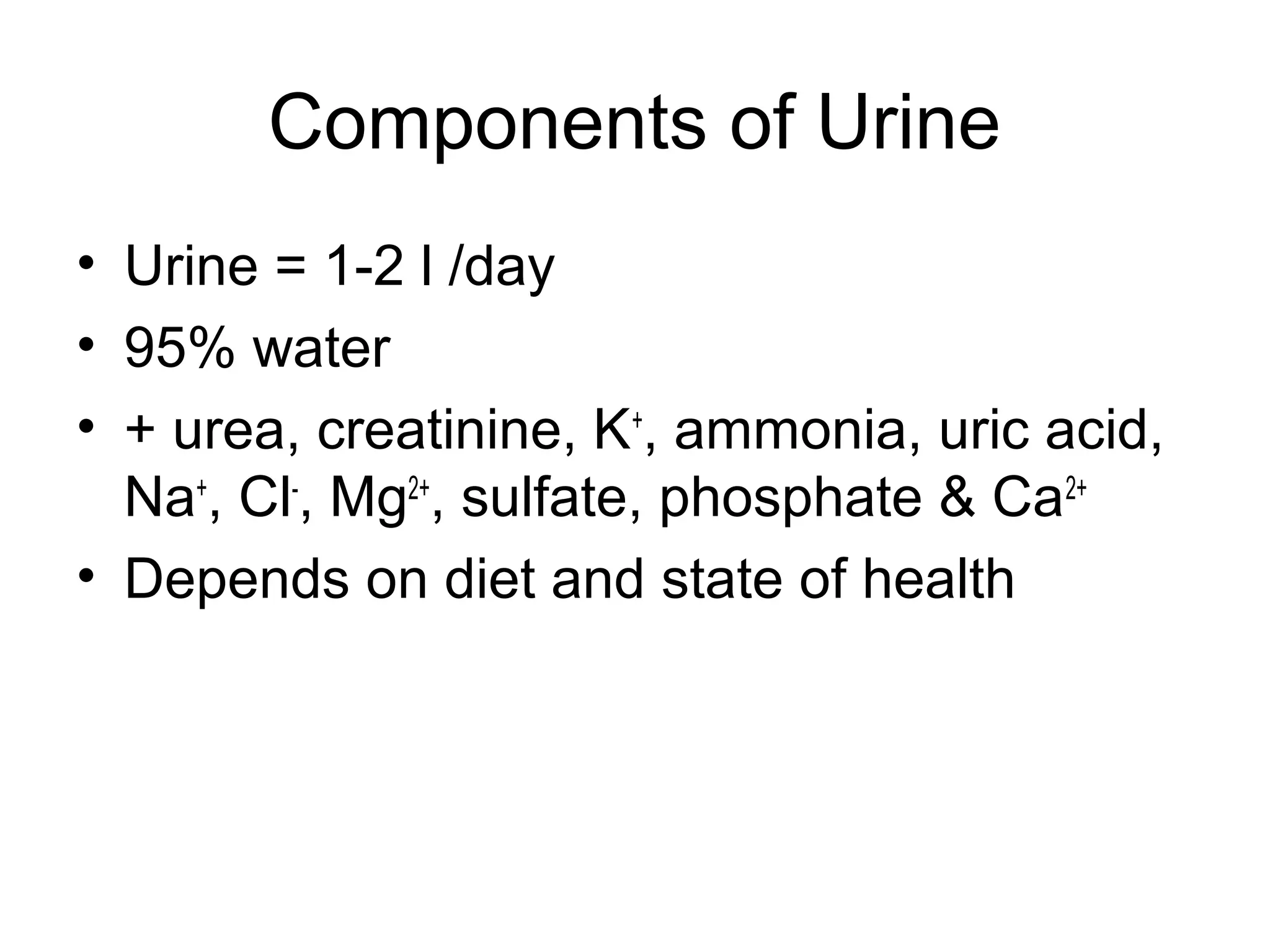
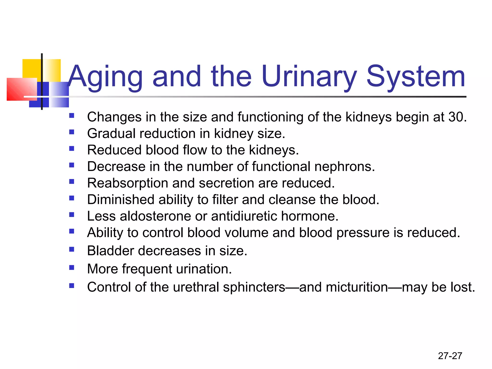
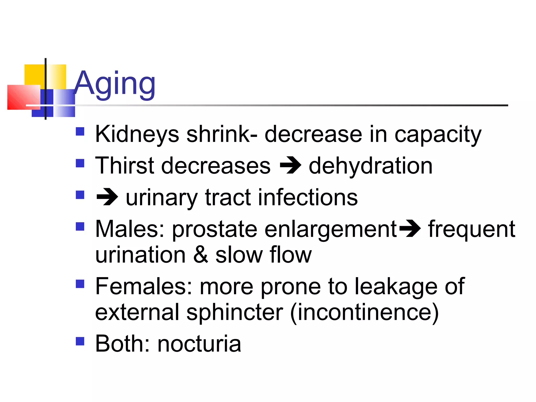
The document discusses the structure and function of the urinary system. It describes the key organs - kidneys, ureters, urinary bladder, and urethra. The kidneys filter waste from the bloodstream and produce urine, which is transported via the ureters and stored in the bladder before being excreted through the urethra. The functions of the urinary system include removing waste, regulating blood volume and pressure, and producing hormones like erythropoietin.
This was done as a Student presentation on the kidney.
Here following topics are covered.
Macroscopic structure of the urinary system
Microscopic anatomy of the urinary system
Functions of the nephron
Renal blood supply
Kidneys and blood pressure regulation
Structure of ureters and urinary bladder to perform its function
Renal failure
gross Anatomy of kidney, description of external and internal structure of kidney, the relation of right and left kidney. difference between right and left kidney, and some clinical abnormalities relate to kidney,
This is not a substitute for Books. Let it just help you understand some concepts in liver anatomy.
Continuation of this work will depend on your feedback. Stay Blessed.
The excretory system is a passive biological system that removes excess, unnecessary materials from the body fluids of an organism, so as to help maintain internal chemical homeostasis and prevent damage to the body.
Earliest Galaxies in the JADES Origins Field: Luminosity Function and Cosmic ...Sérgio Sacani
We characterize the earliest galaxy population in the JADES Origins Field (JOF), the deepest
imaging field observed with JWST. We make use of the ancillary Hubble optical images (5 filters
spanning 0.4−0.9µm) and novel JWST images with 14 filters spanning 0.8−5µm, including 7 mediumband filters, and reaching total exposure times of up to 46 hours per filter. We combine all our data
at > 2.3µm to construct an ultradeep image, reaching as deep as ≈ 31.4 AB mag in the stack and
30.3-31.0 AB mag (5σ, r = 0.1” circular aperture) in individual filters. We measure photometric
redshifts and use robust selection criteria to identify a sample of eight galaxy candidates at redshifts
z = 11.5 − 15. These objects show compact half-light radii of R1/2 ∼ 50 − 200pc, stellar masses of
M⋆ ∼ 107−108M⊙, and star-formation rates of SFR ∼ 0.1−1 M⊙ yr−1
. Our search finds no candidates
at 15 < z < 20, placing upper limits at these redshifts. We develop a forward modeling approach to
infer the properties of the evolving luminosity function without binning in redshift or luminosity that
marginalizes over the photometric redshift uncertainty of our candidate galaxies and incorporates the
impact of non-detections. We find a z = 12 luminosity function in good agreement with prior results,
and that the luminosity function normalization and UV luminosity density decline by a factor of ∼ 2.5
from z = 12 to z = 14. We discuss the possible implications of our results in the context of theoretical
models for evolution of the dark matter halo mass function.
Richard's entangled aventures in wonderlandRichard Gill
Since the loophole-free Bell experiments of 2020 and the Nobel prizes in physics of 2022, critics of Bell's work have retreated to the fortress of super-determinism. Now, super-determinism is a derogatory word - it just means "determinism". Palmer, Hance and Hossenfelder argue that quantum mechanics and determinism are not incompatible, using a sophisticated mathematical construction based on a subtle thinning of allowed states and measurements in quantum mechanics, such that what is left appears to make Bell's argument fail, without altering the empirical predictions of quantum mechanics. I think however that it is a smoke screen, and the slogan "lost in math" comes to my mind. I will discuss some other recent disproofs of Bell's theorem using the language of causality based on causal graphs. Causal thinking is also central to law and justice. I will mention surprising connections to my work on serial killer nurse cases, in particular the Dutch case of Lucia de Berk and the current UK case of Lucy Letby.
This presentation explores a brief idea about the structural and functional attributes of nucleotides, the structure and function of genetic materials along with the impact of UV rays and pH upon them.
Introduction:
RNA interference (RNAi) or Post-Transcriptional Gene Silencing (PTGS) is an important biological process for modulating eukaryotic gene expression.
It is highly conserved process of posttranscriptional gene silencing by which double stranded RNA (dsRNA) causes sequence-specific degradation of mRNA sequences.
dsRNA-induced gene silencing (RNAi) is reported in a wide range of eukaryotes ranging from worms, insects, mammals and plants.
This process mediates resistance to both endogenous parasitic and exogenous pathogenic nucleic acids, and regulates the expression of protein-coding genes.
What are small ncRNAs?
micro RNA (miRNA)
short interfering RNA (siRNA)
Properties of small non-coding RNA:
Involved in silencing mRNA transcripts.
Called “small” because they are usually only about 21-24 nucleotides long.
Synthesized by first cutting up longer precursor sequences (like the 61nt one that Lee discovered).
Silence an mRNA by base pairing with some sequence on the mRNA.
Discovery of siRNA?
The first small RNA:
In 1993 Rosalind Lee (Victor Ambros lab) was studying a non- coding gene in C. elegans, lin-4, that was involved in silencing of another gene, lin-14, at the appropriate time in the
development of the worm C. elegans.
Two small transcripts of lin-4 (22nt and 61nt) were found to be complementary to a sequence in the 3' UTR of lin-14.
Because lin-4 encoded no protein, she deduced that it must be these transcripts that are causing the silencing by RNA-RNA interactions.
Types of RNAi ( non coding RNA)
MiRNA
Length (23-25 nt)
Trans acting
Binds with target MRNA in mismatch
Translation inhibition
Si RNA
Length 21 nt.
Cis acting
Bind with target Mrna in perfect complementary sequence
Piwi-RNA
Length ; 25 to 36 nt.
Expressed in Germ Cells
Regulates trnasposomes activity
MECHANISM OF RNAI:
First the double-stranded RNA teams up with a protein complex named Dicer, which cuts the long RNA into short pieces.
Then another protein complex called RISC (RNA-induced silencing complex) discards one of the two RNA strands.
The RISC-docked, single-stranded RNA then pairs with the homologous mRNA and destroys it.
THE RISC COMPLEX:
RISC is large(>500kD) RNA multi- protein Binding complex which triggers MRNA degradation in response to MRNA
Unwinding of double stranded Si RNA by ATP independent Helicase
Active component of RISC is Ago proteins( ENDONUCLEASE) which cleave target MRNA.
DICER: endonuclease (RNase Family III)
Argonaute: Central Component of the RNA-Induced Silencing Complex (RISC)
One strand of the dsRNA produced by Dicer is retained in the RISC complex in association with Argonaute
ARGONAUTE PROTEIN :
1.PAZ(PIWI/Argonaute/ Zwille)- Recognition of target MRNA
2.PIWI (p-element induced wimpy Testis)- breaks Phosphodiester bond of mRNA.)RNAse H activity.
MiRNA:
The Double-stranded RNAs are naturally produced in eukaryotic cells during development, and they have a key role in regulating gene expression .
Nutraceutical market, scope and growth: Herbal drug technologyLokesh Patil
As consumer awareness of health and wellness rises, the nutraceutical market—which includes goods like functional meals, drinks, and dietary supplements that provide health advantages beyond basic nutrition—is growing significantly. As healthcare expenses rise, the population ages, and people want natural and preventative health solutions more and more, this industry is increasing quickly. Further driving market expansion are product formulation innovations and the use of cutting-edge technology for customized nutrition. With its worldwide reach, the nutraceutical industry is expected to keep growing and provide significant chances for research and investment in a number of categories, including vitamins, minerals, probiotics, and herbal supplements.
Slide 1: Title Slide
Extrachromosomal Inheritance
Slide 2: Introduction to Extrachromosomal Inheritance
Definition: Extrachromosomal inheritance refers to the transmission of genetic material that is not found within the nucleus.
Key Components: Involves genes located in mitochondria, chloroplasts, and plasmids.
Slide 3: Mitochondrial Inheritance
Mitochondria: Organelles responsible for energy production.
Mitochondrial DNA (mtDNA): Circular DNA molecule found in mitochondria.
Inheritance Pattern: Maternally inherited, meaning it is passed from mothers to all their offspring.
Diseases: Examples include Leber’s hereditary optic neuropathy (LHON) and mitochondrial myopathy.
Slide 4: Chloroplast Inheritance
Chloroplasts: Organelles responsible for photosynthesis in plants.
Chloroplast DNA (cpDNA): Circular DNA molecule found in chloroplasts.
Inheritance Pattern: Often maternally inherited in most plants, but can vary in some species.
Examples: Variegation in plants, where leaf color patterns are determined by chloroplast DNA.
Slide 5: Plasmid Inheritance
Plasmids: Small, circular DNA molecules found in bacteria and some eukaryotes.
Features: Can carry antibiotic resistance genes and can be transferred between cells through processes like conjugation.
Significance: Important in biotechnology for gene cloning and genetic engineering.
Slide 6: Mechanisms of Extrachromosomal Inheritance
Non-Mendelian Patterns: Do not follow Mendel’s laws of inheritance.
Cytoplasmic Segregation: During cell division, organelles like mitochondria and chloroplasts are randomly distributed to daughter cells.
Heteroplasmy: Presence of more than one type of organellar genome within a cell, leading to variation in expression.
Slide 7: Examples of Extrachromosomal Inheritance
Four O’clock Plant (Mirabilis jalapa): Shows variegated leaves due to different cpDNA in leaf cells.
Petite Mutants in Yeast: Result from mutations in mitochondrial DNA affecting respiration.
Slide 8: Importance of Extrachromosomal Inheritance
Evolution: Provides insight into the evolution of eukaryotic cells.
Medicine: Understanding mitochondrial inheritance helps in diagnosing and treating mitochondrial diseases.
Agriculture: Chloroplast inheritance can be used in plant breeding and genetic modification.
Slide 9: Recent Research and Advances
Gene Editing: Techniques like CRISPR-Cas9 are being used to edit mitochondrial and chloroplast DNA.
Therapies: Development of mitochondrial replacement therapy (MRT) for preventing mitochondrial diseases.
Slide 10: Conclusion
Summary: Extrachromosomal inheritance involves the transmission of genetic material outside the nucleus and plays a crucial role in genetics, medicine, and biotechnology.
Future Directions: Continued research and technological advancements hold promise for new treatments and applications.
Slide 11: Questions and Discussion
Invite Audience: Open the floor for any questions or further discussion on the topic.
Multi-source connectivity as the driver of solar wind variability in the heli...Sérgio Sacani
The ambient solar wind that flls the heliosphere originates from multiple
sources in the solar corona and is highly structured. It is often described
as high-speed, relatively homogeneous, plasma streams from coronal
holes and slow-speed, highly variable, streams whose source regions are
under debate. A key goal of ESA/NASA’s Solar Orbiter mission is to identify
solar wind sources and understand what drives the complexity seen in the
heliosphere. By combining magnetic feld modelling and spectroscopic
techniques with high-resolution observations and measurements, we show
that the solar wind variability detected in situ by Solar Orbiter in March
2022 is driven by spatio-temporal changes in the magnetic connectivity to
multiple sources in the solar atmosphere. The magnetic feld footpoints
connected to the spacecraft moved from the boundaries of a coronal hole
to one active region (12961) and then across to another region (12957). This
is refected in the in situ measurements, which show the transition from fast
to highly Alfvénic then to slow solar wind that is disrupted by the arrival of
a coronal mass ejection. Our results describe solar wind variability at 0.5 au
but are applicable to near-Earth observatories.
Observation of Io’s Resurfacing via Plume Deposition Using Ground-based Adapt...Sérgio Sacani
Since volcanic activity was first discovered on Io from Voyager images in 1979, changes
on Io’s surface have been monitored from both spacecraft and ground-based telescopes.
Here, we present the highest spatial resolution images of Io ever obtained from a groundbased telescope. These images, acquired by the SHARK-VIS instrument on the Large
Binocular Telescope, show evidence of a major resurfacing event on Io’s trailing hemisphere. When compared to the most recent spacecraft images, the SHARK-VIS images
show that a plume deposit from a powerful eruption at Pillan Patera has covered part
of the long-lived Pele plume deposit. Although this type of resurfacing event may be common on Io, few have been detected due to the rarity of spacecraft visits and the previously low spatial resolution available from Earth-based telescopes. The SHARK-VIS instrument ushers in a new era of high resolution imaging of Io’s surface using adaptive
optics at visible wavelengths.
2. 27-2
General Structure and Functions
of the Urinary System
General Concept:
Waste products accumulate in blood
Are toxic
Must be removed to maintain
homeostasis
Urinary System organs
remove waste products from the blood
then from the body
Major homeostatic system
3. 27-3
General Structure and Functions
of the Urinary System
Organs of the Urinary System:
Kidneys
Ureters
Urinary Bladder
Urethra
Primary organs: kidneys
filter waste products from the bloodstream
convert the filtrate into urine.
The Urinary Tract:
Includes:
ureters
urinary bladder
urethra
Because they transport the urine out of the body.
6. 27-6
Functions of the Urinary System
Removing waste products from the bloodstream.
Storage of urine.
the urinary bladder is an expandable, muscular sac that can
store as much as 1 liter of urine
Excretion of urine.
Blood volume regulation.
the kidneys control the volume of interstitial fluid and blood
under the direction of certain hormones
Regulation of erythrocyte production.
as the kidneys filter the blood, they are also indirectly
measuring the oxygen level in the blood
Erythropoietin (EPO): hormone produced by kidney
Released if blood oxygen levels fall
Stimulates RBC production in red bone marrow
7. 27-7
Kidneys: Gross and Sectional
Anatomy
Retroperitoneal
Anterior surface covered with peritoneum
Posterior surface against posterior
abdominal wall
Superior pole: T-12
Inferior pole: L-3
Right kidney ~ 2cm lower than left
Adrenal gland on superior pole
8. 27-8
Kidneys: Gross and Sectional
Anatomy
Sectioned on a coronal plane:
Renal Cortex - outer
Renal Medulla - inner
Divided into renal pyramids and columns
8 to 15 per kidney
11. 27-11
Blood Supply to the Kidney
Blood plasma is filtered across the
glomerulus into the glomerular space.
12. 27-12
Nephrons
The functional filtration unit in the kidney.
Consists of the following:
Renal corpuscle
Glomerulus
Glomerular capsule (Bowman’s capsule)
Proximal convoluted tubule (PCT)
Nephron loop (loop of Henle)
Ascending loop of Henle
Descending loop of Henle
Distal convoluted tubule (DCT)
collectively called the renal tubule
In both kidneys: approximately 2.5 million nephrons.
Are microscopic: measure about 5 centimeters in
length.
13. 27-13
Urine Formation
Three processes
Filtration
Renal corpuscle: forms filtrate
From blood to tubule
Reabsorption
Mostly PCT
Water and salt: rest of nephron
From tubule to blood
Secretion
From blood to tubule
14. 27-14
Innervation of the Kidney
innervated by a mass of autonomic nervous system
fibers
called the renal plexus.
The renal plexus
accompanies each renal artery
enters the kidney through the hilum.
15. 27-15
Urinary Tract : Ureters
long, fibromuscular tubes
conduct urine from the kidneys to the urinary bladder.
average 25 centimeters in length
retroperitoneal.
ureters originate at the renal pelvis
extend inferiorly to enter the posterolateral wall of the
base of the urinary bladder.
wall is composed of three concentric tunics.
mucosa
muscularis
adventitia.
16. 27-16
Urinary Tract – Urinary Bladder
The urinary bladder:
expandable, muscular container
serves as a reservoir for urine
positioned immediately superior and posterior to the pubic
symphysis.
in females
the urinary bladder is in contact with the uterus posterosuperiorly
and with the vagina posteroinferiorly.
in males
it is in contact with the rectum posterosuperiorly and is immediately
superior to the prostate gland.
is a retroperitoneal organ.
when empty exhibits an upside-down pyramidal shape.
Filling with urine distends it superiorly until it assumes an oval
shape.
17. 27-17
Urinary Tract – Urinary Bladder
Trigone
posteroinferior triangular area of the urinary bladder wall
formed by imaginary lines
connect the two posterior ureteral openings
and the anterior urethral opening.
The trigone remains immovable as the urinary
bladder fills and evacuates.
It functions as a funnel
directs urine into the urethra as the bladder wall contracts
four tunics
mucosa
submucosa
Muscularis: called the detrusor muscle
Adventitia.
19. 27-19
Micturition (Urination)
The expulsion of urine from the bladder.
Initiated by a complex sequence of events called the
micturition reflex.
The bladder is supplied by both parasympathetic and
sympathetic nerve fibers of the autonomic nervous
system.
20. 27-20
Urethra
Fibromuscular tube
exits the urinary bladder through the urethral opening
at anteroinferior surface
conducts urine to the exterior of the body.
Tunica mucosa: is a protective mucous membrane
houses clusters of mucin-producing cells called urethral
glands.
Tunica muscularis: primarily smooth muscle fibers
help propel urine to the outside of the body.
Two urethral sphincters:
Internal urethral sphincter
restrict the release of urine until the pressure within the urinary
bladder is high enough
External urethral sphincter
and voluntary activities needed to release the urine are
activated.
21. 27-21
Urethra
The internal urethral sphincter
involuntary (smooth muscle)
superior sphincter surrounding the neck of the bladder,
where the urethra originates.
a circular thickening of the detrusor muscle
controlled by the autonomic nervous system
The external urethral sphincter
inferior to the internal urethral sphincter
formed by skeletal muscle fibers of the urogenital
diaphragm.
a voluntary sphincter
controlled by the somatic nervous system
this is the muscle children learn to control when they
become “toilet-trained”
22. 27-22
Female Urethra
Has a single function:
to transport urine from the urinary bladder to the vestibule,
an external space immediately internal to the labia minora
3 to 5 centimeters long, and opens to the outside of
the body at the external urethral orifice located in the
female perineum.
24. 27-24
Male Urethra
Urinary and reproductive functions:
passageway for both urine and semen
Approximately 18 to 20 centimeters long.
Partitioned into three segments:
prostatic urethra is approximately 3 to 4 centimeters long and is the most
dilatable portion of the urethra
extends through the prostate gland, immediately inferior to the male
bladder, where multiple small prostatic ducts enter it
membranous urethra is the shortest and least dilatable portion
extends from the inferior surface of the prostate gland through the
urogenital diaphragm
spongy urethra is the longest part (15 centimeters)
encased within a cylinder of erectile tissue in the penis called the
corpus spongiosum
extends to the external urethral orifice
26. Components of Urine
• Urine = 1-2 l /day
• 95% water
• + urea, creatinine, K+
, ammonia, uric acid,
Na+
, Cl-
, Mg2+
, sulfate, phosphate & Ca2+
• Depends on diet and state of health
27. 27-27
Aging and the Urinary System
Changes in the size and functioning of the kidneys begin at 30.
Gradual reduction in kidney size.
Reduced blood flow to the kidneys.
Decrease in the number of functional nephrons.
Reabsorption and secretion are reduced.
Diminished ability to filter and cleanse the blood.
Less aldosterone or antidiuretic hormone.
Ability to control blood volume and blood pressure is reduced.
Bladder decreases in size.
More frequent urination.
Control of the urethral sphincters—and micturition—may be lost.
28. Aging
Kidneys shrink- decrease in capacity
Thirst decreases dehydration
urinary tract infections
Males: prostate enlargement frequent
urination & slow flow
Females: more prone to leakage of
external sphincter (incontinence)
Both: nocturia