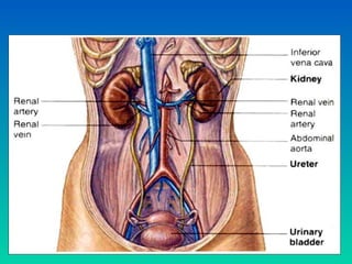
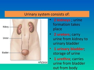
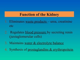
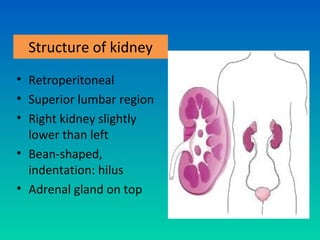
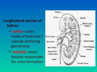
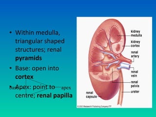
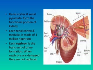
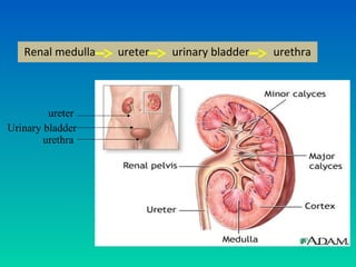
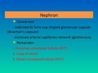
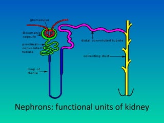
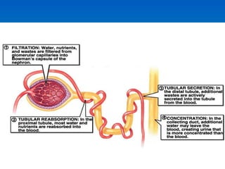
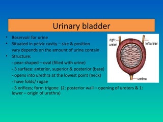
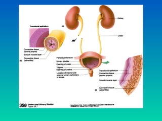
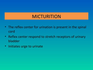
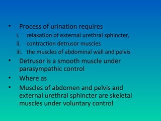
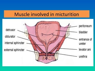
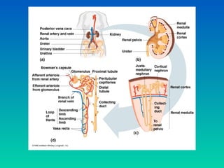
URINARY SYSTEM
    S.S.MOORTHY SEMENCHALAM
 M.Sc. Comm Health (Occ Health) UKM
         B.HSc. Nursing (Aust)
          Dip Med Sc. (Moh)
Learning objectives
At the end of the lesson, students will be able to:
1.State the main components of urinary system
2.Lists the functions of kidney
3.State the path of blood flow through the kidney
4.State the structure of nephrones
5.State and describe 3 main processes in urine formation
6.State the compositions of urine
7.State the structure and specific functions of ureter,
  urinary bladder and urethra
8.Discuss the process of micturition
Urinary system consists of:
              • 2 kidneys ; urine
                formation takes
                place
              • 2 ureters; carry
                urine from kidney to
                urinary bladder
              • 1 urinary bladder;
                storage of urine
              • 1 urethra; carries
                urine from bladder
                out from body
Function of the Kidney
1. Eliminates waste products – urea, creatinine
   etc
2. Regulates blood pressure by secreting renin
   (juxtaglomerular cells)
3. Maintains water & electrolyte balance
4. Synthesis of prostaglandins & erythropoitein
Structure of kidney
• Retroperitoneal
• Superior lumbar region
• Right kidney slightly
  lower than left
• Bean-shaped,
  indentation: hilus
• Adrenal gland on top
Three layers of supportive tissue

• Renal capsule: fibrous
  connective tissue that
  enclosed kidney
• Renal fat pad: adipose
  tissue around renal
  capsule; protects kidney
  from mechanical shock
• Renal fascia: connective
  tissue that anchor kidney
  to abdominal wall
Longitudinal section of
  kidney
    cortex- outer,
     made of bowman’s
     capsule enclosing
     glomerulus
    medulla- inner,
     tubules responsible
     for urine formation
• Within medulla,
  triangular shaped
  structures; renal
  pyramids
• Base: open into
  cortex
• Apex: point to apex
base
  centre; renal papilla
• Renal cortex & renal
  pyramids- form the
  functional portion of
  kidney
• Each renal cortex &
  medulla; is made of 1
  million nephrons
• Each nephron is the
  basic unit of urine
  formation. When
  nephrons are damaged
  they are not replaced
• Urine formed by
  nephron, drains into
  large ducts- papillary
  ducts
• Papillary ducts drain
  their contents into
  minor calyces
• Papillary ducts: 8-18
  per kidney
• Contents of minor
  calyces drain into
  major calyces
• Major calyces: 2-3 per
  kidney
• From major calyces,
  urine drains into renal
  pelvis
• From renal pelvis it
  drains into the ureters
Route taken by the urine in the kidney after its
                 formation:
                                   Nephrons

                         Papillary ducts (renal pyramid)

                                  Minor calyx

                                  Major calyx

                                  Renal pelvis

                                    Ureters
Renal medulla   ureter   urinary bladder   urethra


         ureter
Urinary bladder
        urethra
Nephron: The basic functional unit of kidney

                      • Each kidney made of
                        millions of nephron
                      • Tubule; closed at one
                        end, other end
                        opening to collecting
                        tubule
Nephron
   Closed-end
   - indented to form cup-shaped glomerular capsule
(Bowman’s capsule)
   - enclosed arterial capillaries network (glomerulus)
   Remainder:
   i. Proximal convoluted tubule (PCT)
   ii. Loop of Henle
   ii. Distal convoluted tubule (DCT)
Nephrons: functional units of kidney
Renal artery (at hilum):
• from abdominal aorta,
  enters renal sinus
• Branches to form
  afferent arterioles
• Supply oxygenated
  blood to glomerular
  capillaries of renal
  corpuscles
• Efferent arterioles arise
  from glomerular capillaries
  & carry blood away from
  glomeruli
• These gives rise to plexus
  of capillaries around PCT &
  DCT
• a.k.a peritubular capillary
  system
• Join to form renal vein
• Renal vein: drains
  deoxygenated blood into
  inferior vena cava
Afferent arteriole - brings blood to the glomerulus
Efferent arteriole - brings blood away from the glomerulus
Bowman’s capsule:
• externally layered
  with squamous
  epithelium
• Internal viscera:
  specialized epithelial
  cells (podocytes)
• Basement
  membranes: separate
  endothelial calls of
  glomerular capillaries
  & podocytes
• Capillary
  endothelium
                        *
• Basement
  membrane
• Podocytes
* Made of filtrate
  membrane; major
  role in the first step
  of urine formation
The renal corpuscle opens into:
• Proximal convulated tubule
  (PCT)
  - lined with microvilli in lumen
  - cuboidal epithelium
  - absorption & secretion
  - opens into nephron loop
• Distal convulated tubule (DCT), further end of
  the nephron
  - cuboidal epithelium with fewer microvilli
  - surrounded by smooth muscles of the space
  to form juxta glomerular apparatus
PCT, renal corpuscle & DCT: placed in outer
  kidney cortex
i.     nephron loop connect PCT & DCT
ii.    collecting tubule receives contents from DCT
iii. squamous epithelium protect the tubules against
     abrasion
iv.    cuboidal epithelium: reabsorption of water & ions in
      the process of urine formation
v.    nephron loop, collecting ducts & DCT placed in
      medulla
vi. The DCT opens into collecting tubule carrying urine
    into calyces
      •DCT < microvilli than PCT
Urine production:

In nephron
• 3 processes
   Glomerular
   Filtration
   Tubular
   Reabsorption
   Tubular Secretion
Filtration
• Occurs in Bowman’s
  capsule by filtrate or
  hydrostatic pressure
  from glomerular
  capillaries
• Due to smaller
  diameter of efferent
  arterioles than
  afferent arterioles
Filtration Cont.
• Pressure exerted by plasma & osmotic pressure in
  glomerulus (filteration pressure) -forces substances out of
  glomerulus
• The portion of the plasma entering the nephron is called
  -Filtrate
• Generally, small molecule:
  - diameter <40,000 daltons/ 7nM
  - e.g. water, sugar, ions, aminoacids, ammonia, urea,
  creatine able to pass through
• Large molecules:
  - exit into glomerulus
  - transported through blood into efferent arterioles
Tubular Reabsorption:
           • The filtrate leaves
             Bowman’s capsule &
             flow through proximal
             tubule, nephron loop
             & DCT
           • Substances needed in
             filtrate are reabsorbed
             back into blood
             - to maintain fluid &
             electrolyte, pH
           • Active transport
• These include water, important amino acids,
  nutrients, hormones etc.

  a. Water is reabsorbed by osmosis in PCT


  b. Amino acids, ions(Na+) are reabsorbed by active
  transport in the PCT

  c. limit to glucose reabsorbtion: up to 100
  mg/100ml, then all is reabsorbed (no glucose in
  urine)
d. above 150 mg/100ml glucose, then glucose
    present in urine
e. active ion reabsoption:
  sodium, potassium, calcium,
  magnesium,bicarbonate, phosphate, and sulfate ions
  actively resorbed (selective reabsorption)
f. The small volume of filterate forming a part of
urine are urea, creatine, toxic substances and K+.
Secretion

• Substances that is not required & foreign
  material (e.g. drugs); secreted into tubules to
  be excreted out from body (in urine)
• Tubular secretion: secrete H+ to maintain
  homeostasis of blood pH
• Ammonia is secreted by the epithelial cells of
  nephron and secreted into lumen of nephron by
  passive transport.
• Substances that are toxic to body include drugs,
  hydrogen ions, K+ ions are secreted into PCT, DCT
  by active transport.
• Though the filtrate that enters the proximal
  convoluted tubule is 180 lts,
• only 1% is ultimately removed as urine and
• 99% is reabsorbed along the different regions of
  the nephron
Urine composition
• Colour: clear - Light yellow (presence of
  urobilin)
• Normal volume 1 to 1.5L/day
• pH ≈ 6 (4.5-8) but mostly acidic
• Normal specific gravity- 1.003 to 1.040
Major nitrogen-containing wastes

a. Urea: most abundant organic waste product
   (21g/day), d/t breakdown of amino acids
b. Uric acid: results from breakdown of nucleic
   acids (0.5g/day)
c. Creatinine: generated in muscle tissue from
   breakdown of creatine phosphate (1.8g/day:
   amount depend on muscle mass)
d. Ammonia salt: small amount filtered into
   Bowman’s capsule
Water balance & urine output
•    Regulation of urine formation – regulates
     homeostasis of fluids in the body
•    Hormones:
    i. Antidiuretic hormone (ADH)
    ii. Aldosterone
    iii. Antinatriuretic hormone (ANH)
a. When water concentration is low……….
                        Aldosterone released

Stimulates gene expression of those proteins that involved in Na+ active
                               transport

            Na+ ion concentration in blood (K+ eliminate)

                    Water reabsorbed & conserved

                           Urine formation

               i. Maintain water levels in blood
               ii. Maintain blood pressure
b. When water concentration is high………
           Release of aldosterone is inhibited
Stopping gene expression of those proteins that involved in
                  Na+ active transport

 Na+ ion concentration in blood (reduce ion Na+ uptake)

                Water excretion increases
                    Urine formation
            i. Maintain water levels in blood
            ii. Maintain blood pressure
Ureters
• Tubes; convey urine from
  kidney to urinary bladder
• Continuous with renal
  pelvis; passes obliquely
  through the posterior
  wall of bladder
• Urine accumulates –
  pressure in bladder –
  ureters compressed –
  opening occluded
  - to prevent urine reflux
  back
Ureters: structure & function
•    3 layers of tissue:
    i. fibrous tissue – outer covering
    ii. muscular layer – middle
    iii. Mucosa – inner, transitional epithelium
•    Function: propel urine from kidney into
     bladder by peristaltic contraction of the
     smooth muscle
Urinary bladder
• Reservoir for urine
• Situated in pelvic cavity – size & position
  vary depends on the amount of urine contain
• Structure:
  - pear-shaped – oval (filled with urine)
  - 3 surface: anterior, superior & posterior (base)
  - opens into urethra at the lowest point (neck)
  - have folds/ rugae
  - 3 orifices; form trigone (2: posterior wall – opening of ureters & 1:
  lower – origin of urethra)
Urinary bladder: structure & function
•    3 layers of bladder wall
    i.   outer: loose connective tissue (blood, lymph
         vessels & nerves)
    ii. middle: smooth muscle & elastic tissue;
         dextrusor muscle
    iii. inner: mucosa, transitional epithelium
Urethra

• Urethra opens to out side by external
  sphincter made of skeletal muscles.
• ♂ - opens into penis
• ♀ - opens into vestibule in vagina.
• By parasympathetic stimulation , muscles of
  bladder expel urine.
Male urethra
• 18-20cm long
• 2 curvatures – s-
  shaped
• Extend from internal
  urethral orifice at the
  neck of bladder to
  external urethral
  orrifice at tip of penis
Female Urethra
      • Narrow membranous
        about 4cm long
      • Extend from internal
        to external urethral
        orifice directly in front
        of the vaginal opening
      • Place behind the
        symphysis pubis in the
        anterior wall of vagina
MICTURITION
• The reflex center for urination is present in the spinal
  cord
• Reflex center respond to stretch receptors of urinary
  bladder
• Initiates urge to urinate
•    Process of urination requires
    i. relaxation of external urethral sphincter,
    ii. contraction detrusor muscles
    iii. the muscles of abdominal wall and pelvis
•    Detrusor is a smooth muscle under
     parasympathic control
•    Where as
•    Muscles of abdomen and pelvis and
     external urethral sphincter are skeletal
     muscles under voluntary control
Muscle involved in micturition
Thank you….

Questions please!!

12. urinary system

  • 1.
    URINARY SYSTEM S.S.MOORTHY SEMENCHALAM M.Sc. Comm Health (Occ Health) UKM B.HSc. Nursing (Aust) Dip Med Sc. (Moh)
  • 2.
    Learning objectives At theend of the lesson, students will be able to: 1.State the main components of urinary system 2.Lists the functions of kidney 3.State the path of blood flow through the kidney 4.State the structure of nephrones 5.State and describe 3 main processes in urine formation 6.State the compositions of urine 7.State the structure and specific functions of ureter, urinary bladder and urethra 8.Discuss the process of micturition
  • 4.
    Urinary system consistsof: • 2 kidneys ; urine formation takes place • 2 ureters; carry urine from kidney to urinary bladder • 1 urinary bladder; storage of urine • 1 urethra; carries urine from bladder out from body
  • 5.
    Function of theKidney 1. Eliminates waste products – urea, creatinine etc 2. Regulates blood pressure by secreting renin (juxtaglomerular cells) 3. Maintains water & electrolyte balance 4. Synthesis of prostaglandins & erythropoitein
  • 6.
    Structure of kidney •Retroperitoneal • Superior lumbar region • Right kidney slightly lower than left • Bean-shaped, indentation: hilus • Adrenal gland on top
  • 7.
    Three layers ofsupportive tissue • Renal capsule: fibrous connective tissue that enclosed kidney • Renal fat pad: adipose tissue around renal capsule; protects kidney from mechanical shock • Renal fascia: connective tissue that anchor kidney to abdominal wall
  • 8.
    Longitudinal section of kidney  cortex- outer, made of bowman’s capsule enclosing glomerulus  medulla- inner, tubules responsible for urine formation
  • 9.
    • Within medulla, triangular shaped structures; renal pyramids • Base: open into cortex • Apex: point to apex base centre; renal papilla
  • 10.
    • Renal cortex& renal pyramids- form the functional portion of kidney • Each renal cortex & medulla; is made of 1 million nephrons • Each nephron is the basic unit of urine formation. When nephrons are damaged they are not replaced
  • 11.
    • Urine formedby nephron, drains into large ducts- papillary ducts • Papillary ducts drain their contents into minor calyces • Papillary ducts: 8-18 per kidney
  • 12.
    • Contents ofminor calyces drain into major calyces • Major calyces: 2-3 per kidney • From major calyces, urine drains into renal pelvis • From renal pelvis it drains into the ureters
  • 13.
    Route taken bythe urine in the kidney after its formation: Nephrons Papillary ducts (renal pyramid) Minor calyx Major calyx Renal pelvis Ureters
  • 14.
    Renal medulla ureter urinary bladder urethra ureter Urinary bladder urethra
  • 15.
    Nephron: The basicfunctional unit of kidney • Each kidney made of millions of nephron • Tubule; closed at one end, other end opening to collecting tubule
  • 16.
    Nephron Closed-end - indented to form cup-shaped glomerular capsule (Bowman’s capsule) - enclosed arterial capillaries network (glomerulus) Remainder: i. Proximal convoluted tubule (PCT) ii. Loop of Henle ii. Distal convoluted tubule (DCT)
  • 17.
  • 18.
    Renal artery (athilum): • from abdominal aorta, enters renal sinus • Branches to form afferent arterioles • Supply oxygenated blood to glomerular capillaries of renal corpuscles
  • 19.
    • Efferent arteriolesarise from glomerular capillaries & carry blood away from glomeruli • These gives rise to plexus of capillaries around PCT & DCT • a.k.a peritubular capillary system • Join to form renal vein • Renal vein: drains deoxygenated blood into inferior vena cava
  • 20.
    Afferent arteriole -brings blood to the glomerulus Efferent arteriole - brings blood away from the glomerulus
  • 21.
    Bowman’s capsule: • externallylayered with squamous epithelium • Internal viscera: specialized epithelial cells (podocytes) • Basement membranes: separate endothelial calls of glomerular capillaries & podocytes
  • 22.
    • Capillary endothelium * • Basement membrane • Podocytes * Made of filtrate membrane; major role in the first step of urine formation
  • 23.
    The renal corpuscleopens into: • Proximal convulated tubule (PCT) - lined with microvilli in lumen - cuboidal epithelium - absorption & secretion - opens into nephron loop
  • 24.
    • Distal convulatedtubule (DCT), further end of the nephron - cuboidal epithelium with fewer microvilli - surrounded by smooth muscles of the space to form juxta glomerular apparatus PCT, renal corpuscle & DCT: placed in outer kidney cortex
  • 26.
    i. nephron loop connect PCT & DCT ii. collecting tubule receives contents from DCT iii. squamous epithelium protect the tubules against abrasion iv. cuboidal epithelium: reabsorption of water & ions in the process of urine formation v. nephron loop, collecting ducts & DCT placed in medulla vi. The DCT opens into collecting tubule carrying urine into calyces •DCT < microvilli than PCT
  • 27.
    Urine production: In nephron •3 processes  Glomerular Filtration  Tubular Reabsorption  Tubular Secretion
  • 28.
    Filtration • Occurs inBowman’s capsule by filtrate or hydrostatic pressure from glomerular capillaries • Due to smaller diameter of efferent arterioles than afferent arterioles
  • 29.
    Filtration Cont. • Pressureexerted by plasma & osmotic pressure in glomerulus (filteration pressure) -forces substances out of glomerulus • The portion of the plasma entering the nephron is called -Filtrate • Generally, small molecule: - diameter <40,000 daltons/ 7nM - e.g. water, sugar, ions, aminoacids, ammonia, urea, creatine able to pass through • Large molecules: - exit into glomerulus - transported through blood into efferent arterioles
  • 30.
    Tubular Reabsorption: • The filtrate leaves Bowman’s capsule & flow through proximal tubule, nephron loop & DCT • Substances needed in filtrate are reabsorbed back into blood - to maintain fluid & electrolyte, pH • Active transport
  • 31.
    • These includewater, important amino acids, nutrients, hormones etc. a. Water is reabsorbed by osmosis in PCT b. Amino acids, ions(Na+) are reabsorbed by active transport in the PCT c. limit to glucose reabsorbtion: up to 100 mg/100ml, then all is reabsorbed (no glucose in urine)
  • 32.
    d. above 150mg/100ml glucose, then glucose present in urine e. active ion reabsoption: sodium, potassium, calcium, magnesium,bicarbonate, phosphate, and sulfate ions actively resorbed (selective reabsorption) f. The small volume of filterate forming a part of urine are urea, creatine, toxic substances and K+.
  • 33.
    Secretion • Substances thatis not required & foreign material (e.g. drugs); secreted into tubules to be excreted out from body (in urine) • Tubular secretion: secrete H+ to maintain homeostasis of blood pH
  • 34.
    • Ammonia issecreted by the epithelial cells of nephron and secreted into lumen of nephron by passive transport. • Substances that are toxic to body include drugs, hydrogen ions, K+ ions are secreted into PCT, DCT by active transport. • Though the filtrate that enters the proximal convoluted tubule is 180 lts, • only 1% is ultimately removed as urine and • 99% is reabsorbed along the different regions of the nephron
  • 36.
    Urine composition • Colour:clear - Light yellow (presence of urobilin) • Normal volume 1 to 1.5L/day • pH ≈ 6 (4.5-8) but mostly acidic • Normal specific gravity- 1.003 to 1.040
  • 37.
    Major nitrogen-containing wastes a.Urea: most abundant organic waste product (21g/day), d/t breakdown of amino acids b. Uric acid: results from breakdown of nucleic acids (0.5g/day) c. Creatinine: generated in muscle tissue from breakdown of creatine phosphate (1.8g/day: amount depend on muscle mass) d. Ammonia salt: small amount filtered into Bowman’s capsule
  • 38.
    Water balance &urine output • Regulation of urine formation – regulates homeostasis of fluids in the body • Hormones: i. Antidiuretic hormone (ADH) ii. Aldosterone iii. Antinatriuretic hormone (ANH)
  • 39.
    a. When waterconcentration is low………. Aldosterone released Stimulates gene expression of those proteins that involved in Na+ active transport Na+ ion concentration in blood (K+ eliminate) Water reabsorbed & conserved Urine formation i. Maintain water levels in blood ii. Maintain blood pressure
  • 40.
    b. When waterconcentration is high……… Release of aldosterone is inhibited Stopping gene expression of those proteins that involved in Na+ active transport Na+ ion concentration in blood (reduce ion Na+ uptake) Water excretion increases Urine formation i. Maintain water levels in blood ii. Maintain blood pressure
  • 41.
    Ureters • Tubes; conveyurine from kidney to urinary bladder • Continuous with renal pelvis; passes obliquely through the posterior wall of bladder • Urine accumulates – pressure in bladder – ureters compressed – opening occluded - to prevent urine reflux back
  • 42.
    Ureters: structure &function • 3 layers of tissue: i. fibrous tissue – outer covering ii. muscular layer – middle iii. Mucosa – inner, transitional epithelium • Function: propel urine from kidney into bladder by peristaltic contraction of the smooth muscle
  • 43.
    Urinary bladder • Reservoirfor urine • Situated in pelvic cavity – size & position vary depends on the amount of urine contain • Structure: - pear-shaped – oval (filled with urine) - 3 surface: anterior, superior & posterior (base) - opens into urethra at the lowest point (neck) - have folds/ rugae - 3 orifices; form trigone (2: posterior wall – opening of ureters & 1: lower – origin of urethra)
  • 44.
    Urinary bladder: structure& function • 3 layers of bladder wall i. outer: loose connective tissue (blood, lymph vessels & nerves) ii. middle: smooth muscle & elastic tissue; dextrusor muscle iii. inner: mucosa, transitional epithelium
  • 46.
    Urethra • Urethra opensto out side by external sphincter made of skeletal muscles. • ♂ - opens into penis • ♀ - opens into vestibule in vagina. • By parasympathetic stimulation , muscles of bladder expel urine.
  • 47.
    Male urethra • 18-20cmlong • 2 curvatures – s- shaped • Extend from internal urethral orifice at the neck of bladder to external urethral orrifice at tip of penis
  • 48.
    Female Urethra • Narrow membranous about 4cm long • Extend from internal to external urethral orifice directly in front of the vaginal opening • Place behind the symphysis pubis in the anterior wall of vagina
  • 49.
    MICTURITION • The reflexcenter for urination is present in the spinal cord • Reflex center respond to stretch receptors of urinary bladder • Initiates urge to urinate
  • 50.
    Process of urination requires i. relaxation of external urethral sphincter, ii. contraction detrusor muscles iii. the muscles of abdominal wall and pelvis • Detrusor is a smooth muscle under parasympathic control • Where as • Muscles of abdomen and pelvis and external urethral sphincter are skeletal muscles under voluntary control
  • 51.
    Muscle involved inmicturition
  • 53.