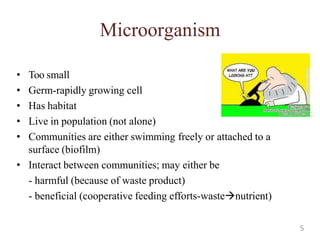
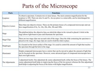
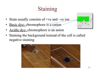
1
Microorganism & Microbiology
Microorganism
• Living things which individually are
too small to be seen with the naked
eye.
• All of the following may be
considered microorganisms:
– bacteria
(eubacteria, archaebacteria)
– fungi (yeasts, molds)
– protozoa
– microscopic algae
– viruses
– various parasitic worms
Microbiology
• Study of microorganisms
• Foundation of modern biotechnology
• Among the many specialized fields of
microbiology
-Virology, Mycology, Bacteriology, Immunology,
Microbial Ecology, Biotechnological
Microbiology, Environmental Microbiology,
Food Microbiology, Forensic Microbiology,
Molecular Biology
2
Microorganism & Microbiology cont’d
Two main themes involved in Microbiology
1- Basic- cellular processes
2-Applied- concerning agriculture, industry and
health
3
Themes in Microbiology and its field
Microbiology
Basic
By organism By process Disease related
Applied
Disease related
Environmentally
related Industrial
Bacteriology
Phycology
Mycology
Virology
Parasitology
Protozoalogy
Microbial metabolism
Microbial genetics
Immunology
Epidemiology
Etiology
Infection
control
Chemotherapy
Environmental
microbiology
Food &
beverage tech
Pharmaceutical
microbiology
Genetic
Engineering
4
Microorganism
• Too small
• Germ-rapidly growing cell
• Has habitat
• Live in population (not alone)
• Communities are either swimming freely or attached to a
surface (biofilm)
• Interact between communities; may either be
- harmful (because of waste product)
- beneficial (cooperative feeding efforts-wastenutrient)
5
Microbes in our lives
• Some are pathogenic (disease-causing)
• Decompose organic waste
• Produces through photosynthesis (e.g.Purple sulphur bacteria
must fix CO2 to live)
• Play role in industry (e.g. fermentation to produce ethanol and
acetone)
• Produce fermented food (vinegar, cheese & bread)
• Produce products used in manufacturing (cellulase) and
treatment (insulin)
6
Microbes in our lives
Food
• Lactococci
• Ferment milk
Water treatment
Such as certain bacteria belonging to the Bacillus species
1.Digest a wide variety of organic material that are present in wastes.
2.Digest waste quickly and completely, without producing significant odors of
noxious gas.
3.Non-pathogenic.
Energy
• Produce methane
Science- laboratories
Warfare -anthrax
7
Microbes and agriculture
• Nitrogen fixation
• Rumen microbes help digest grass and hay in cows,
sheep etc
• Cycles nutrients (C, N and S)
• Causes disease to animals and plants 8
Microorganism and Food
• Microorganism and food
1) Prevent spoilage (tempeh, salted fish)
2) Assist in manufacturing of food
• Microorganisms and energy
1) Natural gas (methane)
2) Ethanol (biofuel)
3) Bioremediation
• Microbes and the future
1)Genetic engineering
9
Microbes and diseases
10
So, how can microbes benefit us?
• In food?
• In environment?
• In preventing disease?
• In agriculture?
• In energy?
• In waste-water treatment?
11
Naming and Classifying
microorganisms
• Linnaeus system for scientific nomenclature
• Each organism has two names:
1) Genus
2) Specific epithet
12
Scientific Names
• Italicized or underlined.
The genus is capitalized, and the specific epithet is with lowercase
• Could be as an honor for the scientist
• ALatin origin
e.g. Escherichia coli (E. coli)
- discoverer: Theodor Escherich
- describes the habitat (colon/intestine)
e.g. Staphylococcus aureus (S. aureus)
- Clustered (staphylo), spherical (cocci)
- Gold colored colonies (aureus)
In intestine
On skin
13
Classification of bacteria
Domain
Bacteria Archaea Eukarya
Plants, animals, Fungi,
Protists
Microorganisms
Also include
fungi, protozoa, algae, viruses, multicellu 14
Bacteria (P)/ Bacterium (S)
• Prokaryotes
• Has peptidoglycan cell walls
• Binary fission
• Utilize organic/inorganic
chemicals, or photosynthesis
to obtain energy
Archaea
• Prokayotic
• Lack peptidoglycan
• Live in extreme
environments
• Include
- Methanogens
- Extreme halophiles
- Extreme thermophiles
15
Types of Eukaryotes
Protozoa
• Unicellular eukaryote
• Absorb or ingest organic
chemicals
• May move using
pseudopods, cilia or flagella
• e.g.Amoeba
Algae
• Unicellular/multicellular
eukaryote
• Has cellulose cell walls
• Gain energy through
photosynthesis
• Produce molecular and
organic compounds
16
Fungi (singular: Fungus)
• Eukaryotes
• Chitin cell walls
• Use organic chemicals for
energy
• Molds and mushrooms are
multicellular, consists of
mycelia (composed of filaments
called hyphae)
• Yeasts are unicellular
17
Viruses
• Too small to be observed with light
microscope
• Consists of DNA/RNAcore
• Core is surrounded by protein coat
• Coat may be enclosed in a lipid
envelope
• Viruses are replicated only when
they are in living host cell
• Bacteriophage-viruses that infect
bacteria
• Viroids-nucleic acid without protein
coating
• Prions- Infectious protenacious
particles
Multicellular animal parasites
• Helminths: flatworms and
roundworms
• Multicellular
18
How do we view microorganisms?
• Units of measurement
When talking about cells and microscopic organisms, you would be
measuring using MICROMETRE (abbreviated: µ --micron ) or stated
as: µm (micrometer).
1 µm = 1 x 10⁻⁶ meters/ 1 x 10⁻³ mm
1 mm= 1 x 10³ nanometers/ 1 x 103 µm
To give you the idea of how small a micro metre is,
1- a human hair is about 100 µm, wide,
2 a red blood cell would be around 8 µm wide
3 typical size of an animal cell would be from 10 - 100 µm
19
Microscope
Light microscope
• Uses light
• Few types
Compound light microscopy
Darkfield microscopy
Phase-contrast microscopy
Differential interference contrast microscopy
Fluorescence microscopy
Confocal microscopy
20
Compound light microscope
•The image is magnified again by ocular lens
Total magnification= objective lens x ocular lens
•Resolution- ability of lenses to distinguish two points
e.g. RP of 0.4 nm can distinguish between 2 points ≥ 0.4 nm
•Shorter light wavelength provides greater resolution
•Refractive index- Light bending ability of a medium
•Light may bend in air sthat it misses the small high-magnification
lens
•Immersion oil is used to keep the air from bending.
21
Light Microscope - found in most schools, use compound lenses and light
to magnify objects. The lenses bend or refract the light, which makes the
object beneath them appear closer.
Stereoscope - this microscope allows for binocular (two eyes) viewing of
larger specimens. (The spinning microscope at the top of this page is a
stereoscope)
Scanning Electron Microscope - allow scientists to view a universe too
small to be seen with a light microscope. SEMs do not use light waves;
they use electrons (negatively charged electrical particles) to magnify
objects up to two million times.
Transmission Electron Microscope - also uses electrons, but instead of
scanning the surface (as with SEM's) electrons are passed through very
thin specimens. Specimens may be stained with heavy metal salts
Types of Microscopes
22
Parts of the Microscope
Parts Functions
Eyepiece
To observe specimen. Contains two or more lenses. The most common magnification for the
eyepiece is 10X. There are also 2x and 5x. An eye piece is a removable, can be interchanged for
different magnification.
Objective Lenses
More than one objective lenses. These are the primary lenses of a compound microscope and can
have magnification of 4x, 5x, 10x, 20x, 40x, 50x and 100x.
Stage
The platform below the objective lens on which the object to be viewed is placed. A hole in the
stage allows light beam to pass and illuminate the specimen.
Stage Clips
There are two stage clips one on each side of the stage. Once the slide containing the specimen is
placed on the stage, the stage clips are used to hold the slide in place.
Diaphragm
It is located on the lower surface of the stage. It is used to control the amount of light that reaches
the specimen through the hole in the stage.
Illuminator
Simple compound microscopes have a mirror that can be moved to adjust the amount of light that
can be focused on the specimen. However, some advanced types of compound microscopes have
their own light source.
The Adjustments
2 adjustment knobs: fine adjustment & coarse adjustment knob; refine the focus of the lenses. The
coarse adjustment knob helps in improving the focus of the low powers whereas the fine adjustment
knob helps in adjusting the focus of the lenses with higher magnification.
23
Total magnification = magnification of eyepiece x magnification of objective lens
Magnification
Your microscope has 3 magnifications:
Scanning, Low and High. Each objective will have written the
magnification. In addition to this, the ocular lens (eyepiece) has a
magnification.
The total magnification is the ocular x objective.
Magnification Ocular lens
Total
Magnification
Scanning 4x 10x 40x
Low Power 10x 10x 100x
High Power 40x 10x 400x
24
Development of microscopy
•(384-322)Aristotle and others believed that living organisms could develop from
non-living materials.
•1590: Hans and Zacharias Janssen (Dutch lens grinders) mounted two lenses in a
tube to produce the first compound microscope.
•1660: Robert Hooke (1635-1703) published "Micrographia“; drawings and
detailed observations of biological materials made with the best compound
microscope and illumination system of the time.
•1676:Anton van Leeuwenhoek (1632-1723) 1st person to observe
microorganisms.
•1883: Carl Zeiss and ErnstAbbe pioneered developments in microscopy (such as
immersion lenses and apochromatic lenses which reduce chromatic aberration)
exist until the present day.
•1931: Ernst Ruska constructed the 1st electron microscope.
A Brief History of Microbiology
25
Van Leeuwenhoek’s description of Bacteria
From his teeth, he observed
(A)& (B)- rod forms
(C) & (D)- motion pathway
(E)- Spherical form
(F)- Longer type of spherical form
(H)- Cluster
-Royal Society letter (Sept 17th, 1683)
The microscope used
Simple microscope (one lens)
26
1688: Francesco Redi (1626-1678) was an
Italian physician who refuted the idea of
spontaneous generation by showing that rotting
meat carefully kept from flies will not
spontaneously produce maggots.
1836: Theodor Schwann (1810-1882) helped develop the cell
theory of living organisms, namely that that all living organisms are
composed of one or more cells and that the cell is the basic
functional unit of living organisms.
1861: Louis Pasteur's (1822-1895) famous experiments with swan-
necked flasks finally proved that microorganisms do not arise by
spontaneous generation.
Spontaneous generation controversy
27
The Golden Age of Microbiology
~1857-1914 (about 50 years)
Beginning with Pasteur’s work, discoveries included relationship between
microbes and disease, immunity, and antimicrobial drugs
Robert Koch a.
b.
Identified a bacterium as cause of anthrax
Introduced agar, inoculating loop to transfer bacteria
and prepare pure cultures.
Introduced “Koch’s Postulates” and the concept that
a disease is caused by a single organism.
c.
Joseph Lister (1865)
a.
b.
Martinus Beijerinck
a.
b.
Introduced the “antiseptic technique”.
Use of phenol (carbolic acid) as disinfectant.
(1884 - 85)
Discovered “viruses” (toxins, poisons).
Infectious agents in tobacco plant fluids.
Paul Ehrlich (1910)
a.
b.
Alexander Fleming
a.
Introduced concept of chemotherapy.
Use of salvarsan for the treatment of syphilis.
(1928)
Discovered the first antibiotic - penicillin. 28
This eventually led to:
•Development of sterilization
•Development of aseptic technique
Louis Pasteur's (1822-1895) famous
experiments with swan-necked flasks
29
1546: Hieronymus Fracastorius (Girolamo Fracastoro) wrote "On Contagion", the 1st known
discussion of the phenomenon of contagious infection.
1835: Agostino Bassi de Lodi showed that a disease affecting silkworms was caused by a
fungus - the first microorganism to be recognized as a contagious agent of animal disease.
1847: Ignaz Semmelweiss (1818-1865), a Hungarian physician- decided that doctors in
Vienna hospitals were spreading childbed fever while delivering babies. He started forcing
doctors under his supervision to wash their hands before touching patients.
1857: Louis Pasteur proposed the “Germ theory of disease”.
- Ancients believed that disease was the result of a divine punishment. Pasteur fought
to convince surgeons that germs existed and carried diseases, and dirty instruments and
hands spread germs and therefore disease. Pasteur's pasteurization process killed germs and
prevented the spread of disease.
1867: Joseph Lister (1827-1912) introduced antiseptics in surgery. By spraying carbolic acid
on surgical instruments, wounds and dressings, he reduced surgical mortality due to bacterial
infection considerably.
1876: Robert Koch (1843-1910). German bacteriologist was the first to cultivate anthrax
bacteria outside the body using blood serum at body temperature.
Proof that microbes cause disease
30
"Koch's postulates" (1884), the critical test for the involvement of
a microorganism in a disease:
1.The agent must be present in every case of the disease.
2.The agent must be isolated and cultured in vitro.
3.The disease must be reproduced when a pure culture of the
agent is inoculated into a susceptible host.
4.The agent must be recoverable from the experimentally-infected
host.
This eventually led to:
•Development of pure culture techniques
•Stains, agar, culture media, petri dishes
Koch's postulates
Robert Koch demonstrated
the first direct role of a
bacterium in disease
31
Preparing smears for staining
• Staining- coloring microbe with a dye to emphasize
certain structure
• Smear- A thin film of a microbe solution on a slide, a
smear is usually fixed to attach microbes to the slide
and kill microbes
32
Staining
• Stain usually consists of +ve and –ve ion
• Basic dye- chromophore is a cation
• Acidic dye- chromophore is an anion
• Staining the background instead of the cell is called
negative staining
33
Staining
Simple stain
- staining with one dye
-mordant may be used to
hold the stain or to coat the
specimen to enlarge it
Gram stain
Distinguish
• Gram +ve and gram -ve
• Gram +ve bacteria are prone to
penicillin and detergents
• Gram –ve are more resistant to
antibiotics
Differential stain
Distinguish
• Gram stain
• Acid-fast stain
Acid-fast stain
Stained waxy cell wall is not
decolorized by acid-alcohol
• Mycobacterium
• Nocardia
34
Distinguish special parts of
cells
• Capsule
• Endospore (Malachite
green and safranin)
• Flagella (carbolfuchsin
simple stain)
• Gram +ve and gram -ve
• Gram +ve bacteria are prone
to penicillin and detergents
• Gram –ve are more resistant
to antibiotics
Gram stain
Distinguish
Special stain
Gram –ve (pink) 35

Microbial world

  • 1.
  • 2.
    Microorganism & Microbiology Microorganism •Living things which individually are too small to be seen with the naked eye. • All of the following may be considered microorganisms: – bacteria (eubacteria, archaebacteria) – fungi (yeasts, molds) – protozoa – microscopic algae – viruses – various parasitic worms Microbiology • Study of microorganisms • Foundation of modern biotechnology • Among the many specialized fields of microbiology -Virology, Mycology, Bacteriology, Immunology, Microbial Ecology, Biotechnological Microbiology, Environmental Microbiology, Food Microbiology, Forensic Microbiology, Molecular Biology 2
  • 3.
    Microorganism & Microbiologycont’d Two main themes involved in Microbiology 1- Basic- cellular processes 2-Applied- concerning agriculture, industry and health 3
  • 4.
    Themes in Microbiologyand its field Microbiology Basic By organism By process Disease related Applied Disease related Environmentally related Industrial Bacteriology Phycology Mycology Virology Parasitology Protozoalogy Microbial metabolism Microbial genetics Immunology Epidemiology Etiology Infection control Chemotherapy Environmental microbiology Food & beverage tech Pharmaceutical microbiology Genetic Engineering 4
  • 5.
    Microorganism • Too small •Germ-rapidly growing cell • Has habitat • Live in population (not alone) • Communities are either swimming freely or attached to a surface (biofilm) • Interact between communities; may either be - harmful (because of waste product) - beneficial (cooperative feeding efforts-wastenutrient) 5
  • 6.
    Microbes in ourlives • Some are pathogenic (disease-causing) • Decompose organic waste • Produces through photosynthesis (e.g.Purple sulphur bacteria must fix CO2 to live) • Play role in industry (e.g. fermentation to produce ethanol and acetone) • Produce fermented food (vinegar, cheese & bread) • Produce products used in manufacturing (cellulase) and treatment (insulin) 6
  • 7.
    Microbes in ourlives Food • Lactococci • Ferment milk Water treatment Such as certain bacteria belonging to the Bacillus species 1.Digest a wide variety of organic material that are present in wastes. 2.Digest waste quickly and completely, without producing significant odors of noxious gas. 3.Non-pathogenic. Energy • Produce methane Science- laboratories Warfare -anthrax 7
  • 8.
    Microbes and agriculture •Nitrogen fixation • Rumen microbes help digest grass and hay in cows, sheep etc • Cycles nutrients (C, N and S) • Causes disease to animals and plants 8
  • 9.
    Microorganism and Food •Microorganism and food 1) Prevent spoilage (tempeh, salted fish) 2) Assist in manufacturing of food • Microorganisms and energy 1) Natural gas (methane) 2) Ethanol (biofuel) 3) Bioremediation • Microbes and the future 1)Genetic engineering 9
  • 10.
  • 11.
    So, how canmicrobes benefit us? • In food? • In environment? • In preventing disease? • In agriculture? • In energy? • In waste-water treatment? 11
  • 12.
    Naming and Classifying microorganisms •Linnaeus system for scientific nomenclature • Each organism has two names: 1) Genus 2) Specific epithet 12
  • 13.
    Scientific Names • Italicizedor underlined. The genus is capitalized, and the specific epithet is with lowercase • Could be as an honor for the scientist • ALatin origin e.g. Escherichia coli (E. coli) - discoverer: Theodor Escherich - describes the habitat (colon/intestine) e.g. Staphylococcus aureus (S. aureus) - Clustered (staphylo), spherical (cocci) - Gold colored colonies (aureus) In intestine On skin 13
  • 14.
    Classification of bacteria Domain BacteriaArchaea Eukarya Plants, animals, Fungi, Protists Microorganisms Also include fungi, protozoa, algae, viruses, multicellu 14
  • 15.
    Bacteria (P)/ Bacterium(S) • Prokaryotes • Has peptidoglycan cell walls • Binary fission • Utilize organic/inorganic chemicals, or photosynthesis to obtain energy Archaea • Prokayotic • Lack peptidoglycan • Live in extreme environments • Include - Methanogens - Extreme halophiles - Extreme thermophiles 15
  • 16.
    Types of Eukaryotes Protozoa •Unicellular eukaryote • Absorb or ingest organic chemicals • May move using pseudopods, cilia or flagella • e.g.Amoeba Algae • Unicellular/multicellular eukaryote • Has cellulose cell walls • Gain energy through photosynthesis • Produce molecular and organic compounds 16
  • 17.
    Fungi (singular: Fungus) •Eukaryotes • Chitin cell walls • Use organic chemicals for energy • Molds and mushrooms are multicellular, consists of mycelia (composed of filaments called hyphae) • Yeasts are unicellular 17
  • 18.
    Viruses • Too smallto be observed with light microscope • Consists of DNA/RNAcore • Core is surrounded by protein coat • Coat may be enclosed in a lipid envelope • Viruses are replicated only when they are in living host cell • Bacteriophage-viruses that infect bacteria • Viroids-nucleic acid without protein coating • Prions- Infectious protenacious particles Multicellular animal parasites • Helminths: flatworms and roundworms • Multicellular 18
  • 19.
    How do weview microorganisms? • Units of measurement When talking about cells and microscopic organisms, you would be measuring using MICROMETRE (abbreviated: µ --micron ) or stated as: µm (micrometer). 1 µm = 1 x 10⁻⁶ meters/ 1 x 10⁻³ mm 1 mm= 1 x 10³ nanometers/ 1 x 103 µm To give you the idea of how small a micro metre is, 1- a human hair is about 100 µm, wide, 2 a red blood cell would be around 8 µm wide 3 typical size of an animal cell would be from 10 - 100 µm 19
  • 20.
    Microscope Light microscope • Useslight • Few types Compound light microscopy Darkfield microscopy Phase-contrast microscopy Differential interference contrast microscopy Fluorescence microscopy Confocal microscopy 20
  • 21.
    Compound light microscope •Theimage is magnified again by ocular lens Total magnification= objective lens x ocular lens •Resolution- ability of lenses to distinguish two points e.g. RP of 0.4 nm can distinguish between 2 points ≥ 0.4 nm •Shorter light wavelength provides greater resolution •Refractive index- Light bending ability of a medium •Light may bend in air sthat it misses the small high-magnification lens •Immersion oil is used to keep the air from bending. 21
  • 22.
    Light Microscope -found in most schools, use compound lenses and light to magnify objects. The lenses bend or refract the light, which makes the object beneath them appear closer. Stereoscope - this microscope allows for binocular (two eyes) viewing of larger specimens. (The spinning microscope at the top of this page is a stereoscope) Scanning Electron Microscope - allow scientists to view a universe too small to be seen with a light microscope. SEMs do not use light waves; they use electrons (negatively charged electrical particles) to magnify objects up to two million times. Transmission Electron Microscope - also uses electrons, but instead of scanning the surface (as with SEM's) electrons are passed through very thin specimens. Specimens may be stained with heavy metal salts Types of Microscopes 22
  • 23.
    Parts of theMicroscope Parts Functions Eyepiece To observe specimen. Contains two or more lenses. The most common magnification for the eyepiece is 10X. There are also 2x and 5x. An eye piece is a removable, can be interchanged for different magnification. Objective Lenses More than one objective lenses. These are the primary lenses of a compound microscope and can have magnification of 4x, 5x, 10x, 20x, 40x, 50x and 100x. Stage The platform below the objective lens on which the object to be viewed is placed. A hole in the stage allows light beam to pass and illuminate the specimen. Stage Clips There are two stage clips one on each side of the stage. Once the slide containing the specimen is placed on the stage, the stage clips are used to hold the slide in place. Diaphragm It is located on the lower surface of the stage. It is used to control the amount of light that reaches the specimen through the hole in the stage. Illuminator Simple compound microscopes have a mirror that can be moved to adjust the amount of light that can be focused on the specimen. However, some advanced types of compound microscopes have their own light source. The Adjustments 2 adjustment knobs: fine adjustment & coarse adjustment knob; refine the focus of the lenses. The coarse adjustment knob helps in improving the focus of the low powers whereas the fine adjustment knob helps in adjusting the focus of the lenses with higher magnification. 23
  • 24.
    Total magnification =magnification of eyepiece x magnification of objective lens Magnification Your microscope has 3 magnifications: Scanning, Low and High. Each objective will have written the magnification. In addition to this, the ocular lens (eyepiece) has a magnification. The total magnification is the ocular x objective. Magnification Ocular lens Total Magnification Scanning 4x 10x 40x Low Power 10x 10x 100x High Power 40x 10x 400x 24
  • 25.
    Development of microscopy •(384-322)Aristotleand others believed that living organisms could develop from non-living materials. •1590: Hans and Zacharias Janssen (Dutch lens grinders) mounted two lenses in a tube to produce the first compound microscope. •1660: Robert Hooke (1635-1703) published "Micrographia“; drawings and detailed observations of biological materials made with the best compound microscope and illumination system of the time. •1676:Anton van Leeuwenhoek (1632-1723) 1st person to observe microorganisms. •1883: Carl Zeiss and ErnstAbbe pioneered developments in microscopy (such as immersion lenses and apochromatic lenses which reduce chromatic aberration) exist until the present day. •1931: Ernst Ruska constructed the 1st electron microscope. A Brief History of Microbiology 25
  • 26.
    Van Leeuwenhoek’s descriptionof Bacteria From his teeth, he observed (A)& (B)- rod forms (C) & (D)- motion pathway (E)- Spherical form (F)- Longer type of spherical form (H)- Cluster -Royal Society letter (Sept 17th, 1683) The microscope used Simple microscope (one lens) 26
  • 27.
    1688: Francesco Redi(1626-1678) was an Italian physician who refuted the idea of spontaneous generation by showing that rotting meat carefully kept from flies will not spontaneously produce maggots. 1836: Theodor Schwann (1810-1882) helped develop the cell theory of living organisms, namely that that all living organisms are composed of one or more cells and that the cell is the basic functional unit of living organisms. 1861: Louis Pasteur's (1822-1895) famous experiments with swan- necked flasks finally proved that microorganisms do not arise by spontaneous generation. Spontaneous generation controversy 27
  • 28.
    The Golden Ageof Microbiology ~1857-1914 (about 50 years) Beginning with Pasteur’s work, discoveries included relationship between microbes and disease, immunity, and antimicrobial drugs Robert Koch a. b. Identified a bacterium as cause of anthrax Introduced agar, inoculating loop to transfer bacteria and prepare pure cultures. Introduced “Koch’s Postulates” and the concept that a disease is caused by a single organism. c. Joseph Lister (1865) a. b. Martinus Beijerinck a. b. Introduced the “antiseptic technique”. Use of phenol (carbolic acid) as disinfectant. (1884 - 85) Discovered “viruses” (toxins, poisons). Infectious agents in tobacco plant fluids. Paul Ehrlich (1910) a. b. Alexander Fleming a. Introduced concept of chemotherapy. Use of salvarsan for the treatment of syphilis. (1928) Discovered the first antibiotic - penicillin. 28
  • 29.
    This eventually ledto: •Development of sterilization •Development of aseptic technique Louis Pasteur's (1822-1895) famous experiments with swan-necked flasks 29
  • 30.
    1546: Hieronymus Fracastorius(Girolamo Fracastoro) wrote "On Contagion", the 1st known discussion of the phenomenon of contagious infection. 1835: Agostino Bassi de Lodi showed that a disease affecting silkworms was caused by a fungus - the first microorganism to be recognized as a contagious agent of animal disease. 1847: Ignaz Semmelweiss (1818-1865), a Hungarian physician- decided that doctors in Vienna hospitals were spreading childbed fever while delivering babies. He started forcing doctors under his supervision to wash their hands before touching patients. 1857: Louis Pasteur proposed the “Germ theory of disease”. - Ancients believed that disease was the result of a divine punishment. Pasteur fought to convince surgeons that germs existed and carried diseases, and dirty instruments and hands spread germs and therefore disease. Pasteur's pasteurization process killed germs and prevented the spread of disease. 1867: Joseph Lister (1827-1912) introduced antiseptics in surgery. By spraying carbolic acid on surgical instruments, wounds and dressings, he reduced surgical mortality due to bacterial infection considerably. 1876: Robert Koch (1843-1910). German bacteriologist was the first to cultivate anthrax bacteria outside the body using blood serum at body temperature. Proof that microbes cause disease 30
  • 31.
    "Koch's postulates" (1884),the critical test for the involvement of a microorganism in a disease: 1.The agent must be present in every case of the disease. 2.The agent must be isolated and cultured in vitro. 3.The disease must be reproduced when a pure culture of the agent is inoculated into a susceptible host. 4.The agent must be recoverable from the experimentally-infected host. This eventually led to: •Development of pure culture techniques •Stains, agar, culture media, petri dishes Koch's postulates Robert Koch demonstrated the first direct role of a bacterium in disease 31
  • 32.
    Preparing smears forstaining • Staining- coloring microbe with a dye to emphasize certain structure • Smear- A thin film of a microbe solution on a slide, a smear is usually fixed to attach microbes to the slide and kill microbes 32
  • 33.
    Staining • Stain usuallyconsists of +ve and –ve ion • Basic dye- chromophore is a cation • Acidic dye- chromophore is an anion • Staining the background instead of the cell is called negative staining 33
  • 34.
    Staining Simple stain - stainingwith one dye -mordant may be used to hold the stain or to coat the specimen to enlarge it Gram stain Distinguish • Gram +ve and gram -ve • Gram +ve bacteria are prone to penicillin and detergents • Gram –ve are more resistant to antibiotics Differential stain Distinguish • Gram stain • Acid-fast stain Acid-fast stain Stained waxy cell wall is not decolorized by acid-alcohol • Mycobacterium • Nocardia 34
  • 35.
    Distinguish special partsof cells • Capsule • Endospore (Malachite green and safranin) • Flagella (carbolfuchsin simple stain) • Gram +ve and gram -ve • Gram +ve bacteria are prone to penicillin and detergents • Gram –ve are more resistant to antibiotics Gram stain Distinguish Special stain Gram –ve (pink) 35