Downloaded 109 times

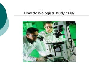
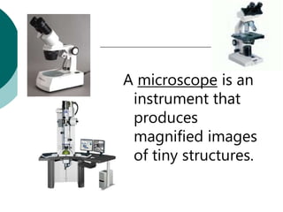
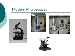
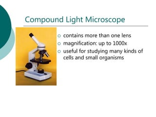
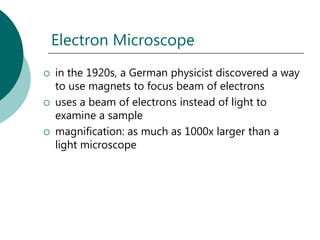
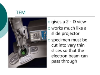
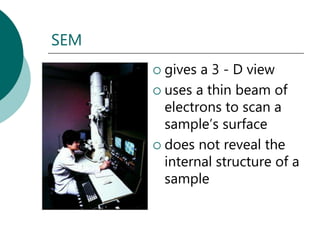
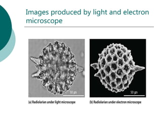
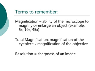
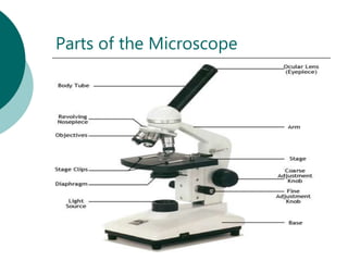


The document discusses different types of microscopes, including compound light microscopes which can magnify up to 1000x and electron microscopes which use beams of electrons instead of light to examine samples at magnifications over 1000x. It describes the key parts of microscopes like the objectives, eyepiece, stage, and mechanical components used to focus and move the microscope. The document also provides information on how biologists use microscopes to study cells and small organisms.