Downloaded 288 times


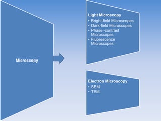


The document summarizes the history and types of microscopes. It describes how Antony van Leeuwenhoek invented the first microscope in the 17th century and how modern microscopes work. There are two main types of microscopes - light microscopes, which use visible light and lenses, and electron microscopes, which use electron beams for higher resolution. Light microscopes can be further divided into brightfield, darkfield, phase contrast, and fluorescence microscopes. Electron microscopes include SEM and TEM. The document also discusses resolution, staining, and other techniques used in microscopy.


