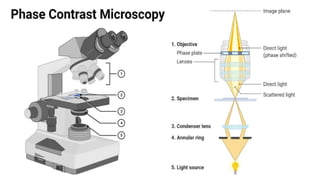
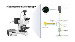
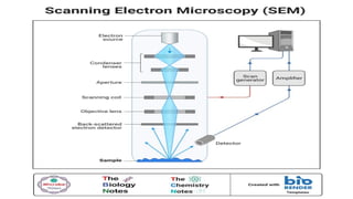
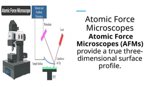
Chapter II:
Viewing the
Microbial
World
REGIE L. MAGALLANES, LPT, M.ED
Microbiology and Parasitology -
Instructor
At the end of the unit, the student
should be able to:
1. State the metric units used to express the
sizes of bacteria, protozoa and viruses.
2. Compare and contrast the various types of
microscopes.
Microbes are so tiny. But how small
are they? Most of the time, some
microscopes are required to view
them; thus, microbes are considered
to be microscopic. We will learn the
various types of microscopes in this
lesson.
The metric system units of length
will also be discussed because
they are used to express the sizes
of microbes and the resolving
power of microscopes.
At the end of the unit, the student
should be able to:
1. State the metric units used to
express the sizes of bacteria,
protozoa and viruses.
2. Compare and contrast the
various types of microscopes.
USING THE METRIC
SYSTEM TO EXPRESS
THE SIZES OF
MICROBES
In microbiology,
metric units
(primarily
micrometers and
nanometer) are
used to express
the sizes of
microbes.
A meter can be divided into 10 (101
)
equally spaced units called
decimeters; or 100 (102
) equally
spaced units called centimeters; 1,000
(103
) equally spaced units called
millimeters, or 1 million (106
) equally
spaced units called micrometers; or 1
billion (109
) equally spaced units called
It should be noted that the old terms
micron (μ) and millimicron (mμ) have
been replaced by the terms
micrometer (μm) and nanometer (nm),
respectively. An angstrom (Å) is 0.1 nm.
Using this scale,
human red blood
cells are
approximately
about 7 μm in
diameter.
The sizes of bacteria
and protozoa are
usually expressed in
terms of micrometers.
For example, a typical
spherical bacterium
(coccus; pl., cocci) is
approximately 1 μm in
diameter.
A typical rod-shaped
bacterium (bacillus; pl.,
bacilli) is about 1 μm wide
3 3 μm long, although
some bacilli are shorter,
and some form very long
filaments. The sizes of
viruses are expressed in
terms of nanometers.
Most of the viruses that cause human
disease range in size from about 10 to
300 nm, although some (e.g., Ebola
virus, a cause of hemorrhagic fever)
can be as long as 1,000 nm (1 μm).
Some very large protozoa reach a
length of 2,000 μm (2 mm).
In the microbiology
laboratory, the sizes
of cellular microbes
are measured using
an ocular
micrometer, a tiny
ruler within the
eyepiece (ocular) of
the compound light
microscope.
Before it can be
utilized to measure
objects, however,
the ocular
micrometer must
first be calibrated,
using a measuring
device called a
stage micrometer.
Calibration must be done for each of
the objective lenses to determine the
distance between the marks on the
ocular micrometer.
The ocular micrometer can then be
used to measure lengths and widths of
microbes and other objects on the
specimen slide
MICROSCOPES
A microscope is
an optical
instrument that
is used to
observe tiny
organisms that
cannot be seen
with the unaided
Each optical instrument has a limit
as to what can be seen using that
instrument.
This limit is referred to as the
resolving power or resolution of
the instrument.
TYPES
OF
MICROSCOPES
Simple Microscopes
A simple microscope contains only
one magnifying lens.
Simple Microscopes
During the late 1600s, Anton van
Leeuwenhoek, used simple
microscopes to observe many tiny
objects, including bacteria and
protozoa. His simple microscopes had
a maximum magnifying power of
about x100 (300 times).
Compound Microscopes
A compound microscope
contains more than one
magnifying lens which usually
magnifies objects about 1,000
times.
Compound
Microscopes
Photographs taken
through the lens
system of compound
microscopes are
called
photomicrographs.
Compound Microscopes
It is the wavelength of visible light
(approximately 0.45 μm) that
limits the size of objects that can
be
seen using the compound light
microscope.
Compound Microscopes
When using the light microscope,
objects cannot be seen if they are
smaller than about 0.225 μm.
Compound
Microscopes
The compound light
microscopes usually
contain two magnifying
lens systems. Within the
eyepiece is the ocular lens
which has a magnifying
power of x10.
Compound Microscopes
The other magnifying lens system
is found in the objective which is
situated immediately above the
object to be viewed.
Compound Microscopes
The four objectives used in most
laboratory compound light
microscopes are X4, X10, X40, and
X100 objectives.
Compound Microscopes
The higher the magnification, the
more light that is needed. Hence,
as magnification is increased, the
amount of light striking the
specimen to be studied must also
be increased.
Compound Microscopes
Image clarity depends on the
microscope’s resolving power or
resolution which is the ability of
the
lens system to distinguish
between two adjacent objects.
Compound
Microscopes
When objects are
observed against a
bright background,
that microscope is
referred to as the
brightfield
microscope.
Compound
Microscopes
In the laboratory,
darkfield
microscopy is
used to diagnose
syphilis where the
causative agent is
the spiral-shaped
bacterium,
Treponema
Compound Microscopes
Phase-contrast microscopes can be used to
view unstained living microorganisms.
Because the
light refracted by living cells is different from
the light refracted by the surrounding
medium, contrast is
therefore increased, and the specimens are
clearly viewed.
Compound Microscopes
Fluorescence microscopes,
however, contain a built-in
ultraviolet light source which emit
longer
wavelength light, causing them to
glow against dark background.
Electron Microscopes
Electron microscopes utilize an electron beam as
a source of illumination and magnets to focus the
beam. Because the wavelength of electrons
traveling in a vacuum is much shorter than the
wavelength of visible light, electron microscopes
have a much greater resolving power than
compound microscopes.
Electron Microscopes
There are two types of electron
microscopes: transmission electron
microscopes (TEMs) and
scanning electron microscopes (SEMs).
Electron Microscopes
A transmission electron microscope
(TEM) has a tall column at the top of which
an electron gun
fires a beam of electrons downward.
Viruses can be observed using a TEM. A
TEM reveals the
internal structure of cells.
Electron Microscopes
A scanning electron microscope (SEM), on the
other hand, has a shorter column, where the
specimen is placed at the bottom of the column.
Electrons that bounce off the surface of the specimen
are captured by detectors, and an image of the
specimen appears on a monitor. Hence, SEMs are
used to observe the outer surfaces of specimens.
Atomic Force
Microscopes
Atomic Force
Microscopes (AFMs)
provide a true three-
dimensional surface
profile.
Atomic Force Microscopes
AFMs have silicon or silicon nitride having
a sharp tip or probe at its end which is
used to scan the specimen surface.
Atomic Force Microscopes
When the tip is brought in
proximity to a sample surface,
forces between the tip and the
sample lead to a deflection of the
cantilever.
Atomic Force Microscopes
The deflection is measured using
a laser spot reflected from the top
surface of the cantilever into an
array of photodiodes creating an
image on a monitor screen.
Let’s
Recap!
A compound microscope contains
more than one magnifying lens
which usually magnifies objects
about 1,000 times.
When objects are observed
against a bright background, that
microscope is referred to as the
brightfield microscope.
Phase-contrast microscopes can be
used to view unstained living
microorganisms. Because the light
refracted by living cells is different
from the light refracted by the
surrounding medium, contrast is
therefore increased, and the
specimens are clearly viewed.
Fluorescence microscopes,
however, contain a built-in
ultraviolet light source which emit
longer
wavelength light, causing them to
glow against dark background.
A transmission electron microscope
(TEM) has a tall column at the top of
which an electron gun fires
a beam of electrons downward.
Viruses can be observed using a
TEM.
An scanning electron microscope
(SEM), on the other hand, has a
shorter column, where the
specimen is placed at the bottom
of the column. SEMs are used to
observe the outer surfaces of
specimens.
Atomic Force Microscopes (AFMs)
provide a true three-dimensional
surface profile. AFMs have
silicon or silicon nitride having a
sharp tip or probe at its end which
is used to scan the specimen
surface.
Chapter II Viewing the Microbial World.pptx

Chapter II Viewing the Microbial World.pptx

  • 1.
    Chapter II: Viewing the Microbial World REGIEL. MAGALLANES, LPT, M.ED Microbiology and Parasitology - Instructor
  • 2.
    At the endof the unit, the student should be able to: 1. State the metric units used to express the sizes of bacteria, protozoa and viruses. 2. Compare and contrast the various types of microscopes.
  • 3.
    Microbes are sotiny. But how small are they? Most of the time, some microscopes are required to view them; thus, microbes are considered to be microscopic. We will learn the various types of microscopes in this lesson.
  • 4.
    The metric systemunits of length will also be discussed because they are used to express the sizes of microbes and the resolving power of microscopes.
  • 5.
    At the endof the unit, the student should be able to: 1. State the metric units used to express the sizes of bacteria, protozoa and viruses. 2. Compare and contrast the various types of microscopes.
  • 6.
    USING THE METRIC SYSTEMTO EXPRESS THE SIZES OF MICROBES
  • 7.
    In microbiology, metric units (primarily micrometersand nanometer) are used to express the sizes of microbes.
  • 8.
    A meter canbe divided into 10 (101 ) equally spaced units called decimeters; or 100 (102 ) equally spaced units called centimeters; 1,000 (103 ) equally spaced units called millimeters, or 1 million (106 ) equally spaced units called micrometers; or 1 billion (109 ) equally spaced units called
  • 9.
    It should benoted that the old terms micron (μ) and millimicron (mμ) have been replaced by the terms micrometer (μm) and nanometer (nm), respectively. An angstrom (Å) is 0.1 nm.
  • 10.
    Using this scale, humanred blood cells are approximately about 7 μm in diameter.
  • 11.
    The sizes ofbacteria and protozoa are usually expressed in terms of micrometers. For example, a typical spherical bacterium (coccus; pl., cocci) is approximately 1 μm in diameter.
  • 12.
    A typical rod-shaped bacterium(bacillus; pl., bacilli) is about 1 μm wide 3 3 μm long, although some bacilli are shorter, and some form very long filaments. The sizes of viruses are expressed in terms of nanometers.
  • 13.
    Most of theviruses that cause human disease range in size from about 10 to 300 nm, although some (e.g., Ebola virus, a cause of hemorrhagic fever) can be as long as 1,000 nm (1 μm). Some very large protozoa reach a length of 2,000 μm (2 mm).
  • 14.
    In the microbiology laboratory,the sizes of cellular microbes are measured using an ocular micrometer, a tiny ruler within the eyepiece (ocular) of the compound light microscope.
  • 15.
    Before it canbe utilized to measure objects, however, the ocular micrometer must first be calibrated, using a measuring device called a stage micrometer.
  • 17.
    Calibration must bedone for each of the objective lenses to determine the distance between the marks on the ocular micrometer.
  • 18.
    The ocular micrometercan then be used to measure lengths and widths of microbes and other objects on the specimen slide
  • 19.
  • 20.
    A microscope is anoptical instrument that is used to observe tiny organisms that cannot be seen with the unaided
  • 21.
    Each optical instrumenthas a limit as to what can be seen using that instrument.
  • 22.
    This limit isreferred to as the resolving power or resolution of the instrument.
  • 23.
  • 24.
    Simple Microscopes A simplemicroscope contains only one magnifying lens.
  • 25.
    Simple Microscopes During thelate 1600s, Anton van Leeuwenhoek, used simple microscopes to observe many tiny objects, including bacteria and protozoa. His simple microscopes had a maximum magnifying power of about x100 (300 times).
  • 27.
    Compound Microscopes A compoundmicroscope contains more than one magnifying lens which usually magnifies objects about 1,000 times.
  • 28.
    Compound Microscopes Photographs taken through thelens system of compound microscopes are called photomicrographs.
  • 29.
    Compound Microscopes It isthe wavelength of visible light (approximately 0.45 μm) that limits the size of objects that can be seen using the compound light microscope.
  • 30.
    Compound Microscopes When usingthe light microscope, objects cannot be seen if they are smaller than about 0.225 μm.
  • 31.
    Compound Microscopes The compound light microscopesusually contain two magnifying lens systems. Within the eyepiece is the ocular lens which has a magnifying power of x10.
  • 33.
    Compound Microscopes The othermagnifying lens system is found in the objective which is situated immediately above the object to be viewed.
  • 34.
    Compound Microscopes The fourobjectives used in most laboratory compound light microscopes are X4, X10, X40, and X100 objectives.
  • 35.
    Compound Microscopes The higherthe magnification, the more light that is needed. Hence, as magnification is increased, the amount of light striking the specimen to be studied must also be increased.
  • 36.
    Compound Microscopes Image claritydepends on the microscope’s resolving power or resolution which is the ability of the lens system to distinguish between two adjacent objects.
  • 37.
    Compound Microscopes When objects are observedagainst a bright background, that microscope is referred to as the brightfield microscope.
  • 38.
    Compound Microscopes In the laboratory, darkfield microscopyis used to diagnose syphilis where the causative agent is the spiral-shaped bacterium, Treponema
  • 40.
    Compound Microscopes Phase-contrast microscopescan be used to view unstained living microorganisms. Because the light refracted by living cells is different from the light refracted by the surrounding medium, contrast is therefore increased, and the specimens are clearly viewed.
  • 42.
    Compound Microscopes Fluorescence microscopes, however,contain a built-in ultraviolet light source which emit longer wavelength light, causing them to glow against dark background.
  • 44.
    Electron Microscopes Electron microscopesutilize an electron beam as a source of illumination and magnets to focus the beam. Because the wavelength of electrons traveling in a vacuum is much shorter than the wavelength of visible light, electron microscopes have a much greater resolving power than compound microscopes.
  • 45.
    Electron Microscopes There aretwo types of electron microscopes: transmission electron microscopes (TEMs) and scanning electron microscopes (SEMs).
  • 46.
    Electron Microscopes A transmissionelectron microscope (TEM) has a tall column at the top of which an electron gun fires a beam of electrons downward. Viruses can be observed using a TEM. A TEM reveals the internal structure of cells.
  • 48.
    Electron Microscopes A scanningelectron microscope (SEM), on the other hand, has a shorter column, where the specimen is placed at the bottom of the column. Electrons that bounce off the surface of the specimen are captured by detectors, and an image of the specimen appears on a monitor. Hence, SEMs are used to observe the outer surfaces of specimens.
  • 50.
    Atomic Force Microscopes Atomic Force Microscopes(AFMs) provide a true three- dimensional surface profile.
  • 51.
    Atomic Force Microscopes AFMshave silicon or silicon nitride having a sharp tip or probe at its end which is used to scan the specimen surface.
  • 52.
    Atomic Force Microscopes Whenthe tip is brought in proximity to a sample surface, forces between the tip and the sample lead to a deflection of the cantilever.
  • 53.
    Atomic Force Microscopes Thedeflection is measured using a laser spot reflected from the top surface of the cantilever into an array of photodiodes creating an image on a monitor screen.
  • 54.
  • 55.
    A compound microscopecontains more than one magnifying lens which usually magnifies objects about 1,000 times.
  • 56.
    When objects areobserved against a bright background, that microscope is referred to as the brightfield microscope.
  • 57.
    Phase-contrast microscopes canbe used to view unstained living microorganisms. Because the light refracted by living cells is different from the light refracted by the surrounding medium, contrast is therefore increased, and the specimens are clearly viewed.
  • 58.
    Fluorescence microscopes, however, containa built-in ultraviolet light source which emit longer wavelength light, causing them to glow against dark background.
  • 59.
    A transmission electronmicroscope (TEM) has a tall column at the top of which an electron gun fires a beam of electrons downward. Viruses can be observed using a TEM.
  • 60.
    An scanning electronmicroscope (SEM), on the other hand, has a shorter column, where the specimen is placed at the bottom of the column. SEMs are used to observe the outer surfaces of specimens.
  • 61.
    Atomic Force Microscopes(AFMs) provide a true three-dimensional surface profile. AFMs have silicon or silicon nitride having a sharp tip or probe at its end which is used to scan the specimen surface.

Editor's Notes

  • #9 Angstrom (Å), unit of length, equal to 10−10 metre, or 0.1 nanometre. It is used chiefly in measuring wavelengths of light. (Visible light stretches from 4000 to 7000 Å.) It is named for the 19th-century Swedish physicist Anders Jonas Ångström.
  • #28 Photomicrographs of root cross sections in the late stage of primary development (A-B) and early stage of secondary development (C-D) of Achillea sivasica. Abbreviations: cacambium, co -cortex, en -endodermis, lu -lumen, pd -phelloderma, pg -phellogen, phphloem, pl -phellem, pm -periderm, pr -perisicle, sc -secretory cell, sd -secretory duct, xy -xylem.
  • #55 What is Microbiology And Parasitology?
  • #56 What is Microbiology And Parasitology?
  • #57 What is Microbiology And Parasitology?
  • #58 What is Microbiology And Parasitology?
  • #59 What is Microbiology And Parasitology?
  • #60 What is Microbiology And Parasitology?
  • #61 What is Microbiology And Parasitology?