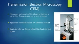
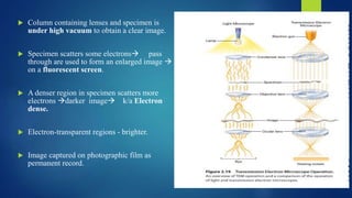
SlideShare a Scribd company logo
ELECTRON MICROSCOPY
 Dr. AMEENA THAHA
 23rd JULY 2020
HISTORY
 DERIVED FROM 2 GREEK WORDS
 MIKROS meaning small
 SKOPEO meaning look at
 Electron Microscope was invented by two Germans
 ERNST RUSKA & MAX KNOLL in 1931
 ERNST RUSKA –original model in 1933
 NOBEL PRIZE -1986
Uses a beam of accelerated electrons as a source of illumination
As the wavelength of electrons can be up to 1,00,000 times shorter
than that of visible light photons,
the electron microscope has a much better resolving power than a
light microscope.
It can reveal details of:
flagella
fimbriae
intra-cellular structures of cells
TYPES OF ELECTRON MICROSCOPE
• Transmission Electron Microscopy (TEM)
• Scanning Electron Microscopy (SEM)
Transmission Electron Microscopy
(TEM)
 Microscopy technique in which a beam of electrons is
transmitted through a specimen to form an image.
 Specimen - ultrathin section 20 -100 nm is viewed.
 Bacterial cells are thicker. Should be sliced into thin
layers
SPECIMEN PREPARATION
➢ Fixation - Glutaraldehyde or osmium tetroxide to stabilize cell structure.
➢ Dehydrated - Organic solvents (e.g., acetone or ethanol).*
➢ Embedding - Specimen is embedded in plastic polymer and is then hardened to form
a solid block.
➢ Slicing -Thin sections are cut from this block with a glass or diamond knife (
ultramicrotome)
➢ Staining
➢ Mounted on Metal Slide (Copper)
BASIC PRINCIPLES
 A heated tungsten filament in electron gun generates a beam of
electrons.
 Travels at high Speed  Bombarded on thin slice specimen.
 Electrons cannot pass through glass lens, therefore doughnut shaped
electromagnets called magnetic lenses are used.
 Column containing lenses and specimen is
under high vacuum to obtain a clear image.
 Specimen scatters some electrons pass
through are used to form an enlarged image 
on a fluorescent screen.
 A denser region in specimen scatters more
electrons darker image k/a Electron
dense.
 Electron-transparent regions - brighter.
 Image captured on photographic film as
permanent record.
Measures to increase contrast
 STAINING of specimen:
Increases contrast
Lead citrate and uranyl acetate
Heavy osmium atoms from osmium tetroxide fixative.
➢NEGATIVE STAINING
➢SHADOWING
NEGATIVE STAINING
• Specimen is spread out in a thin film
with heavy metals like phosphotungstic
acid or uranyl acetate.
• Heavy metals do not penetrate
specimen but render background dark
• Specimen appears bright.
• Excellent method to study structure of
viruses & bacterial gas vacuoles.
SHADOWING
• Specimen coated with a thin film
platinum or other heavy metal at 45
degree angle , so that the metal strikes the
micro-organism on only one side.
• Area coated with metal appears light in
photographs
• Uncoated side and the shadow region
created by the object appears dark.
• Useful in studying virus morphology,
prokaryotic flagella, and DNA.
Freeze-etching
 Cells are rapidly frozen in liquid
nitrogen & then warmed to -100°C
vacuum chamber.
 Knife precooled with liquid nitrogen
(-196°C) fractures the frozen cells
 The specimen left in high vacuum for
a minute or more.
 Ice sublimate away and uncover more
structural detail.
 Exposed surfaces are shadowed and
coated with layers of platinum and
carbon to form a replica of the surface.
 After the specimen removed
chemically replica studied in TEM
 3D view of intracellular structure.
TEM
Advantages
• Very powerful magnification and
resolution.
• Wide-range of applications - utilized in a
variety of different scientific, educational
and industrial fields
• Provide information on element and
compound structure .
• Images are high-quality and detailed.
Disadvantages
• Large and very expensive.
• Laborious sample preparation.
• Operation and analysis requires special
training.
• Samples are limited to those that are electron
transparent.
• Require special housing and maintenance.
• Images are black and white .
APPLICATION of TEM
 In medicine as a diagnostic tool – important in renal biopsies.
 Cellular tomography
– Used to obtaining detailed 3D structures of subcellular macromolecular objects.
 Cancer research – study of tumor cell ultrastructure .
 Toxicology – study the impacts of environmental pollution on the different
levels of biological organization.
SCANNING ELECTRON MICROSCOPE
 Produces an image from electrons released from
atoms on object’s surface.
 Used examine the surfaces of microorganisms in
great detail.
 Have resolution of 7 nm or less.
 Specimen preparation is relatively easy.
 Some cases , air-dried material can be examined
directly.
PRINCIPLE
 Incoming (primary) electrons
– can be “reflected” (backscattered) from bulk
specimen.
– can release secondary electrons.
 Primary electrons focused into small diameter electron
probe that scans across the specimen.
 Electrostatic or magnetic fields, applied at right angles
to the beam, used to change its direction of travel.
 By scanning simultaneously in two perpendicular
directions square or rectangular area of specimen (k/a
raster) covered.
 Image can be formed by collecting secondary electrons
from each point on the specimen.
SPECIMEN PREPRATION
 Sample coated with a thin layer of conductive material.
 Done using device called "sputter coater.”
 Sample placed in small chamber that is at a vacuum .
 Gold foil is placed in the instrument.
 Argon gas and electric field, electron to be removed from the argon  makes the
atoms positively charged.
 Argon ions gets attracted to negatively charged gold foil  knock gold atoms
from surface of gold foil.
 These gold atoms fall and settle onto the surface of the sample producing a thin
gold coating.
WORKING
 When the beam strikes a particular area,
surface atoms discharge  electrons called
secondary electrons trapped by a special
detector.
 Secondary electrons entering the detector
strike a scintillator emit light flashes 
photomultiplier converts to an electrical
current and amplifies.
 The signal sent to a cathode-ray tube
produces an image like television
picture  viewed or photographed.
 The number of secondary electrons
reaching the detector depends on the nature
of the specimen’s surface.
 Raised areas appear lighter on the
screen
 Depressions are darker.
 A realistic 3D image of the
microorganism’s surface results.
 Actual in situ location of
microorganisms in ecological
niches such as the human skin and
the lining of the gut also can be
examined.
SEM
Advantages
 Detailed 3D and topographical imaging
and the versatile information.
 Works very fast.
 Modern SEMs allow generation of data
in digital form.
 Most SEM samples require minimal
preparation actions.
.
DISADVANTAGES
• Expensive and large.
• Special training is required to operate.
• Preparation of samples can result in
artifacts.
• Limited to solid samples.
• Carry small risk of radiation exposure
APPLICATIONS of SEM
 Virology - for investigations of virus structure
 Cryo-electron microscopy – Images can be made of the surface of
frozen materials.
 3D tissue imaging
– Helps to know how cells are organized in a 3D network
– Their organization determines how cells can interact.
 Forensics - SEM reveals the presence of materials on evidences
that is otherwise undetectable
 SEM renders detailed 3-D images
– extremely small microorganisms
– anatomical pictures of insect, worm, spore, or other organic
Structures.
Comparison
TEM SEM
1. Transmitted electrons Scattered electrons
2. Electrons directly point towards sample Scattered electrons produce the image of sample after
microscope collects & counts the scattered electron
3. Seeks to see what is inside or beyond the surface Focuses on samples surface &its composition
4. Shows sample as a whole Shows sample bit by bit
5. Delivers a 2D picture Provides a 3D image
6. Has a 50 million magnification. Offers 2 million as max level of magnification
7. Resolution- 0.5 A° Resolution- 0.4m
SCANNING PROBE MICROSCOPY
 Most powerful new microscope
 Measure surface features by moving a sharp probe over the object’s
surface.
 The scanning tunneling microscope, invented in 1980.
 It can achieve magnifications of 100 million times
 Allow scientists to view atoms on the surface of a solid.
2 types:
1. Scanning Tunnelling Microscope
2. Atomic Force Microscope
SCANNING TUNNELING MICROSCOPE
 Gerd Binnig and Heinrich Rohrer invented in 1980.
 Won Nobel prize for physics with Ernst ruska.
 Needle like probe with point so sharp that often only one atom
at its tip.
 Probe lowered toward the specimen surface until its electron
cloud just touches the surface atoms.
 Small voltage is applied between the tip and specimen
 Electrons flow through a narrow channel in the electron
cloudsTunnelling current extraordinarily sensitive to
distance
 Arrangement of atoms on specimen surface
determined  moving probe tip back and
forth over the surface.
 Probe height constant above the specimen to
maintain a steady tunneling current.
 Its motion recorded and analyzed by a
computer  creates accurate 3D image of
the surface atoms.
 The surface map displayed on computer
screen or plotted on paper.
 The resolution is so great that individual
atoms are observed easily.
 Have major impact in biology.
 Used to directly view DNA and other biological molecules.
 Examine objects when they are immersed in water, used study
biological molecules(DNA).
ATOMIC FORCE MICROSCOPE
 Second type of scanning probe microscope.
 Moves a sharp probe over the specimen surface while
keeping the distance between the probe tip and the surface
constant.
 Exerting a very small amount of force on the tip enough
to maintain a constant distance but not enough force to
damage the surface.
 The vertical motion of the tip usually is followed by
measuring the deflection of a laser beam that strikes the
lever holding the probe.
Tip used to probe specimen
attached to cantilever.
As probe passes over surface
,cantilever deflected
vertically.
Laser beam directed towards
cantilever used to monitor
vertical movements.
Light reflected by cantilever
 detected by photodiode
 generates image.
USES
 Study surfaces that do not conduct
electricity well.
 Study interactions between the E. coli
GroES and GroEL chaperone proteins.
 To map plasmids by locating restriction
enzymes bound to specific sites.
 To follow behavior of living bacteria and
other cells.
 To visualize membrane proteins.
MICROSCOPE IMPORTANT FEATURES
Visible Light as source of illumination
1. Bright-field microscope Common,multi purpose, for live and preserved stained specimen.
Specimen -dark field-bright
2 Dark field microscope Best for observing live unstained specimens, specimen - bright field dark,
provides outline of specimen with reduced internal cellular details.
3. Phase contrast Used for live specimens, specimen is dark against bright background excellent
for internal cellular details.
UV light as source of illumination
1. Fluorescent microscope Specimen stained with fluorescent dye or combined with fluorescent
antibodies, emit visible light; specificity makes this microscope an excellent
diagnostic tool.
Electron beam forms image of specimen
1 TEM Section of specimen viewed under very high magnification ,finest detail
structure of cells and viruses shown, used only on preserved material.
2 SEM Scans and magnifies external surface of specimen ,produces striking 3D
images
References
 Prescott’s microbiology 8th edition.
 Bailey & scotts Diagnostic microbiology 14th edition.
 Manual of clinical Microbiology by Pfaller & James.H.Jorgensen 11th edition.
Electron microscopy ameena

More Related Content

What's hot

Confocal microscope presentation pt
Confocal microscope presentation  ptConfocal microscope presentation  pt
Confocal microscope presentation ptShrigouri4605
 
ELECTRON MICROSCOPY (TEM & SEM)
ELECTRON MICROSCOPY (TEM & SEM)ELECTRON MICROSCOPY (TEM & SEM)
ELECTRON MICROSCOPY (TEM & SEM)ADITYA ARYA
 
Transmission electron microscope
Transmission electron microscopeTransmission electron microscope
Transmission electron microscopeSenthil Arasan
 
Transmission Electron Microscope (TEM) for research (Full version)
Transmission Electron Microscope (TEM) for research (Full version)Transmission Electron Microscope (TEM) for research (Full version)
Transmission Electron Microscope (TEM) for research (Full version)Mohit Rajput
 
Electron microscopy
Electron microscopyElectron microscopy
Electron microscopyGanga Huvin
 
Electron microscope, principle and application
Electron microscope, principle and applicationElectron microscope, principle and application
Electron microscope, principle and applicationKAUSHAL SAHU
 
transmission electron microscopy
transmission electron microscopytransmission electron microscopy
transmission electron microscopyJessa Ariño
 
Electron Microscopy - Scanning electron microscope, Transmission Electron Mic...
Electron Microscopy - Scanning electron microscope, Transmission Electron Mic...Electron Microscopy - Scanning electron microscope, Transmission Electron Mic...
Electron Microscopy - Scanning electron microscope, Transmission Electron Mic...Sumer Pankaj
 
transmission Electron Microscopy (Tem)
transmission Electron Microscopy (Tem)transmission Electron Microscopy (Tem)
transmission Electron Microscopy (Tem)Sivasangari Shanmugam
 
2018 HM-scanning electron microscope
2018 HM-scanning electron microscope2018 HM-scanning electron microscope
2018 HM-scanning electron microscopeHarsh Mohan
 

What's hot (20)

Electron microscope
Electron microscopeElectron microscope
Electron microscope
 
Confocal microscope presentation pt
Confocal microscope presentation  ptConfocal microscope presentation  pt
Confocal microscope presentation pt
 
ELECTRON MICROSCOPY (TEM & SEM)
ELECTRON MICROSCOPY (TEM & SEM)ELECTRON MICROSCOPY (TEM & SEM)
ELECTRON MICROSCOPY (TEM & SEM)
 
Transmission electron microscope
Transmission electron microscopeTransmission electron microscope
Transmission electron microscope
 
Electron microscope
Electron microscopeElectron microscope
Electron microscope
 
Transmission Electron Microscope (TEM) for research (Full version)
Transmission Electron Microscope (TEM) for research (Full version)Transmission Electron Microscope (TEM) for research (Full version)
Transmission Electron Microscope (TEM) for research (Full version)
 
Electron microscopy
Electron microscopyElectron microscopy
Electron microscopy
 
Electron microscope, principle and application
Electron microscope, principle and applicationElectron microscope, principle and application
Electron microscope, principle and application
 
Transmission electron
Transmission        electronTransmission        electron
Transmission electron
 
Sem and tem
Sem and temSem and tem
Sem and tem
 
transmission electron microscopy
transmission electron microscopytransmission electron microscopy
transmission electron microscopy
 
Electron Microscopy - Scanning electron microscope, Transmission Electron Mic...
Electron Microscopy - Scanning electron microscope, Transmission Electron Mic...Electron Microscopy - Scanning electron microscope, Transmission Electron Mic...
Electron Microscopy - Scanning electron microscope, Transmission Electron Mic...
 
electron microscopy
electron microscopyelectron microscopy
electron microscopy
 
Electron microscope
Electron microscopeElectron microscope
Electron microscope
 
Confocal microscopy
Confocal microscopyConfocal microscopy
Confocal microscopy
 
Bright field microscope
Bright field microscopeBright field microscope
Bright field microscope
 
Stem
StemStem
Stem
 
transmission Electron Microscopy (Tem)
transmission Electron Microscopy (Tem)transmission Electron Microscopy (Tem)
transmission Electron Microscopy (Tem)
 
Electron microscopy (EM)
Electron microscopy (EM) Electron microscopy (EM)
Electron microscopy (EM)
 
2018 HM-scanning electron microscope
2018 HM-scanning electron microscope2018 HM-scanning electron microscope
2018 HM-scanning electron microscope
 

Similar to Electron microscopy ameena

Fluorescence and electron Microscope.pptx
Fluorescence  and electron Microscope.pptxFluorescence  and electron Microscope.pptx
Fluorescence and electron Microscope.pptxsaraso888
 
Electron microscope
Electron microscopeElectron microscope
Electron microscopeReena Rai
 
Transmission electron microscopeppt
Transmission electron microscopepptTransmission electron microscopeppt
Transmission electron microscopepptGenevia Vincent
 
Scanning Electron Microscopy (SEM)
Scanning Electron Microscopy (SEM)Scanning Electron Microscopy (SEM)
Scanning Electron Microscopy (SEM)Junaid Ahmad
 
scanning electron microscope
scanning electron microscopescanning electron microscope
scanning electron microscopeDrAkhilaCNV
 
Scanning Electron Microscopy Advantages, Application and Disadvantages
Scanning Electron Microscopy Advantages, Application and DisadvantagesScanning Electron Microscopy Advantages, Application and Disadvantages
Scanning Electron Microscopy Advantages, Application and DisadvantagesBhanu Krishan
 
Scanning and transmission electroscopy.pptx
Scanning and transmission electroscopy.pptxScanning and transmission electroscopy.pptx
Scanning and transmission electroscopy.pptxNirmal P J
 
5. Microsocope ELECTRON MICROSCOPE (TEM & SEM ) - Basics
5. Microsocope   ELECTRON MICROSCOPE (TEM & SEM ) - Basics5. Microsocope   ELECTRON MICROSCOPE (TEM & SEM ) - Basics
5. Microsocope ELECTRON MICROSCOPE (TEM & SEM ) - BasicsNethravathi Siri
 
Scanning electron microscope (sem)
Scanning electron microscope (sem)Scanning electron microscope (sem)
Scanning electron microscope (sem)Mary Theresa
 
Lec 3; Microscopy ALI.pptx
Lec 3; Microscopy ALI.pptxLec 3; Microscopy ALI.pptx
Lec 3; Microscopy ALI.pptxalihaider64675
 
Scanning electron microscope
Scanning electron microscopeScanning electron microscope
Scanning electron microscopeSuganyaPaulraj
 
ELECTRON MICROSCOPE AND ITS APPLICATION.pptx
ELECTRON MICROSCOPE AND ITS APPLICATION.pptxELECTRON MICROSCOPE AND ITS APPLICATION.pptx
ELECTRON MICROSCOPE AND ITS APPLICATION.pptxPallaviKumari112
 
Scanning electron microscopy
Scanning electron microscopyScanning electron microscopy
Scanning electron microscopyJessa Ariño
 
Scanning Electron Microscope
Scanning Electron MicroscopeScanning Electron Microscope
Scanning Electron MicroscopeMarian L
 

Similar to Electron microscopy ameena (20)

Electron microscope
Electron microscopeElectron microscope
Electron microscope
 
Fluorescence and electron Microscope.pptx
Fluorescence  and electron Microscope.pptxFluorescence  and electron Microscope.pptx
Fluorescence and electron Microscope.pptx
 
Electron microscope
Electron microscopeElectron microscope
Electron microscope
 
Transmission electron microscopeppt
Transmission electron microscopepptTransmission electron microscopeppt
Transmission electron microscopeppt
 
Tem ppt
Tem ppt Tem ppt
Tem ppt
 
Scanning Electron Microscopy (SEM)
Scanning Electron Microscopy (SEM)Scanning Electron Microscopy (SEM)
Scanning Electron Microscopy (SEM)
 
Sem n tem
Sem n temSem n tem
Sem n tem
 
Electron Microscope
Electron MicroscopeElectron Microscope
Electron Microscope
 
scanning electron microscope
scanning electron microscopescanning electron microscope
scanning electron microscope
 
Scanning Electron Microscopy Advantages, Application and Disadvantages
Scanning Electron Microscopy Advantages, Application and DisadvantagesScanning Electron Microscopy Advantages, Application and Disadvantages
Scanning Electron Microscopy Advantages, Application and Disadvantages
 
Scanning and transmission electroscopy.pptx
Scanning and transmission electroscopy.pptxScanning and transmission electroscopy.pptx
Scanning and transmission electroscopy.pptx
 
SEM- scanning electron microscope
SEM- scanning electron microscope SEM- scanning electron microscope
SEM- scanning electron microscope
 
5. Microsocope ELECTRON MICROSCOPE (TEM & SEM ) - Basics
5. Microsocope   ELECTRON MICROSCOPE (TEM & SEM ) - Basics5. Microsocope   ELECTRON MICROSCOPE (TEM & SEM ) - Basics
5. Microsocope ELECTRON MICROSCOPE (TEM & SEM ) - Basics
 
Scanning electron microscope (sem)
Scanning electron microscope (sem)Scanning electron microscope (sem)
Scanning electron microscope (sem)
 
Lec 3; Microscopy ALI.pptx
Lec 3; Microscopy ALI.pptxLec 3; Microscopy ALI.pptx
Lec 3; Microscopy ALI.pptx
 
Scanning electron microscope
Scanning electron microscopeScanning electron microscope
Scanning electron microscope
 
ELECTRON MICROSCOPE AND ITS APPLICATION.pptx
ELECTRON MICROSCOPE AND ITS APPLICATION.pptxELECTRON MICROSCOPE AND ITS APPLICATION.pptx
ELECTRON MICROSCOPE AND ITS APPLICATION.pptx
 
Scanning electron microscopy
Scanning electron microscopyScanning electron microscopy
Scanning electron microscopy
 
Electron microscope
Electron microscopeElectron microscope
Electron microscope
 
Scanning Electron Microscope
Scanning Electron MicroscopeScanning Electron Microscope
Scanning Electron Microscope
 

Recently uploaded

Anuman- An inference for helpful in diagnosis and treatment
Anuman- An inference for helpful in diagnosis and treatmentAnuman- An inference for helpful in diagnosis and treatment
Anuman- An inference for helpful in diagnosis and treatmentabdeli bhadarva
 
Antiplatelets in IHD, Dose Duration, DAPT vs SAPT
Antiplatelets in IHD, Dose Duration, DAPT vs SAPTAntiplatelets in IHD, Dose Duration, DAPT vs SAPT
Antiplatelets in IHD, Dose Duration, DAPT vs SAPTAkashGanganePatil1
 
ANATOMY OF THE LOWER URINARY TRACT AND MALE [Autosaved] [Autosaved].pptx
ANATOMY OF THE LOWER URINARY TRACT AND MALE [Autosaved] [Autosaved].pptxANATOMY OF THE LOWER URINARY TRACT AND MALE [Autosaved] [Autosaved].pptx
ANATOMY OF THE LOWER URINARY TRACT AND MALE [Autosaved] [Autosaved].pptxBright Chipili
 
Relationship between vascular system disfunction, neurofluid flow and Alzheim...
Relationship between vascular system disfunction, neurofluid flow and Alzheim...Relationship between vascular system disfunction, neurofluid flow and Alzheim...
Relationship between vascular system disfunction, neurofluid flow and Alzheim...Catherine Liao
 
A thorough review of supernormal conduction.pptx
A thorough review of supernormal conduction.pptxA thorough review of supernormal conduction.pptx
A thorough review of supernormal conduction.pptxSergio Pinski
 
The hemodynamic and autonomic determinants of elevated blood pressure in obes...
The hemodynamic and autonomic determinants of elevated blood pressure in obes...The hemodynamic and autonomic determinants of elevated blood pressure in obes...
The hemodynamic and autonomic determinants of elevated blood pressure in obes...Catherine Liao
 
Temporal, Infratemporal & Pterygopalatine BY Dr.RIG.pptx
Temporal, Infratemporal & Pterygopalatine BY Dr.RIG.pptxTemporal, Infratemporal & Pterygopalatine BY Dr.RIG.pptx
Temporal, Infratemporal & Pterygopalatine BY Dr.RIG.pptxDr. Rabia Inam Gandapore
 
Effects of vaping e-cigarettes on arterial health
Effects of vaping e-cigarettes on arterial healthEffects of vaping e-cigarettes on arterial health
Effects of vaping e-cigarettes on arterial healthCatherine Liao
 
𝕔ALL #𝕘IRLS Service in Jaipur %(8901183002)# Jaipur #𝕔ALL #𝕘IRLS
𝕔ALL #𝕘IRLS Service in Jaipur %(8901183002)#  Jaipur #𝕔ALL #𝕘IRLS𝕔ALL #𝕘IRLS Service in Jaipur %(8901183002)#  Jaipur #𝕔ALL #𝕘IRLS
𝕔ALL #𝕘IRLS Service in Jaipur %(8901183002)# Jaipur #𝕔ALL #𝕘IRLSlakehe2738
 
Cardiac Impulse: Rhythmical Excitation and Conduction in the Heart
Cardiac Impulse: Rhythmical Excitation and Conduction in the HeartCardiac Impulse: Rhythmical Excitation and Conduction in the Heart
Cardiac Impulse: Rhythmical Excitation and Conduction in the HeartMedicoseAcademics
 
Factors Affecting child behavior in Pediatric Dentistry
Factors Affecting child behavior in Pediatric DentistryFactors Affecting child behavior in Pediatric Dentistry
Factors Affecting child behavior in Pediatric DentistryDr Simran Deepak Vangani
 
Hemodialysis: Chapter 2, Extracorporeal Blood Circuit - Dr.Gawad
Hemodialysis: Chapter 2, Extracorporeal Blood Circuit - Dr.GawadHemodialysis: Chapter 2, Extracorporeal Blood Circuit - Dr.Gawad
Hemodialysis: Chapter 2, Extracorporeal Blood Circuit - Dr.GawadNephroTube - Dr.Gawad
 
5cl adbb 5cladba cheap and fine Telegram: +85297504341
5cl adbb 5cladba cheap and fine Telegram: +852975043415cl adbb 5cladba cheap and fine Telegram: +85297504341
5cl adbb 5cladba cheap and fine Telegram: +85297504341Sherrylee83
 
Scientificity and feasibility study of non-invasive central arterial pressure...
Scientificity and feasibility study of non-invasive central arterial pressure...Scientificity and feasibility study of non-invasive central arterial pressure...
Scientificity and feasibility study of non-invasive central arterial pressure...Catherine Liao
 
End Feel -joint end feel - Normal and Abnormal end feel
End Feel -joint end feel - Normal and Abnormal end feelEnd Feel -joint end feel - Normal and Abnormal end feel
End Feel -joint end feel - Normal and Abnormal end feeldranji1
 
Impact of cancers therapies on the loss in cardiac function, myocardial fffic...
Impact of cancers therapies on the loss in cardiac function, myocardial fffic...Impact of cancers therapies on the loss in cardiac function, myocardial fffic...
Impact of cancers therapies on the loss in cardiac function, myocardial fffic...Catherine Liao
 
Arterial health throughout cancer treatment and exercise rehabilitation in wo...
Arterial health throughout cancer treatment and exercise rehabilitation in wo...Arterial health throughout cancer treatment and exercise rehabilitation in wo...
Arterial health throughout cancer treatment and exercise rehabilitation in wo...Catherine Liao
 
Non-Invasive assessment of arterial stiffness in advanced heart failure patie...
Non-Invasive assessment of arterial stiffness in advanced heart failure patie...Non-Invasive assessment of arterial stiffness in advanced heart failure patie...
Non-Invasive assessment of arterial stiffness in advanced heart failure patie...Catherine Liao
 
Is preeclampsia and spontaneous preterm delivery associate with vascular and ...
Is preeclampsia and spontaneous preterm delivery associate with vascular and ...Is preeclampsia and spontaneous preterm delivery associate with vascular and ...
Is preeclampsia and spontaneous preterm delivery associate with vascular and ...Catherine Liao
 
THORACOTOMY . SURGICAL PERSPECTIVES VOL 1
THORACOTOMY . SURGICAL PERSPECTIVES VOL 1THORACOTOMY . SURGICAL PERSPECTIVES VOL 1
THORACOTOMY . SURGICAL PERSPECTIVES VOL 1DR SETH JOTHAM
 

Recently uploaded (20)

Anuman- An inference for helpful in diagnosis and treatment
Anuman- An inference for helpful in diagnosis and treatmentAnuman- An inference for helpful in diagnosis and treatment
Anuman- An inference for helpful in diagnosis and treatment
 
Antiplatelets in IHD, Dose Duration, DAPT vs SAPT
Antiplatelets in IHD, Dose Duration, DAPT vs SAPTAntiplatelets in IHD, Dose Duration, DAPT vs SAPT
Antiplatelets in IHD, Dose Duration, DAPT vs SAPT
 
ANATOMY OF THE LOWER URINARY TRACT AND MALE [Autosaved] [Autosaved].pptx
ANATOMY OF THE LOWER URINARY TRACT AND MALE [Autosaved] [Autosaved].pptxANATOMY OF THE LOWER URINARY TRACT AND MALE [Autosaved] [Autosaved].pptx
ANATOMY OF THE LOWER URINARY TRACT AND MALE [Autosaved] [Autosaved].pptx
 
Relationship between vascular system disfunction, neurofluid flow and Alzheim...
Relationship between vascular system disfunction, neurofluid flow and Alzheim...Relationship between vascular system disfunction, neurofluid flow and Alzheim...
Relationship between vascular system disfunction, neurofluid flow and Alzheim...
 
A thorough review of supernormal conduction.pptx
A thorough review of supernormal conduction.pptxA thorough review of supernormal conduction.pptx
A thorough review of supernormal conduction.pptx
 
The hemodynamic and autonomic determinants of elevated blood pressure in obes...
The hemodynamic and autonomic determinants of elevated blood pressure in obes...The hemodynamic and autonomic determinants of elevated blood pressure in obes...
The hemodynamic and autonomic determinants of elevated blood pressure in obes...
 
Temporal, Infratemporal & Pterygopalatine BY Dr.RIG.pptx
Temporal, Infratemporal & Pterygopalatine BY Dr.RIG.pptxTemporal, Infratemporal & Pterygopalatine BY Dr.RIG.pptx
Temporal, Infratemporal & Pterygopalatine BY Dr.RIG.pptx
 
Effects of vaping e-cigarettes on arterial health
Effects of vaping e-cigarettes on arterial healthEffects of vaping e-cigarettes on arterial health
Effects of vaping e-cigarettes on arterial health
 
𝕔ALL #𝕘IRLS Service in Jaipur %(8901183002)# Jaipur #𝕔ALL #𝕘IRLS
𝕔ALL #𝕘IRLS Service in Jaipur %(8901183002)#  Jaipur #𝕔ALL #𝕘IRLS𝕔ALL #𝕘IRLS Service in Jaipur %(8901183002)#  Jaipur #𝕔ALL #𝕘IRLS
𝕔ALL #𝕘IRLS Service in Jaipur %(8901183002)# Jaipur #𝕔ALL #𝕘IRLS
 
Cardiac Impulse: Rhythmical Excitation and Conduction in the Heart
Cardiac Impulse: Rhythmical Excitation and Conduction in the HeartCardiac Impulse: Rhythmical Excitation and Conduction in the Heart
Cardiac Impulse: Rhythmical Excitation and Conduction in the Heart
 
Factors Affecting child behavior in Pediatric Dentistry
Factors Affecting child behavior in Pediatric DentistryFactors Affecting child behavior in Pediatric Dentistry
Factors Affecting child behavior in Pediatric Dentistry
 
Hemodialysis: Chapter 2, Extracorporeal Blood Circuit - Dr.Gawad
Hemodialysis: Chapter 2, Extracorporeal Blood Circuit - Dr.GawadHemodialysis: Chapter 2, Extracorporeal Blood Circuit - Dr.Gawad
Hemodialysis: Chapter 2, Extracorporeal Blood Circuit - Dr.Gawad
 
5cl adbb 5cladba cheap and fine Telegram: +85297504341
5cl adbb 5cladba cheap and fine Telegram: +852975043415cl adbb 5cladba cheap and fine Telegram: +85297504341
5cl adbb 5cladba cheap and fine Telegram: +85297504341
 
Scientificity and feasibility study of non-invasive central arterial pressure...
Scientificity and feasibility study of non-invasive central arterial pressure...Scientificity and feasibility study of non-invasive central arterial pressure...
Scientificity and feasibility study of non-invasive central arterial pressure...
 
End Feel -joint end feel - Normal and Abnormal end feel
End Feel -joint end feel - Normal and Abnormal end feelEnd Feel -joint end feel - Normal and Abnormal end feel
End Feel -joint end feel - Normal and Abnormal end feel
 
Impact of cancers therapies on the loss in cardiac function, myocardial fffic...
Impact of cancers therapies on the loss in cardiac function, myocardial fffic...Impact of cancers therapies on the loss in cardiac function, myocardial fffic...
Impact of cancers therapies on the loss in cardiac function, myocardial fffic...
 
Arterial health throughout cancer treatment and exercise rehabilitation in wo...
Arterial health throughout cancer treatment and exercise rehabilitation in wo...Arterial health throughout cancer treatment and exercise rehabilitation in wo...
Arterial health throughout cancer treatment and exercise rehabilitation in wo...
 
Non-Invasive assessment of arterial stiffness in advanced heart failure patie...
Non-Invasive assessment of arterial stiffness in advanced heart failure patie...Non-Invasive assessment of arterial stiffness in advanced heart failure patie...
Non-Invasive assessment of arterial stiffness in advanced heart failure patie...
 
Is preeclampsia and spontaneous preterm delivery associate with vascular and ...
Is preeclampsia and spontaneous preterm delivery associate with vascular and ...Is preeclampsia and spontaneous preterm delivery associate with vascular and ...
Is preeclampsia and spontaneous preterm delivery associate with vascular and ...
 
THORACOTOMY . SURGICAL PERSPECTIVES VOL 1
THORACOTOMY . SURGICAL PERSPECTIVES VOL 1THORACOTOMY . SURGICAL PERSPECTIVES VOL 1
THORACOTOMY . SURGICAL PERSPECTIVES VOL 1
 

Electron microscopy ameena

  • 1. ELECTRON MICROSCOPY  Dr. AMEENA THAHA  23rd JULY 2020
  • 2. HISTORY  DERIVED FROM 2 GREEK WORDS  MIKROS meaning small  SKOPEO meaning look at  Electron Microscope was invented by two Germans  ERNST RUSKA & MAX KNOLL in 1931  ERNST RUSKA –original model in 1933  NOBEL PRIZE -1986
  • 3. Uses a beam of accelerated electrons as a source of illumination As the wavelength of electrons can be up to 1,00,000 times shorter than that of visible light photons, the electron microscope has a much better resolving power than a light microscope. It can reveal details of: flagella fimbriae intra-cellular structures of cells
  • 4. TYPES OF ELECTRON MICROSCOPE • Transmission Electron Microscopy (TEM) • Scanning Electron Microscopy (SEM)
  • 5. Transmission Electron Microscopy (TEM)  Microscopy technique in which a beam of electrons is transmitted through a specimen to form an image.  Specimen - ultrathin section 20 -100 nm is viewed.  Bacterial cells are thicker. Should be sliced into thin layers
  • 6. SPECIMEN PREPARATION ➢ Fixation - Glutaraldehyde or osmium tetroxide to stabilize cell structure. ➢ Dehydrated - Organic solvents (e.g., acetone or ethanol).* ➢ Embedding - Specimen is embedded in plastic polymer and is then hardened to form a solid block. ➢ Slicing -Thin sections are cut from this block with a glass or diamond knife ( ultramicrotome) ➢ Staining ➢ Mounted on Metal Slide (Copper)
  • 7. BASIC PRINCIPLES  A heated tungsten filament in electron gun generates a beam of electrons.  Travels at high Speed  Bombarded on thin slice specimen.  Electrons cannot pass through glass lens, therefore doughnut shaped electromagnets called magnetic lenses are used.
  • 8.  Column containing lenses and specimen is under high vacuum to obtain a clear image.  Specimen scatters some electrons pass through are used to form an enlarged image  on a fluorescent screen.  A denser region in specimen scatters more electrons darker image k/a Electron dense.  Electron-transparent regions - brighter.  Image captured on photographic film as permanent record.
  • 9. Measures to increase contrast  STAINING of specimen: Increases contrast Lead citrate and uranyl acetate Heavy osmium atoms from osmium tetroxide fixative. ➢NEGATIVE STAINING ➢SHADOWING
  • 10. NEGATIVE STAINING • Specimen is spread out in a thin film with heavy metals like phosphotungstic acid or uranyl acetate. • Heavy metals do not penetrate specimen but render background dark • Specimen appears bright. • Excellent method to study structure of viruses & bacterial gas vacuoles. SHADOWING • Specimen coated with a thin film platinum or other heavy metal at 45 degree angle , so that the metal strikes the micro-organism on only one side. • Area coated with metal appears light in photographs • Uncoated side and the shadow region created by the object appears dark. • Useful in studying virus morphology, prokaryotic flagella, and DNA.
  • 11. Freeze-etching  Cells are rapidly frozen in liquid nitrogen & then warmed to -100°C vacuum chamber.  Knife precooled with liquid nitrogen (-196°C) fractures the frozen cells  The specimen left in high vacuum for a minute or more.  Ice sublimate away and uncover more structural detail.  Exposed surfaces are shadowed and coated with layers of platinum and carbon to form a replica of the surface.  After the specimen removed chemically replica studied in TEM  3D view of intracellular structure.
  • 12.
  • 13. TEM Advantages • Very powerful magnification and resolution. • Wide-range of applications - utilized in a variety of different scientific, educational and industrial fields • Provide information on element and compound structure . • Images are high-quality and detailed. Disadvantages • Large and very expensive. • Laborious sample preparation. • Operation and analysis requires special training. • Samples are limited to those that are electron transparent. • Require special housing and maintenance. • Images are black and white .
  • 14. APPLICATION of TEM  In medicine as a diagnostic tool – important in renal biopsies.  Cellular tomography – Used to obtaining detailed 3D structures of subcellular macromolecular objects.  Cancer research – study of tumor cell ultrastructure .  Toxicology – study the impacts of environmental pollution on the different levels of biological organization.
  • 15. SCANNING ELECTRON MICROSCOPE  Produces an image from electrons released from atoms on object’s surface.  Used examine the surfaces of microorganisms in great detail.  Have resolution of 7 nm or less.  Specimen preparation is relatively easy.  Some cases , air-dried material can be examined directly.
  • 16. PRINCIPLE  Incoming (primary) electrons – can be “reflected” (backscattered) from bulk specimen. – can release secondary electrons.  Primary electrons focused into small diameter electron probe that scans across the specimen.  Electrostatic or magnetic fields, applied at right angles to the beam, used to change its direction of travel.  By scanning simultaneously in two perpendicular directions square or rectangular area of specimen (k/a raster) covered.  Image can be formed by collecting secondary electrons from each point on the specimen.
  • 17. SPECIMEN PREPRATION  Sample coated with a thin layer of conductive material.  Done using device called "sputter coater.”  Sample placed in small chamber that is at a vacuum .  Gold foil is placed in the instrument.  Argon gas and electric field, electron to be removed from the argon  makes the atoms positively charged.  Argon ions gets attracted to negatively charged gold foil  knock gold atoms from surface of gold foil.  These gold atoms fall and settle onto the surface of the sample producing a thin gold coating.
  • 18. WORKING  When the beam strikes a particular area, surface atoms discharge  electrons called secondary electrons trapped by a special detector.  Secondary electrons entering the detector strike a scintillator emit light flashes  photomultiplier converts to an electrical current and amplifies.  The signal sent to a cathode-ray tube produces an image like television picture  viewed or photographed.  The number of secondary electrons reaching the detector depends on the nature of the specimen’s surface.
  • 19.  Raised areas appear lighter on the screen  Depressions are darker.  A realistic 3D image of the microorganism’s surface results.  Actual in situ location of microorganisms in ecological niches such as the human skin and the lining of the gut also can be examined.
  • 20. SEM Advantages  Detailed 3D and topographical imaging and the versatile information.  Works very fast.  Modern SEMs allow generation of data in digital form.  Most SEM samples require minimal preparation actions. . DISADVANTAGES • Expensive and large. • Special training is required to operate. • Preparation of samples can result in artifacts. • Limited to solid samples. • Carry small risk of radiation exposure
  • 21. APPLICATIONS of SEM  Virology - for investigations of virus structure  Cryo-electron microscopy – Images can be made of the surface of frozen materials.  3D tissue imaging – Helps to know how cells are organized in a 3D network – Their organization determines how cells can interact.  Forensics - SEM reveals the presence of materials on evidences that is otherwise undetectable  SEM renders detailed 3-D images – extremely small microorganisms – anatomical pictures of insect, worm, spore, or other organic Structures.
  • 22. Comparison TEM SEM 1. Transmitted electrons Scattered electrons 2. Electrons directly point towards sample Scattered electrons produce the image of sample after microscope collects & counts the scattered electron 3. Seeks to see what is inside or beyond the surface Focuses on samples surface &its composition 4. Shows sample as a whole Shows sample bit by bit 5. Delivers a 2D picture Provides a 3D image 6. Has a 50 million magnification. Offers 2 million as max level of magnification 7. Resolution- 0.5 A° Resolution- 0.4m
  • 23. SCANNING PROBE MICROSCOPY  Most powerful new microscope  Measure surface features by moving a sharp probe over the object’s surface.  The scanning tunneling microscope, invented in 1980.  It can achieve magnifications of 100 million times  Allow scientists to view atoms on the surface of a solid. 2 types: 1. Scanning Tunnelling Microscope 2. Atomic Force Microscope
  • 24. SCANNING TUNNELING MICROSCOPE  Gerd Binnig and Heinrich Rohrer invented in 1980.  Won Nobel prize for physics with Ernst ruska.  Needle like probe with point so sharp that often only one atom at its tip.  Probe lowered toward the specimen surface until its electron cloud just touches the surface atoms.  Small voltage is applied between the tip and specimen  Electrons flow through a narrow channel in the electron cloudsTunnelling current extraordinarily sensitive to distance
  • 25.  Arrangement of atoms on specimen surface determined  moving probe tip back and forth over the surface.  Probe height constant above the specimen to maintain a steady tunneling current.  Its motion recorded and analyzed by a computer  creates accurate 3D image of the surface atoms.  The surface map displayed on computer screen or plotted on paper.  The resolution is so great that individual atoms are observed easily.
  • 26.  Have major impact in biology.  Used to directly view DNA and other biological molecules.  Examine objects when they are immersed in water, used study biological molecules(DNA).
  • 27. ATOMIC FORCE MICROSCOPE  Second type of scanning probe microscope.  Moves a sharp probe over the specimen surface while keeping the distance between the probe tip and the surface constant.  Exerting a very small amount of force on the tip enough to maintain a constant distance but not enough force to damage the surface.  The vertical motion of the tip usually is followed by measuring the deflection of a laser beam that strikes the lever holding the probe.
  • 28. Tip used to probe specimen attached to cantilever. As probe passes over surface ,cantilever deflected vertically. Laser beam directed towards cantilever used to monitor vertical movements. Light reflected by cantilever  detected by photodiode  generates image.
  • 29. USES  Study surfaces that do not conduct electricity well.  Study interactions between the E. coli GroES and GroEL chaperone proteins.  To map plasmids by locating restriction enzymes bound to specific sites.  To follow behavior of living bacteria and other cells.  To visualize membrane proteins.
  • 30. MICROSCOPE IMPORTANT FEATURES Visible Light as source of illumination 1. Bright-field microscope Common,multi purpose, for live and preserved stained specimen. Specimen -dark field-bright 2 Dark field microscope Best for observing live unstained specimens, specimen - bright field dark, provides outline of specimen with reduced internal cellular details. 3. Phase contrast Used for live specimens, specimen is dark against bright background excellent for internal cellular details. UV light as source of illumination 1. Fluorescent microscope Specimen stained with fluorescent dye or combined with fluorescent antibodies, emit visible light; specificity makes this microscope an excellent diagnostic tool. Electron beam forms image of specimen 1 TEM Section of specimen viewed under very high magnification ,finest detail structure of cells and viruses shown, used only on preserved material. 2 SEM Scans and magnifies external surface of specimen ,produces striking 3D images
  • 31. References  Prescott’s microbiology 8th edition.  Bailey & scotts Diagnostic microbiology 14th edition.  Manual of clinical Microbiology by Pfaller & James.H.Jorgensen 11th edition.

Editor's Notes

  1. Lead and uranium ions - bind to cell structures make them electron opaque.
  2. Lead and uranium ions - bind to cell structures make them electron opaque.
  3. since fewer electrons strike that area of the screen; these regions are said to be “electron dense.” *Both for viewing and prep of specimen ** place harsh restriction on nature of sample both viewing and preparing the specimen Such a thin slice cannot be cut unless the specimen has support of some kind; the necessary support is provided by plastic
  4. Cells are very brittle and break along lines of greatest weakness, usually down the middle of internal membranes
  5. Adv: minimizes the danger of artifacts  b/c frozen quickly rather than being subjected to chemical fixation, dehydration, and plastic embedding
  6. Tomography refers to imaging by sectioning, through the use of anykind of penetrating wave. – Information is collected and used to assemble a three dimensional image of the target.
  7. Transmission electron microscopes form an image from radiation that has passed through a specimen. fixed, dehydrated, and dried to preserve surface structure and prevent collapse of the cells when they are exposed to the SEM’s high vacuum. Before viewing, dried samples are mounted and coated with a thin layer of metal to prevent the build up of an electrical charge on the surface and to give a better image. To create an image, the SEM scans a narrow, tapered electron beam back and forth over the specimen.
  8. The electrons surrounding surface atoms tunnel or project out from the surface boundary a very short distance.
  9. Tunneling currentwill decrease about a thousand fold if the probe is moved away from the surface by a distance equivalent to the diameter of an atom The microscope’s inventors, Gerd Binnig and Heinrich Rohrer, shared the 1986 Nobel Prize in Physics for their work, together with Ernst Ruska, the designer of the first transmission electron microscope