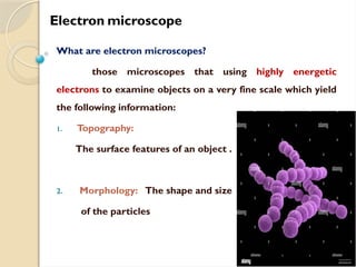
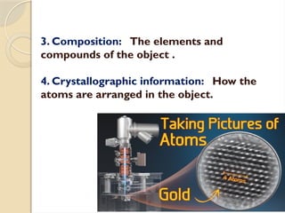
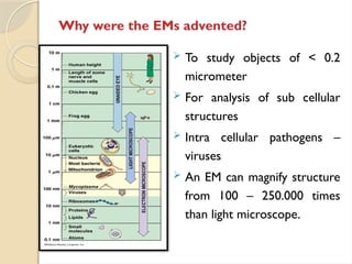
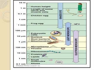
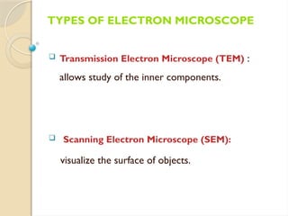
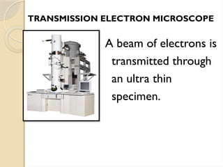
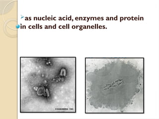
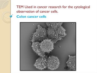
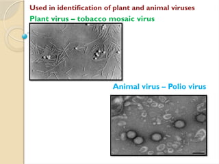
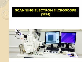
ELECTRON MICROSCOPE
Electron microscope
What are electron microscopes?
those microscopes that using highly energetic
electrons to examine objects on a very fine scale which yield
the following information:
1. Topography:
The surface features of an object .
2. Morphology: The shape and size
of the particles
3. Composition: The elements and
compounds of the object .
4. Crystallographic information: How the
atoms are arranged in the object.
Why were the EMs advented?
 To study objects of < 0.2
micrometer
 For analysis of sub cellular
structures
 Intra cellular pathogens –
viruses
 An EM can magnify structure
from 100 – 250.000 times
than light microscope.
TYPES OF ELECTRON MICROSCOPE
 Transmission Electron Microscope (TEM) :
allows study of the inner components.
 Scanning Electron Microscope (SEM):
visualize the surface of objects.
TRANSMISSION ELECTRON MICROSCOPE
A beam of electrons is
transmitted through
an ultra thin
specimen.
TheTEM Components
1. Electron gun
2. Condenser Lenses
3. Objective Lenses
4. Projector lenses and
5. Fluorescent screen
Electrons are emitted by
the electron gun
on a fluorescent screen
or a computer monitor.
SAMPLE PREPARATION
DEHYDRATION
Using increasing concentration of ethanol
FIXATION
By fixative as formalin
EMBEDDING
• The specimen is embedded in a hard
embedding medium like Plastic medium
 The embedded specimen is cut into thin
section of 50-100nm thickness using a glass
or diamond knife fixed in an
ultramicrotome.
Ultramicrotome
STAINING
• The specimen is kept dipped in a solution
containing heavy metal ions for metallic
staining.
• The image of the selected section is then
viewed on the fluorescent screen.
APPLICATIONS OF TEM
Ideal tool for the study of ultra structure of
cells
Chloroplast
Plant cell
as nucleic acid, enzymes and protein
in cells and cell organelles.
virus release DNA Uncoiled DNA
TEM Used in cancer research for the cytological
observation of cancer cells.
Colon cancer cells
Used in identification of plant and animal viruses
Plant virus – tobacco mosaic virus
Animal virus – Polio virus
ADAVANTAGES
 TEM offer the most powerful magnification,
potentially over one million times or more
 TEMs provide information on element and
compound structure
 Images are high- quality and detailed
 They are easy to operate with proper training
SCANNING ELECTRON MICROSCOPE
(SEM)
 Electrons interact
with electrons in
sample and convey
information in form of
signals to detectors.
SEM COMPONENTS
1) Electron gun :
2) Condenser lens –
collect and
concentrate the
electrons .
3) Reflection coil –
change the direction of
electron path helps for
focussing the e- beam
on the specimen
4) Specimen stage
Vacuum tube
The entire setup is placed in a vacuum tube because electrons
can move in a straight line only in a vacuum. Vacuum pressure
of 10-7 to 10-9 Pa is applied in the vacuum tube.
Cooling System
While TEM is working, a large amount of heat is produced. To
keep the apparatus at a low temperature cooling water is
circulated through a cooling system around the TEM.
Blood cells Head of bee
Metallic Coating
Dry materials such as wood, bone, feathers,
insect’s wings and shells are coated with electronically
conducting materials.
They are metallic gold, platinum, tungsten,
chromium and graphite.
Metallic coating prevents the accumulation of
electrostatic charges on the specimen.
Penguin feather Peacock feather
SPECIMEN PREPARATION - DRY SAMPLE
Fixation
Fixation is done by immersing the specimen in
chemical preservatives. It stabilize the molecular
organization.
Common fixatives are potassium permanganate,
formalin etc.
Dehydration
After fixation the specimen is dehydrated by
keeping it in concentrated ethanol or acetone.
Staining.
WET SAMPLE PREPARATION
APPLICATIONS OF SEM
 SEM is very useful to view the surface.
 SEM gives 3-D structure of objects.
SEM is employed in the analysis of structural features
of compound eyes of insects.
Eye of black garden ant
Mosquito eyes
Eyes of yellow dung fly
Animation

electron microscopes ppt lecture 4.pptx

  • 1.
  • 3.
    Electron microscope What areelectron microscopes? those microscopes that using highly energetic electrons to examine objects on a very fine scale which yield the following information: 1. Topography: The surface features of an object . 2. Morphology: The shape and size of the particles
  • 4.
    3. Composition: Theelements and compounds of the object . 4. Crystallographic information: How the atoms are arranged in the object.
  • 5.
    Why were theEMs advented?  To study objects of < 0.2 micrometer  For analysis of sub cellular structures  Intra cellular pathogens – viruses  An EM can magnify structure from 100 – 250.000 times than light microscope.
  • 7.
    TYPES OF ELECTRONMICROSCOPE  Transmission Electron Microscope (TEM) : allows study of the inner components.  Scanning Electron Microscope (SEM): visualize the surface of objects.
  • 8.
    TRANSMISSION ELECTRON MICROSCOPE Abeam of electrons is transmitted through an ultra thin specimen.
  • 9.
    TheTEM Components 1. Electrongun 2. Condenser Lenses 3. Objective Lenses 4. Projector lenses and 5. Fluorescent screen Electrons are emitted by the electron gun on a fluorescent screen or a computer monitor.
  • 10.
    SAMPLE PREPARATION DEHYDRATION Using increasingconcentration of ethanol FIXATION By fixative as formalin
  • 11.
    EMBEDDING • The specimenis embedded in a hard embedding medium like Plastic medium  The embedded specimen is cut into thin section of 50-100nm thickness using a glass or diamond knife fixed in an ultramicrotome. Ultramicrotome
  • 12.
    STAINING • The specimenis kept dipped in a solution containing heavy metal ions for metallic staining. • The image of the selected section is then viewed on the fluorescent screen.
  • 13.
    APPLICATIONS OF TEM Idealtool for the study of ultra structure of cells Chloroplast Plant cell
  • 14.
    as nucleic acid,enzymes and protein in cells and cell organelles. virus release DNA Uncoiled DNA
  • 15.
    TEM Used incancer research for the cytological observation of cancer cells. Colon cancer cells
  • 16.
    Used in identificationof plant and animal viruses Plant virus – tobacco mosaic virus Animal virus – Polio virus
  • 17.
    ADAVANTAGES  TEM offerthe most powerful magnification, potentially over one million times or more  TEMs provide information on element and compound structure  Images are high- quality and detailed  They are easy to operate with proper training
  • 18.
  • 19.
     Electrons interact withelectrons in sample and convey information in form of signals to detectors.
  • 20.
    SEM COMPONENTS 1) Electrongun : 2) Condenser lens – collect and concentrate the electrons . 3) Reflection coil – change the direction of electron path helps for focussing the e- beam on the specimen 4) Specimen stage
  • 22.
    Vacuum tube The entiresetup is placed in a vacuum tube because electrons can move in a straight line only in a vacuum. Vacuum pressure of 10-7 to 10-9 Pa is applied in the vacuum tube. Cooling System While TEM is working, a large amount of heat is produced. To keep the apparatus at a low temperature cooling water is circulated through a cooling system around the TEM. Blood cells Head of bee
  • 23.
    Metallic Coating Dry materialssuch as wood, bone, feathers, insect’s wings and shells are coated with electronically conducting materials. They are metallic gold, platinum, tungsten, chromium and graphite. Metallic coating prevents the accumulation of electrostatic charges on the specimen. Penguin feather Peacock feather SPECIMEN PREPARATION - DRY SAMPLE
  • 24.
    Fixation Fixation is doneby immersing the specimen in chemical preservatives. It stabilize the molecular organization. Common fixatives are potassium permanganate, formalin etc. Dehydration After fixation the specimen is dehydrated by keeping it in concentrated ethanol or acetone. Staining. WET SAMPLE PREPARATION
  • 25.
    APPLICATIONS OF SEM SEM is very useful to view the surface.  SEM gives 3-D structure of objects.
  • 26.
    SEM is employedin the analysis of structural features of compound eyes of insects. Eye of black garden ant
  • 27.
    Mosquito eyes Eyes ofyellow dung fly
  • 28.