

![Light Microscope and Electron Microscope [Best one]](https://image.slidesharecdn.com/presentation-170404212835/85/Light-Microscope-and-Electron-Microscope-Best-one-23-320.jpg)
![Light Microscope and Electron Microscope [Best one]](https://image.slidesharecdn.com/presentation-170404212835/85/Light-Microscope-and-Electron-Microscope-Best-one-24-320.jpg)

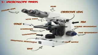
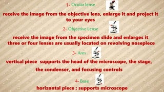
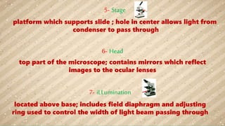
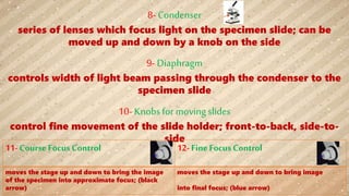
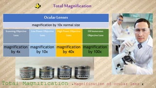
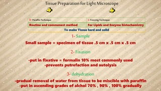
The document describes the main components and functions of a light microscope. It includes: 1. Objective lenses that enlarge the specimen image and project it to the ocular lenses. 2. An arm that supports the microscope head and stage. 3. A stage that holds the specimen slide. 4. Various controls for illumination, focus, and slide movement. 5. Ocular lenses that further magnify the enlarged specimen image for viewing.


