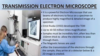
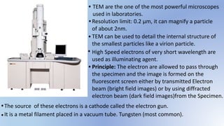
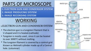
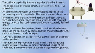
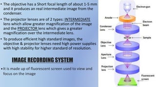
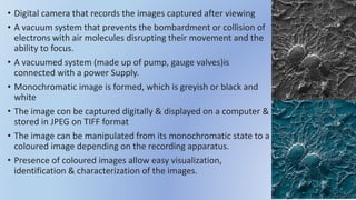
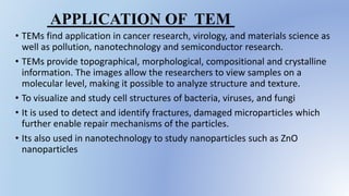
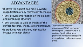
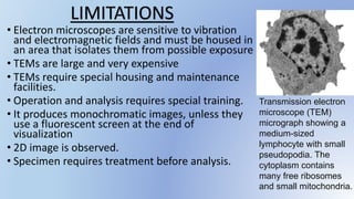
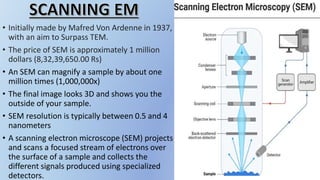
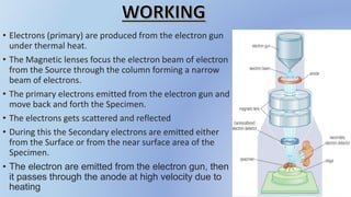
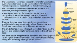
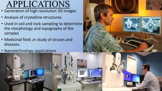
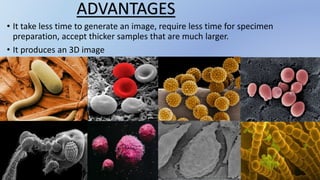
JAPAN: ORGANISATION OF PMDA, PHARMACEUTICAL LAWS & REGULATIONS, TYPES OF REGI...
Scanning and transmission electroscopy.pptx
1.
2. It is a powerful Electron Microscope that use
beams of electrons to focus on a specimen to
produce highly magnified & detailed image of a
specimen
Up to 10-50 million times magnification
Samples must be incredibly thin ,often less than
150mm thick to allow the electrons to pass
through them.
After the transmission of the electrons through
the sample, they arrive at a detecter below & a
2D image is created.
The Magnetic lenses are used
Ernst Ruska (1933) developed the TEM
.
.
.
.
.
.
3. Principle: The electron are allowed to pass through
the specimen and the image is formed on the
fluorescent screen either by transmitted Electron
beam (bright field images) or by using diffracted
electron beam (dark field images)from the Specimen.
High Speed electrons of very short wavelength are
used as illuminating agent.
TEM can be used to detail the internal structure of
the smallest particles like a virion particle.
Resolution limit: 0.2 µm, it can magnify a particle
of about 2nm.
TEM are the one of the most powerful microscopes
used in laboratories.
The source of these electrons is a cathode called the electron gun.
.
.
.
.
.
.
.It is a metal filament placed in a vacuum tube. Tungsten (most common).
4. Enlarged image of cotton phloem tissue
showing a sieve element (top cell) and a
companion cell (bottom cell), TEM
Cotton stem; area in the circle is the
phloem tissue. Light microscope x250.
Photo by K. Esau.
5. PARTS OF MICROSCOPE
1.ELECTRON GUN AND CONDENSOR SYSTEM
The electron gun is a tungsten Filament that is
V-shaped and it is heated.(cathode)
Tungsten is mostly used , since it can be heated
to over 3000°C without melting.
The tungsten filament is covered by a control grid
Known as Wehnelt cylinder made up of a Central
hole. (columnar)
.
.
.
6. • The cathode cap is slightly more negative than the filament.
• The anode is a disk shaped structure with on axial hole. (+ve
potential).
• An accelerating voltage (-ve High voltage) is applied to the
surrounding cap,it is applied to filament→ release of electrons.
• When electrons are transmitted from the cathode, they pass
through the columnar aperture at high voltage with constant
energy, to focus the specimen to produce an accurately defined
image.
• It has a condensor lens system, it works to focus the electron
beam on the Specimen by controlling the energy intensity & the
columnar hole of the electron gun.
• TEM has 2 condensor lenses to converge the beam of electrons
to the specimen.
• They function to produce an image, i.e first lens has Strong
magnification, it produces a smaller (reduced) image of the
specimen, & the second lens direct the image to the objectives.
7. • Second lens is used for this arrangement is
economical of Space between the electron
gun and the objective stage.
• The reduction in the size of the image of the
source may be varied widely by controlling
with the first lens; it also reduces
disturbances due to heating & irradiation.
• It is made up of the objective lens, a
movable stage, intermediate lens &
projector lenses.
• They function by focusing the passing
electrons through the Specimen forming a
highly magnified image.
IMAGE PRODUCING SYSTEM
8. • The objective has a Short focal length of about 1-5 mm
and it produces an real intermediate image from the
condenser.
• The projector lenses are of 2 types: INTERMEDIATE
lens which allow greater magnification of the image
and the PROJECTOR lens which gives a greater
magnification over the intermediate lens.
• To produce efficient high standard images, the
objective & projector lenses need high power supplies
with high stability for higher standard of resolution.
IMAGE RECORDING SYSTEM
It is made up of fluorescent screen used to view and
focus on the image
.
9. • Digital camera that records the images captured after viewing
• A vacuum system that prevents the bombardment or collision of
electrons with air molecules disrupting their movement and the
ability to focus.
• A vacuumed system (made up of pump, gauge valves)is
connected with a power Supply.
• Monochromatic image is formed, which is greyish or black and
white
• The image con be captured digitally & displayed on a computer &
stored in JPEG on TIFF format
• The image can be manipulated from its monochromatic state to a
coloured image depending on the recording apparatus.
• Presence of coloured images allow easy visualization,
identification & characterization of the images.
10. • TEMs find application in cancer research, virology, and materials science as
well as pollution, nanotechnology and semiconductor research.
• TEMs provide topographical, morphological, compositional and crystalline
information. The images allow the researchers to view samples on a
molecular level, making it possible to analyze structure and texture.
• To visualize and study cell structures of bacteria, viruses, and fungi
• It is used to detect and identify fractures, damaged microparticles which
further enable repair mechanisms of the particles.
• Its also used in nanotechnology to study nanoparticles such as ZnO
nanoparticles
APPLICATION OF TEM
11. .
ADVANTAGES
• It offers the highest and most powerful
magnification of any microscopy technique.
• TEMs provide information on the element
and compound structure
• TEMs are able to yield an insight of the
surface features, shape, size and structure.
• It produces very efficient, high-quality
images with high clarity.
Transmission electron
microscope (TEM) micrograph
showing the ultrastructure of a
nucleus (gold) with a very
prominent nucleolus (blue) and
a remarkable nuclear envelope
(red).
12. • Electron microscopes are sensitive to vibration
and electromagnetic fields and must be housed in
an area that isolates them from possible exposure
• TEMs are large and very expensive
• TEMs require special housing and maintenance
facilities.
• Operation and analysis requires special training.
• It produces monochromatic images, unless they
use a fluorescent screen at the end of
visualization
• 2D image is observed.
• Specimen requires treatment before analysis.
Transmission electron
microscope (TEM)
micrograph showing a
medium-sized
lymphocyte with small
pseudopodia. The
cytoplasm contains
many free ribosomes
and small mitochondria.
LIMITATIONS
13. • Initially made by Mafred Von Ardenne in 1937,
with an aim to Surpass TEM.
• The price of SEM is approximately 1 million
dollars (8,32,39,650.00 Rs)
• An SEM can magnify a sample by about one
million times (1,000,000x)
• The final image looks 3D and shows you the
outside of your sample.
• SEM resolution is typically between 0.5 and 4
nanometers
• A scanning electron microscope (SEM) projects
and scans a focused stream of electrons over
the surface of a sample and collects the
different signals produced using specialized
detectors.
14. PARTS OF SEM
1. ELECTRON GUN
2. MAGNETIC LENSES
3. SCANNING COIL
4. DETECTOR
5. DISPLAY DEVICE
6. VACUUM SYSTEM
15. • Electrons (primary) are produced from the electron gun
under thermal heat.
• The Magnetic lenses focus the electron beam of electron
from the Source through the column forming a narrow
beam of electrons.
• The primary electrons emitted from the electron gun and
move back and forth the Specimen.
• The electrons gets scattered and reflected
• During this the Secondary electrons are emitted either
from the Surface or from the near surface area of the
Specimen.
• The electron are emitted from the electron gun, then
it passes through the anode at high velocity due to
heating
16. • The Specimen doesn't need special treatment for visualization,
even air dried samples can be examined directly. However,
microbial specimens needs fixation, dehydration & drying
• The Secondary electrons interact with the atoms of the
Specimen, eliciting detectable Signals
• These signals provide information about the surface
topography, external morphology (texture), chemical
composition, electrical conductivity and Other aspects of the
Sample.
• They are detected by an detector device, they strike a
scintillator (a luminescence material that fluoresces when
Struck by a charged particle or high energy photon)
• This emits flashes of light which get converted into an electric
current by a photomultipler sending a signal to the cathode ray
tube.
• This produces an image that can be viewed and photographed,
i.e the signals are transferred to a television tube(cathode ray
tube), thus producing an image on the Screen.
17. • The quantity of the secondary electrons, that enter the
detector is highly defined by the nature of the specimen, i.e
the raised surfaces receive high quantities of electron
entering the detector, while depressed surface have fewer
electrons reaching the surface and hence fewer electrons
enter The detector.
• The raised Surface will appear brighter on the screen, while
the depressed Surface appear darker in colour.
(Kidney stone under SEM)
18. • Generation of high resolution 3D images
• Analysis of crystalline structures.
• Used in soil and rock sampling to determine
the morphology and topography of the
samples
• Medicinal field ,in study of viruses and
diseases.
• Nanotechnology applications
APPLICATIONS
19. • It take less time to generate an image, require less time for specimen
preparation, accept thicker samples that are much larger.
• It produces an 3D image
ADVANTAGES
POLLEN GRAINS
BULLET ANT CANCER CELLS