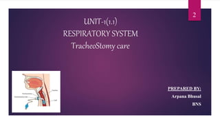
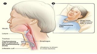
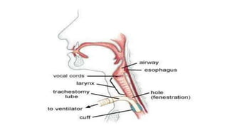
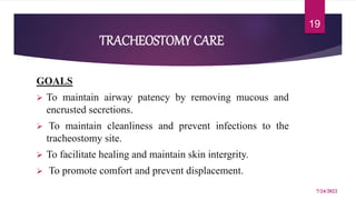
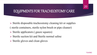
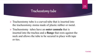
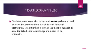
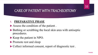
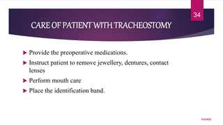
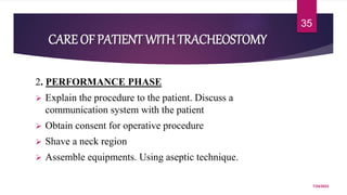
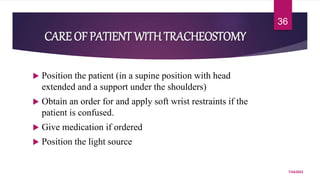
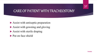
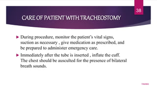
A tracheostomy is a surgical opening made in the neck into the trachea to allow direct access to the breathing tube. It is done to relieve upper airway obstruction, facilitate mechanical ventilation, or remove secretions. Key aspects of tracheostomy care include cleaning and suctioning the tube regularly to keep the airway clear, checking for complications like infection or bleeding, and maintaining proper tube placement and function. Daily care involves monitoring the patient's breathing status and suctioning as needed to prevent obstruction and promote comfort.
Endotracheal suctioning involves mechanically aspirating pulmonary secretions from patients with an artificial airway. It is done to maintain a clear airway, improve oxygenation, stimulate coughing, and prevent infections. Signs that suctioning is needed include abnormal breath sounds, increased pressures during ventilation, inability to cough effectively, or deteriorating blood gases. Risks include hypoxemia, infection, and trauma, so nurses assess patients' tolerance of the procedure and position them comfortably before carefully performing suctioning with sterile technique.
The intensive care unit (ICU) provides specialized monitoring and treatment for critically ill patients. There are various types of ICUs depending on the specific medical needs, such as surgical ICU, cardiac ICU, and pediatric ICU. The ICU is equipped to provide life support and closely monitor vital functions through equipment like cardiac monitors, ventilators, and invasive pressure monitors. Patients admitted to the ICU typically have critical illnesses, organ failures, or require major surgery and post-operative care. The ICU aims to optimize life support and adequate monitoring through the use of specialized equipment, monitoring devices, catheters, drains, and medical staff expertise.
This document discusses various complications that can occur during or as a result of critical care unit (CCU) treatment and care. It outlines common complications such as ventilator-associated pneumonia, bloodstream infections, delirium, weakness, pressure ulcers, and kidney or liver failure. It provides details on prevention and treatment strategies for each complication, with a focus on minimizing the risks through careful monitoring, following best practices and bundles of care, and considering patients' long-term prognosis and quality of life beyond their acute illness.
Central venous pressure (CVP) is the pressure measured in the central veins close to the heart and indicates right atrial pressure. CVP is measured using a catheter placed in a central vein that is connected to a manometer or pressure transducer. Normal CVP ranges from 1-7 mmHg or 5-10 cm H2O. CVP monitoring provides information about cardiac function and volume status and is used to guide fluid administration and assess patients' hemodynamic status. Complications of CVP monitoring include hemorrhage, pneumothorax, infection, and thrombosis.
Critical care involves close monitoring and treatment of seriously ill patients in an intensive care unit (ICU). It aims to support failing organ systems and prevent further damage through evidence-based interventions and a multidisciplinary team approach. Key aspects of ICU care include recognizing severity of illness, initiating early goal-directed therapy to stabilize physiological parameters, and implementing bundles of best practices for issues like ventilation, central lines, and sepsis to improve outcomes. Bundles comprise proven practices that must be reliably performed together to achieve benefits greater than their individual elements alone.
CVP monitoring involves inserting a catheter into a large central vein and connecting it to a pressure monitoring device to measure central venous pressure. CVP provides information about right ventricular function and intravascular volume status. It is used to guide fluid resuscitation and assess the effectiveness of treatments for conditions like heart failure. Key steps in CVP monitoring include positioning the patient supine, zeroing the monitoring device at the level of the right atrium, and observing pressure waveforms and readings to evaluate volume status and cardiac function. Nurses are responsible for assessing the catheter site for complications and maintaining the sterility and function of the CVP monitoring system.
What type of procedure is suctioning?
Suctioning is 'the mechanical aspiration of pulmonary secretions from a patient with an artificial airway in place'. The procedure involves patient preparation, the suctioning event(s) and follow-up care.
Mechanical ventilator for nurses 08.02.19Johny Wilbert
A mechanical ventilator is a device that provides mechanical ventilation by delivering oxygen-enriched air into and out of the lungs. It is used to treat conditions that cause inadequate breathing, such as respiratory failure. There are different types of ventilators that deliver breaths via positive or negative pressure. Modes of ventilation include controlled, assist-control, and synchronized intermittent mandatory ventilation. Nursing care for a patient on a ventilator involves frequent monitoring of vital signs and lung sounds, suctioning as needed, ensuring proper ventilator settings and alarms, turning/mobilizing the patient, and assessing readiness for weaning from the ventilator.
This document discusses the monitoring of critically ill patients. It explains that critically ill patients are difficult to diagnose and manage in general wards due to incomplete histories, inconclusive exams, and disappearing signs near death. Intensive care units are better equipped with monitoring technologies and expert staff to continuously monitor patients. The document outlines various physiological parameters to monitor in critically ill patients, including cardiovascular, respiratory, renal, central nervous, coagulation systems, and hematological factors. Continuous monitoring in ICUs allows for assessment of patients' physiological reserves and the effectiveness of treatments.
This document provides information about a seminar on hemodynamic monitoring presented by UMAdevi.k. It discusses the purpose of hemodynamic monitoring in critically ill patients, which is to continuously assess the cardiovascular system and diagnose/manage complex medical conditions. Specific techniques covered include arterial blood pressure monitoring, central venous pressure monitoring, and pulmonary artery catheter pressure monitoring. Key aspects of each technique like indications, equipment, procedures, nursing responsibilities, and potential complications are defined. Normal hemodynamic values are also provided.
OXYGEN DELIVERY DEVICES - Dr ADIL FAROOQAdil Farooq
Dr. Adil Farooq presented on oxygen therapy and different devices used to deliver oxygen to patients. There are many ways to prescribe oxygen that are often not followed properly. The goal of oxygen therapy is to treat hypoxemia, decrease work of breathing, and decrease myocardial work. The appropriate oxygen delivery method depends on factors like the needed FiO2 level, humidification needs, and patient comfort. Common devices include nasal cannulas, masks, Venturi masks, hoods and tents. Proper prescription and monitoring are important to provide benefits while avoiding risks like hypoventilation.
Central venous catheters and other intravenous (IV) lines are inserted into large veins to administer medications and fluids in critically ill patients. They are used when peripheral veins are inadequate or for medications that cannot be given elsewhere. Potential complications include infection, bleeding, collapsed lungs, and clots. Nurses monitor for complications, ensure patency and sterility of lines, and record indwelling catheter lengths. Mechanical ventilators, pulse oximeters, and other devices are also described. Intensive care units are equipped with advanced monitoring and life support devices operated by specialized healthcare teams to care for critically ill patients.
1. The trauma protocol outlines the assessment and management of critically injured patients according to ATLS guidelines, with a focus on the ABCs - airway, breathing, circulation, disability and exposure.
2. Interventions include securing the airway with bag-valve-mask ventilation or intubation, assessing breathing with pulse oximetry and chest x-rays, supporting circulation with IV access and fluid boluses, evaluating neurologic status via GCS, and conducting a full secondary survey and spinal precautions.
3. Additional tests such as FAST scan, pelvic films and head/neck CTs help guide management, which may involve mechanical ventilation, chest tube insertion, blood transfusion, pelvic binding, wound
1. The document discusses the assessment and management of critically ill patients using the ABCDE approach. It outlines the objectives, definitions of critical illness, principles of management, and scoring systems used to evaluate severity of illness.
2. The ABCDE approach involves assessing the airway, breathing, circulation, disability, and exposure/environment. Initial assessment involves stabilizing the patient and identifying life-threatening problems. Further examination is then conducted once the patient is stabilized.
3. Severity of illness scoring systems like APACHE II and SOFA are used to predict outcomes, guide resource allocation, and evaluate quality of care over time. They assess physiological variables and degree of organ dysfunction to determine illness severity.
The document presents information about a seminar on Acute Respiratory Distress Syndrome (ARDS). The seminar aims to provide in-depth knowledge of ARDS including defining it, describing the pathophysiology and management. ARDS is a life-threatening condition that prevents enough oxygen from entering the blood. It occurs when the lungs become severely inflamed and fluid builds up in the tiny air sacs of the lungs. The seminar will discuss etiology, risk factors, clinical manifestations, diagnostic evaluation, complications, and the nurse's role in management.
1. The document provides guidelines for the care of patients requiring mechanical ventilation including indications for ventilation, modes of ventilation, troubleshooting alarms, weaning criteria and processes, and complications.
2. Mechanical ventilation is used to support breathing for those unable to maintain adequate oxygen or CO2 levels spontaneously, including those with respiratory failure, neuromuscular diseases, or trauma/illnesses affecting breathing.
3. Modes of ventilation include controlled, assisted, intermittent mandatory, and pressure support modes. Nurses must monitor for alarms related to pressures, volumes, and apnea and address causes such as tubing issues or secretions.
Nosocomial or hospital-acquired infections are a major problem, especially in intensive care units. The document discusses the definition, incidence, common types of infections, causative organisms, risk factors, modes of transmission, prevention methods, and the roles and responsibilities of nurses and infection control programs. It emphasizes the importance of hand hygiene, use of personal protective equipment, surveillance, policies, training, and guidelines to reduce infection rates in critical care units.
Endotracheal intubation involves inserting a tube into the trachea through the mouth or nose. It is done to administer oxygen, remove secretions, promote airway patency, or assist with breathing difficulties. The document discusses indications for intubation, equipment needed, steps for performing intubation, post-intubation care, complications, and the importance of documentation.
A lung abscess is a localized cavity filled with pus in the lung, usually caused by a bacterial infection. Common causes include infections by anaerobic bacteria, aspiration of foreign materials, and complications from conditions like pneumonia or tumors. Symptoms include cough with foul smelling sputum, chest pain, fever, and dyspnea. Diagnosis involves chest x-ray, CT scan, and sputum culture. Treatment consists of antibiotics based on culture results, drainage procedures, and surgery if complications develop.
This document discusses nasogastric tube feeding, including:
- The purpose is to remove fluid/gas from the GI tract, prevent/relieve nausea/vomiting after surgery, and administer medications/feedings directly into the GI tract.
- The procedure involves measuring and lubricating the tube, inserting it through the nose and down the esophagus into the stomach, and checking placement by aspirating contents.
- Potential complications include pulmonary aspiration, diarrhea, tube occlusion, constipation, abdominal issues, and tube displacement. Nursing care focuses on monitoring outputs, tube care, and skin assessment.
This document provides information about arterial blood gas (ABG) analysis, including the nurse's role in ABG testing. It discusses the importance of ABG interpretation for critically ill patients. The document outlines the six-step process for ABG interpretation and lists common acid-base disorders. It also defines an ABG test, describes normal ABG values, indications for testing, sample collection points and techniques, equipment used, and potential complications of ABG sampling. The overall goal is to help nurses understand ABGs and properly perform arterial punctures to obtain blood samples.
This document outlines the care of critically ill patients in the intensive care unit (ICU). It discusses the levels of care in the ICU from general ward care to intensive care. It describes the comprehensive management of critically ill patients which includes monitoring, respiratory, cardiovascular, gastrointestinal, nutritional, infection control and psychological support. Key aspects of care include pain management, reducing anxiety, preventing complications like delirium, sleep disturbances, and meeting the needs of family members of critically ill patients. The overall aim is to provide optimal care, support rehabilitation and improve the patient and family experience in the ICU.
1) The document discusses nursing management of critically ill patients, defining critical care nursing, critically ill patients, and critical care units.
2) It outlines the admission process and assessments nurses perform on patients in critical care units, including checking airway, breathing, circulation, and performing full physical assessments.
3) The document details aspects of nursing management in critical care units, which includes continuous monitoring, respiratory care, cardiovascular care, nutritional care, infection control, and communication with patients and relatives.
The document discusses critical care, describing the intensive care team, critical care nursing, the seven Cs of critical care, and the roles of critical care nurses, units, and physicians. It outlines staffing requirements for critical care units, including nurses, respiratory therapists, and physician subspecialists who should be available to treat critically ill patients.
This document provides information on the care of patients on ventilators and weaning. It begins with definitions of ventilator terminology and then describes the types of ventilators, modes of ventilation, indications for ventilation, initial settings, complications and nursing care considerations. Positive pressure ventilators require an artificial airway while negative pressure ventilators do not. Modes include controlled, assist-control, SIMV, pressure support and others. Nurses must carefully monitor patients, ventilator settings and alarms, suction airways as needed and meet other physiological needs.
ET TUBE intubation and it's nursing management
especially useful for BNS students (Adult)as well as for medical students.: MBBS, Staff Nurse, BDS, Lab Technician etc...
This document discusses current concepts in managing the difficult airway. It summarizes several alternative airway devices and techniques including lighted stylets, video laryngoscopes, rigid and flexible fiber-optic laryngoscopes, supraglottic airway devices, awake intubation techniques using topical anesthesia, flexible fiber-optic intubation, retrograde intubation, transtracheal jet ventilation, cricothyrotomy, and tracheostomy. It provides tables describing many new airway devices and concludes that clinical experience is crucial for applying these techniques and devices to solve most airway problems.
The document discusses various emergency surgical airway techniques including needle cricothyrotomy, percutaneous cricothyrotomy, and surgical cricothyrotomy. It provides indications for when a surgical airway is needed such as airway obstruction or trauma. The steps for performing a surgical cricothyrotomy are outlined which involve locating and incising the cricothyroid membrane to access the trachea. Complications are discussed. Other emergency airway techniques like retrograde intubation, jet ventilation, and open tracheotomy are also mentioned.
The document discusses tracheostomy, including:
1. Tracheostomy is a surgical procedure that creates an opening in the trachea to serve as an airway.
2. Complications include tracheal stenosis, swallowing difficulty, voice changes, breathing issues, and hemorrhage.
3. Post-op care involves having a spare tube available, communicating with patients and caregivers, and determining decannulation based on cough strength.
The document provides information on the history, indications, anatomy, procedures, tube types, and complications of tracheostomy.
This document provides information on tracheostomy care including:
1) Tracheostomies are surgical openings in the neck to facilitate breathing by bypassing the upper airway.
2) Tracheostomy tubes come in several types including single cannula, double cannula, cuffed, and fenestrated.
3) Proper tracheostomy care includes cleaning the inner cannula, suctioning secretions, and changing the dressing around the stoma opening.
This document provides an overview of chest tube insertion including definitions, indications, contraindications, equipment, preparation, techniques, and potential complications. It discusses in detail the appropriate tube size for different clinical scenarios such as pneumothorax, hemothorax, and various pleural effusions. Safe sites for tube insertion are outlined. The standard technique and Seldinger technique for chest tube placement are described.
1. Video-assisted thoracoscopic surgery (VATS) is a minimally invasive procedure used to diagnose and treat illnesses of the lungs and chest cavity.
2. VATS involves making small incisions and inserting surgical instruments and a camera to allow the surgeon to see inside the chest. This avoids the need for large incisions.
3. VATS is used for procedures like lung biopsies, removal of parts of the lung, treatment of collapsed lungs, and draining fluid from the chest cavity. It offers benefits like less pain, shorter recovery time, and smaller scars compared to traditional open chest surgery.
An endotracheal tube is a flexible plastic tube inserted through the mouth into the trachea to allow a patient to breathe with a ventilator when they are unable to do so independently. It may be used during surgery, for respiratory issues like pneumonia, or after lung cancer surgery. Inserting the tube is called intubation and requires preparing equipment like a laryngoscope, securing and positioning the patient, confirming proper tube placement, and attaching the patient to a ventilator. Nurses play an important role in intubation by preparing equipment and medications, assisting the physician, monitoring the patient, and managing care of the endotracheal tube and ventilated patient.
A tracheostomy is an opening (made by an incision) through the neck into the trachea (windpipe). A tracheostomy opens the airway and aids breathing.
A tracheostomy may be done in an emergency, at the patient’s bedside or in an operating room. Anesthesia (pain relief medication) may be used before the procedure. Depending on the person’s condition, the tracheostomy may be temporary or permanent.
A tracheostomy is a surgically created opening in the trachea (windpipe) through which a tracheostomy tube is inserted to provide an airway. There are several types including elective, emergency, and percutaneous tracheostomy. Indications include upper airway obstruction, need for prolonged mechanical ventilation, and inability to clear secretions. The procedure involves making an incision through the neck into the trachea and inserting a tracheostomy tube. Complications can include bleeding, infection, and tracheal stenosis. Ongoing care involves cleaning the stoma, suctioning secretions, monitoring tube placement, and providing nutrition and communication methods.
This document provides information on tracheostomy care and management. It defines key terms, outlines the indications for tracheostomy placement, potential complications, proper placement according to anatomy, and the steps for tracheostomy care and management. The goal of tracheostomy care is to maintain airway patency, cleanliness, comfort, and prevent displacement. It describes assessing the patient's respiratory status, secretions, and dressing before performing tracheostomy tube suctioning, cleaning, and dressing changes with the aim of removing secretions and maintaining a clean tracheostomy site.
Cricothyroidotomy and tracheostomy are emergency procedures used to establish an airway when traditional methods like intubation are ineffective or contraindicated. Cricothyroidotomy involves creating an opening through the cricothyroid membrane below the larynx. It is indicated when upper airway obstruction prevents intubation. Tracheostomy involves surgically creating an opening directly into the trachea below the larynx. Both procedures allow for passage of air and can be life-saving but also carry risks of complications if not performed correctly. The document discusses techniques, indications, contraindications and complications for each procedure.
This document provides information about tracheostomy including:
- A brief history and current uses of the procedure
- Indications and contraindications
- Anatomy of the trachea
- Surgical steps for performing an open tracheostomy
- Types of tracheostomy tubes and their uses
- Post-operative care considerations
The document serves as an educational guide for performing tracheostomies and tracheostomy tube selection and management.
The document discusses tracheostomy, including its definition as a surgical opening into the trachea to place an indwelling tube to manage airway obstruction or facilitate ventilation. It covers the history, indications, types, parts of tracheostomy tubes, advantages, pre-operative workup, surgical techniques, post-operative care, complications, and recent research. The purpose is to provide information about tracheostomy to a nursing professor and students.
This document provides information on tracheostomy care including the purpose, equipment, nursing actions, and responsibilities. A tracheostomy is an opening in the neck into the trachea that aids breathing. Tracheostomy care includes cleaning the site and changing inner tubes and dressings. The purposes are to maintain an open airway, prevent infection, facilitate healing, and assess the condition of the stoma. Required equipment includes tracheostomy kits, gloves, masks, and cleaning solutions. Key nursing actions are examining the site, explaining the procedure, cleaning and changing the inner tube and dressings while maintaining sterility. Nurses are responsible for ensuring proper care, hand hygiene, tube changes, and monitoring for signs of infection.
This document discusses different surgical airway techniques that can be used when endotracheal intubation fails or is not possible. It describes needle cricothyrotomy with translaryngeal jet ventilation, cricothyrotomy, and tracheostomy. In emergency situations, cricothyrotomy is preferred over tracheostomy due to its faster speed and lower risks. However, cricothyrotomy should be avoided in children younger than 10-12 years due to the small size of the cricoid cartilage and higher complication rates. The document provides details on how to perform each technique and notes important pediatric considerations for surgical airways.
UNIT 5.2 PHYSIOLOGICAL CONCEPTS AND PHYSICAL CHARACTERISTICS: Estrogens replacement therapy is commenced on the basis of dependent or previously estrogen sensitive women.
Low dose estrogens for short period to improve metabolic state. Estrogens therapy relieves unpleasant symptoms such as hot flashes and vaginal dryness and also appeared to protect against postmenopausal conditions such as osteoporosis and heart disease
A wound is a break or cut in the continuity of any body structure, internal or external caused by physical means.
A wound is a type of injury which happens relatively quickly in which skin is torn, cut, or punctured (an open wound), or where blunt force trauma causes a contusion (a closed wound).
“Trauma” = Injury of one or more systems,that results in excessive bleeding and mayaffect the normal body functioning.
Defined as cellular disruption caused by anexchange with environmental energy that isbeyond the body's resilience.
This document discusses substance abuse, focusing on alcohol overdose and cannabis intoxication. It defines substance abuse and lists the most common substances abused, including alcohol and cannabis. For alcohol overdose, it describes the lethal dose, signs and symptoms, diagnosis involving breathalyzer or blood tests, and emergency management following CAB principles. For cannabis intoxication, it discusses lethal doses, causes of dependency and overdose, signs of dependency and overdose, diagnosis using urine tests, and general emergency management involving supportive care.
Poisoning is injury or death due to swallowing, inhalation, touching or injecting various drugs, chemical, venoms or gases.
Many substances such as drugs, carbon monoxide, food poisoning, organo-phosphorus are poison.
Poisoning can be an accident or a planned action.
Organophosphate poisoning is poisoning due to organophosphates (OPs). Organophosphates are used as insecticides, medications, and nerve agents.
Symptoms include increased saliva and tear production, diarrhea, vomiting, small pupils, sweating, muscle tremors, and confusion.
Other names: Organophosphate toxicity
Causes: organophosphates
Hypovolemic shock is a life-threatening emergency in which severe blood or other fluid loss makes the heart unable to pump enough blood to the body. This type of shock can cause many organs to stop working.
Hypovolemic shock is a dangerous condition that happens when suddenly lose a lot of blood or fluids from body. This drops blood volume, the amount of blood circulating in body. That’s why it’s also known as low-volume shock.
Heat stroke a core temperature ≥40°C accompanied by CNS dysfunction in patients with environmental heat exposure. This condition represents a failure of the body's ability to maintain thermoregulatory homeostasis.
Hemorrhage is the loss of blood escaping from the circulatory system.
Bleeding can occur internally, where blood leaks from blood vessels inside the body, or externally either through a natural opening such as mouth, nose, ear, urethra or anus or through a break in the skin.
Uncontrolled bleeding can rapidly lead to shock and death.
Excessive or uncontrollable bleeding, often caused by trauma, surgical or obstetrical complications, or the advanced stages of certain illnesses such as cirrhosis and peptic ulcer disease.
An airway obstruction is a blockage in any part of the airway.
The airway is a complex system of tubes that conveys inhaled air from nose and mouth into the lungs.
An obstruction may partially or totally prevent air from getting into lungs.
Acute upper airway obstruction is a life-threatening medical emergency.
This document provides information on emergency care and triage. It discusses the principles of emergency care which include providing care without delay and using triage to prioritize patients. Triage involves sorting patients into categories of emergent, urgent, and non-urgent based on the seriousness of their conditions. The document then describes the triage process in more detail, including the different color codes used to categorize patients and the criteria for each category. It also discusses the roles of triage team members and how to set up an effective triage system.
Emergency nursing is a nursing specialty in which nurses care for patients in the emergency or critical phase of their illness or injury.
While this is common to many nursing specialties, the key difference is that an emergency nurse is skilled at dealing with people in the phase when a diagnosis has not yet been made and the cause of the problem is not known.
The document provides information on the care of patients undergoing traction. It defines traction as applying a pulling force to part of the body. Traction is used to provide alignment, reduce muscle spasms, prevent deformities, provide immobilization, and increase space between opposing surfaces. The document outlines different types of traction including skin, skeletal, and manual traction. It discusses nursing responsibilities like ensuring comfort, preventing complications, educating patients, and promoting exercise and mobility.
Rheumatoid arthritis (RA) facts
Rheumatoid arthritis is an autoimmune disease that can cause chronic inflammation of the joints and other areas of the body.
It can affect people of all ages.
The cause of rheumatoid arthritis is not known.
In rheumatoid arthritis, multiple joints are usually, affected in a symmetrical pattern.
Paget disease is a chronic bone disorder that typically results in enlarged, deformed bones due to excessive breakdown and formation of bone tissue that can cause bones to weaken and may result in bone pain, arthritis deformities or fractures.
Osteomalacia is a bone condition caused by vitamin D deficiency or impaired mineralization. It results in soft, weakened bones due to incomplete mineralization of bone matrix. Symptoms include bone pain, tenderness, fractures, and muscle weakness. Diagnosis involves x-rays showing pseudofractures and bone biopsy showing excess osteoid tissue. Treatment focuses on calcium and vitamin D supplementation to promote bone mineralization. Nursing care includes education on diet, sunlight exposure, pain management, and monitoring for treatment effectiveness.
Osteoporosis is a chronic, progressive disease of multifactorial etiology.
It is most frequently recognized in particularly in elderly people and does occur in sexes, all races, and all age groups.
Osteoporosis is a preventable disease that can result in disturbing physical, psychosocial, and economic consequences.
Osteoporosis is a systemic skeletal disease characterized by low bone mass and micro architectural deterioration of bone tissue.
This document provides information about osteomyelitis, including:
1) Osteomyelitis is an inflammation of bone caused by an infecting organism that may remain localized or spread through the bone. Common causes are bacteria or fungi entering through a break in the skin or spreading via blood.
2) It can be classified as acute (less than 2 weeks), subacute (2-6 weeks), or chronic (over 6 weeks) based on duration of symptoms. It can also be classified based on mechanism of infection such as exogenous (from outside trauma/surgery) or hematogenous (from another infectious site).
3) Staphylococcus aureus is the most common pathogen. Risk factors
Cancer is a disease of the cells in the body. The body is made up from millions of tiny cells. There are several types of oral cancers, but around 90% are squamous cell carcinomas originating in the tissues that line the mouth and lips.
Oral or mouth cancer most commonly involves the tongue.
Pictorial and detailed description of patellar instability with sign and symptoms and how to diagnose , what investigations you should go with and how to approach with treatment options . I have presented this slide in my 2nd year junior residency in orthopedics at LLRM medical college Meerut and got good reviews for it
After getting it read you will definitely understand the topic.
STUDIES IN SUPPORT OF SPECIAL POPULATIONS: GERIATRICS E7shruti jagirdar
Unit 4: MRA 103T Regulatory affairs
This guideline is directed principally toward new Molecular Entities that are
likely to have significant use in the elderly, either because the disease intended
to be treated is characteristically a disease of aging ( e.g., Alzheimer's disease) or
because the population to be treated is known to include substantial numbers of
geriatric patients (e.g., hypertension).
Computer in pharmaceutical research and development-Mpharm(Pharmaceutics)MuskanShingari
Statistics- Statistics is the science of collecting, organizing, presenting, analyzing and interpreting numerical data to assist in making more effective decisions.
A statistics is a measure which is used to estimate the population parameter
Parameters-It is used to describe the properties of an entire population.
Examples-Measures of central tendency Dispersion, Variance, Standard Deviation (SD), Absolute Error, Mean Absolute Error (MAE), Eigen Value
Nutritional deficiency Disorder are problems in india.
It is very important to learn about Indian child's nutritional parameters as well the Disease related to alteration in their Nutrition.
Dr. Tan's Balance Method.pdf (From Academy of Oriental Medicine at Austin)GeorgeKieling1
Home
Organization
Academy of Oriental Medicine at Austin
About AOMA: The Academy of Oriental Medicine at Austin offers a masters-level graduate program in acupuncture and Oriental medicine, preparing its students for careers as skilled, professional practitioners. AOMA is known for its internationally recognized faculty, award-winning student clinical internship program, and herbal medicine program. Since its founding in 1993, AOMA has grown rapidly in size and reputation, drawing students from around the nation and faculty from around the world. AOMA also conducts more than 20,000 patient visits annually in its student and professional clinics. AOMA collaborates with Western healthcare institutions including the Seton Family of Hospitals, and gives back to the community through partnerships with nonprofit organizations and by providing free and reduced price treatments to people who cannot afford them. The Academy of Oriental Medicine at Austin is located at 2700 West Anderson Lane. AOMA also serves patients and retail customers at its south Austin location, 4701 West Gate Blvd. For more information see www.aoma.edu or call 512-492-303434.
Travel Clinic Cardiff: Health Advice for International TravelersNX Healthcare
Travel Clinic Cardiff offers comprehensive travel health services, including vaccinations, travel advice, and preventive care for international travelers. Our expert team ensures you are well-prepared and protected for your journey, providing personalized consultations tailored to your destination. Conveniently located in Cardiff, we help you travel with confidence and peace of mind. Visit us: www.nxhealthcare.co.uk
Discover the benefits of homeopathic medicine for irregular periods with our guide on 5 common remedies. Learn how these natural treatments can help regulate menstrual cycles and improve overall menstrual health.
Visit Us: https://drdeepikashomeopathy.com/service/irregular-periods-treatment/
Breast cancer: Post menopausal endocrine therapyDr. Sumit KUMAR
Breast cancer in postmenopausal women with hormone receptor-positive (HR+) status is a common and complex condition that necessitates a multifaceted approach to management. HR+ breast cancer means that the cancer cells grow in response to hormones such as estrogen and progesterone. This subtype is prevalent among postmenopausal women and typically exhibits a more indolent course compared to other forms of breast cancer, which allows for a variety of treatment options.
Diagnosis and Staging
The diagnosis of HR+ breast cancer begins with clinical evaluation, imaging, and biopsy. Imaging modalities such as mammography, ultrasound, and MRI help in assessing the extent of the disease. Histopathological examination and immunohistochemical staining of the biopsy sample confirm the diagnosis and hormone receptor status by identifying the presence of estrogen receptors (ER) and progesterone receptors (PR) on the tumor cells.
Staging involves determining the size of the tumor (T), the involvement of regional lymph nodes (N), and the presence of distant metastasis (M). The American Joint Committee on Cancer (AJCC) staging system is commonly used. Accurate staging is critical as it guides treatment decisions.
Treatment Options
Endocrine Therapy
Endocrine therapy is the cornerstone of treatment for HR+ breast cancer in postmenopausal women. The primary goal is to reduce the levels of estrogen or block its effects on cancer cells. Commonly used agents include:
Selective Estrogen Receptor Modulators (SERMs): Tamoxifen is a SERM that binds to estrogen receptors, blocking estrogen from stimulating breast cancer cells. It is effective but may have side effects such as increased risk of endometrial cancer and thromboembolic events.
Aromatase Inhibitors (AIs): These drugs, including anastrozole, letrozole, and exemestane, lower estrogen levels by inhibiting the aromatase enzyme, which converts androgens to estrogen in peripheral tissues. AIs are generally preferred in postmenopausal women due to their efficacy and safety profile compared to tamoxifen.
Selective Estrogen Receptor Downregulators (SERDs): Fulvestrant is a SERD that degrades estrogen receptors and is used in cases where resistance to other endocrine therapies develops.
Combination Therapies
Combining endocrine therapy with other treatments enhances efficacy. Examples include:
Endocrine Therapy with CDK4/6 Inhibitors: Palbociclib, ribociclib, and abemaciclib are CDK4/6 inhibitors that, when combined with endocrine therapy, significantly improve progression-free survival in advanced HR+ breast cancer.
Endocrine Therapy with mTOR Inhibitors: Everolimus, an mTOR inhibitor, can be added to endocrine therapy for patients who have developed resistance to aromatase inhibitors.
Chemotherapy
Chemotherapy is generally reserved for patients with high-risk features, such as large tumor size, high-grade histology, or extensive lymph node involvement. Regimens often include anthracyclines and taxanes.
Giloy in Ayurveda - Classical Categorization and SynonymsPlanet Ayurveda
Giloy, also known as Guduchi or Amrita in classical Ayurvedic texts, is a revered herb renowned for its myriad health benefits. It is categorized as a Rasayana, meaning it has rejuvenating properties that enhance vitality and longevity. Giloy is celebrated for its ability to boost the immune system, detoxify the body, and promote overall wellness. Its anti-inflammatory, antipyretic, and antioxidant properties make it a staple in managing conditions like fever, diabetes, and stress. The versatility and efficacy of Giloy in supporting health naturally highlight its importance in Ayurveda. At Planet Ayurveda, we provide a comprehensive range of health services and 100% herbal supplements that harness the power of natural ingredients like Giloy. Our products are globally available and affordable, ensuring that everyone can benefit from the ancient wisdom of Ayurveda. If you or your loved ones are dealing with health issues, contact Planet Ayurveda at 01725214040 to book an online video consultation with our professional doctors. Let us help you achieve optimal health and wellness naturally.
Gene therapy can be broadly defined as the transfer of genetic material to cure a disease or at least to improve the clinical status of a patient.
One of the basic concepts of gene therapy is to transform viruses into genetic shuttles, which will deliver the gene of interest into the target cells.
Safe methods have been devised to do this, using several viral and non-viral vectors.
In the future, this technique may allow doctors to treat a disorder by inserting a gene into a patient's cells instead of using drugs or surgery.
The biggest hurdle faced by medical research in gene therapy is the availability of effective gene-carrying vectors that meet all of the following criteria:
Protection of transgene or genetic cargo from degradative action of systemic and endonucleases,
Delivery of genetic material to the target site, i.e., either cell cytoplasm or nucleus,
Low potential of triggering unwanted immune responses or genotoxicity,
Economical and feasible availability for patients .
Viruses are naturally evolved vehicles that efficiently transfer their genes into host cells.
Choice of viral vector is dependent on gene transfer efficiency, capacity to carry foreign genes, toxicity, stability, immune responses towards viral antigens and potential viral recombination.
There are a wide variety of vectors used to deliver DNA or oligo nucleotides into mammalian cells, either in vitro or in vivo.
The most common vector system based on retroviruses, adenoviruses, herpes simplex viruses, adeno associated viruses.
3. Content layout
Definition
Purpose
Indication
Contraindication
Equipments for tracheostomy insertion.
Goals of tracheostomy care.
Equipments for tracheostomy care.
7/24/2022
3
4. Content Layout
Components and Types of tracheostomy tube
Care of patient with tracheostomy
Daily care of patient with tracheostomy.
Nursing management
Complication.
Preventing complication associated with tracheostomy
tube
7/24/2022
4
5. DEFINITION
Trachea = windpipe
Ostomy = surgical opening in the body
Tracheostomy is a surgical procedure which consist of
making an artificial opening on the anterior aspect of neck
and opening a direct airway through an incision in the
trachea(2-3/3-4 tracheal rings).
7/24/2022
5
6. DEFINITION (1/4)
A tracheostomy is a surgical opening into the trachea
below the larynx through which an indwelling tube is
placed to overcome upper airway obstruction, facilitate
mechanical ventilatory support and/ or the removal of
tracheobronchial secretions.
7/24/2022
6
9. PURPOSE
a) To maintain the airways to facilitate the therapeutic
exchanges of gases.
b) To remove trachea bronchial secretion.
c) To maintain optimum physical comfort.
d) To decrease airway resistance.
7/24/2022
9
10. PURPOSE
e) To provide a method of mechanical ventilation.
f) To improve respiratory insufficiency.
g) To prevent from aspiration and transmission of
pathogenic micro-organisms.
7/24/2022
10
11. INDICATION
1. Airway obstruction
Hemorrhage after thyroid surgery or upper airway
bleeding.
Foreign bodies impacted in larynx.
Trauma to larynx or pharynx.
Acute edema of epiglottis e.g. diphtheria,
Facial burns
7/24/2022
11
12.
13. indication
2. Congenital cause
Laryngeal weakness stenosis
Inflammatory disease conditions
Tracheal laryngeal fracture
Need for continuous mechanical ventilation
Tumor in the respiratory airway
7/24/2022
13
14. indication
3. Retained secretions in tracheo-bronchial tree
Unconscious patient following head injury and poisoning.
Chest injury patient who is unable to cough.
Paralysis of the muscles of respiration.
Tetanus
For radical surgery in the neck e.g. laryngectomy.
7/24/2022
14
15. INDICATION
4. Others
To protect / minimize risk of aspiration in the patients
with poor or absent cough reflex
Neurologic conditions (Amyotrophic lateral sclerosis)
Severe sleep apnea
Laryngeal hypoplasia
7/24/2022
15
16. CONTRAINDICATION
No absolute contraindications exist to tracheostomy.
Relative contraindication is Laryngeal CA.
Tracheo-esophageal fistula.
Cancer in upper GI or respiratory tract
7/24/2022
16
17. EQUIPMENTS FOR TRACHEOSTOMY
INSERTION
Tracheostomy tube (size 6-9 mm for most adults)
Sterile instruments: blade, forceps, suture material,
scissors
Sterile gown and gloves
Cap and face shield
Antiseptic preparation solution
Gauze pads
7/24/2022
17
18. EQUIPMENTS FOR TRACHEOSTOMY
INSERTION
Shave preparation kit
Sedation
Local anesthetic and syringe
Resuscitation bag and mask with oxygen source
Suction source and catheters
Syringe for cuff inflation
Respiratory support available for post
tracheostomy(mechanical ventilation, tracheal oxygen mask,
CPAP)
7/24/2022
18
19. TRACHEOSTOMY CARE
GOALS
To maintain airway patency by removing mucous and
encrusted secretions.
To maintain cleanliness and prevent infections to the
tracheostomy site.
To facilitate healing and maintain skin intergrity.
To promote comfort and prevent displacement.
7/24/2022
19
20. EQUIPMENTS FOR TRACHEOSTOMY CARE
Sterile disposable tracheostomy cleaning kit or supplies
( sterile containers, sterile nylon brush or pipe cleaner)
Sterile applicators ( gauze squares)
Sterile suction kit and Sterile normal saline
Sterile gloves and clean gloves
7/24/2022
20
21. EQUIPMENTS
Towel to drape to protect bed linens
Sterile gauze dressing, cotton twill ties and clean scissors
Moisture proof bag
7/24/2022
21
23. BED SIDE EQUIPMENTS
Spare tracheostomy tubes same size and type as the patient is
wearing
Tracheal dilator
Suctioning equipment
Oxygen equipment with humidification
Gloves (non sterile)
Gloves (sterile) for suctioning
Infectious waste bag
7/24/2022
23
25. Tracheostomy tube
Tracheostomy tube is a curved tube that is inserted into
the tracheostomy stoma made of plastic rubber or metal.
Tracheostomy tubes have an outer cannula that is
inserted into the trachea and a flange that rests against the
neck and allows the tube to be secured in place with tape
or ties.
7/24/2022
25
26. TRACHEOSTOMY TUBE
Tracheostomy tubes also have an obturator which is used
to insert the outer cannula which is then removed
afterwards. The obturator is kept at the client's bedside in
case the tube becomes dislodge and needs to be
reinserted.
7/24/2022
26
27. COMPONENTS OF TRACHEOSTOMY TUBE
Outer cannula
Inner cannula: fits snugly into outer tube, can be easily
removed for cleaning
Flange: Flat plastic plate attached to outer tube –lies flush
against the patient’s neck
15mm outer diameter termination: Fits all ventilator and
respiratory equipment.
7/24/2022
27
33. CARE OF PATIENT WITHTRACHEOSTOMY
1. PREPARATIVE PHASE
Assess the condition of the patient .
Bathing or scrubbing the local skin area with antiseptic
procedures.
Keep the patient in NPO.
Promote rest and sleep
Collect informed consent, report of diagnostic test .
7/24/2022
33
34. CARE OF PATIENT WITHTRACHEOSTOMY
Provide the preoperative medications.
Instruct patient to remove jewellery, dentures, contact
lenses
Perform mouth care
Place the identification band.
7/24/2022
34
35. CARE OF PATIENT WITHTRACHEOSTOMY
2. PERFORMANCE PHASE
Explain the procedure to the patient. Discuss a
communication system with the patient
Obtain consent for operative procedure
Shave a neck region
Assemble equipments. Using aseptic technique.
7/24/2022
35
36. CARE OF PATIENT WITHTRACHEOSTOMY
Position the patient (in a supine position with head
extended and a support under the shoulders)
Obtain an order for and apply soft wrist restraints if the
patient is confused.
Give medication if ordered
Position the light source
7/24/2022
36
37. CARE OF PATIENT WITHTRACHEOSTOMY
Assist with antiseptic preparation
Assist with gowning and gloving
Assist with sterile draping
Put on face shield
7/24/2022
37
38. During procedure, monitor the patient’s vital signs,
suction as necessary , give medication as prescribed, and
be prepared to administer emergency care.
Immediately after the tube is inserted , inflate the cuff.
The chest should be ausculted for the presence of bilateral
breath sounds.
7/24/2022
38
CARE OF PATIENT WITHTRACHEOSTOMY
39. CARE OF PATIENT WITHTRACHEOSTOMY
Secure the tracheostomy tube with tapes or other securing
device and apply dressing
Apply appropriate respiratory assistive device(mechanical
ventilation. Tracheostomy, oxygen mask, CPAP)
Check the tracheostomy tube cuff pressure
7/24/2022
39
40. 3. Follow up phase/ Post-operative phase
Check the symmetry of chest expansion.
Auscultate the breathe sound of anterior and the lateral chest
bilaterally .
Obtain order for chest x-ray to verify proper tube
placement.
Check cuff pressure every 8-12 hrs
7/24/2022
40
CARE OF PATIENT WITHTRACHEOSTOMY
41. CARE OF PATIENT WITHTRACHEOSTOMY
Monitor the sign and symptoms of aspiration
Assess vitals signs and breath sounds; note tube size
used, physician performing procedure, type, dose, and
route of medications given.
Assess and chart condition of stoma: bleeding, swelling,
subcutaneous air
7/24/2022
41
42. CARE OF PATIENT WITHTRACHEOSTOMY
Administer oxygen concentration as prescribed by
physician
Secure the tube to the patient face with the tape, and
mark the proximal end for position maintenance.
7/24/2022
42
43. Use the sterile suction technique and airway care to
prevent iatrogenic contamination and infection.
Continue to reposition patient every 2 hours and as needed
to prevent atelectasis and to optimize lung expansion.
Provide oral hygiene and suction the oropharynx whenever
required.
7/24/2022
43
CARE OF PATIENT WITHTRACHEOSTOMY
44. CARE OF PATIENT WITHTRACHEOSTOMY
Maintain patency of tracheostomy tube and airway
Frequent atraumatic suction.
Humidification of inspired air and oxygen
Fowler’s position to aid in breathing
Maintain adequate fluid intake
7/24/2022
44
45. CARE OF PATIENT WITHTRACHEOSTOMY
Provide frequent mouth wash
Mucolytic agents
Coughing and physiotherapy
7/24/2022
45
46. CARE OF PATIENT WITHTRACHEOSTOMY
Prevent infection and complication:
Aseptic tube suction, handling and tube changing
Prophylactic antibiotics
An extra tube, obturator, and tracheostomy kit should be
kept at the bedside. In the event of tube dislodgement,
reinsertion of a new tube mat be necessary .
7/24/2022
46
47. DAILY CARE OF PATIENT WITH
TRACHEOSTOMY
1.Assessment
An increased monitoring of the patient’s blood pressure,
respiratory rate, pulse and color is necessary
An increased in the respiratory rate, wheezes ,an
increased pulse rate may indicate the need for suction
7/24/2022
47
48. DAILY CARE OF PATIENT WITH
TRACHEOSTOMY
Cyanosis and distress not relieved by suctioning should
be reported promptly
Increasing restlessness with a rapid pulse rate may
indicate hypoxia or bleeding
Observe the wound for bleeding and then check daily for
signs of infection
7/24/2022
48
49. DAILY CARE OF PATIENT WITH
TRACHEOSTOMY
2.Maintain an open airway:
Assess the patient regularly for excess secretions and
suction and clean the tube as indicated.
The trachea is suctioned using a sterile glove and a sterile
suction catheter moisture in sterile water or normal saline
with aseptic technique.
7/24/2022
49
50. DAILY CARE OF PATIENT WITH
TRACHEOSTOMY
Frequent repositioning of the patient
3. Suctioning
7/24/2022
50
51. DAILY CARE OF PATIENT WITH
TRACHEOSTOMY
Suctioning raises the risk of hypoxemia, bronchospasm,
and other adverse reactions, so suction only when needed,
not on a set schedule, and suction for the shortest time
necessary to clear secretions.
Suctioning can be an uncomfortable and scary experience
for the patient, so thoroughly explain the procedure to
him before start.
7/24/2022
51
52. Indications for suctioning include coughing, secretions in
the airway, respiratory distress, presence of rhonchi on
auscultation, increased peak airway pressures on the
ventilator, and decreasing SaO2 or PaO2.
Comfortable position of the patient.
Maintain aseptic technique .
7/24/2022
52
DAILY CARE OF PATIENT WITH
TRACHEOSTOMY
53. DAILY CARE OF PATIENT WITH
TRACHEOSTOMY
4.Cleaning of the tracheostomy tube and wound care:
Tracheostomy care includes cleaning or changing the
inner cannula, changing the dressing and tracheostomy
tube holder, and suctioning if needed.
Never clean and reuse a disposable cannula.
7/24/2022
53
54. DAILY CARE OF PATIENT WITH
TRACHEOSTOMY
Unlock and remove the inner cannula and place it in a solution
of equal parts hydrogen peroxide and 0.9% sodium chloride
unless the manufacturer directs otherwise.
The inner cannula of the tracheostomy tube is carefully
removed, cleansed every 3-5 hours or as often as
necessary(perhaps even hourly)
7/24/2022
54
55. Remove encrusted secretions from the lumen with sterile
pipe cleaners.
After cleaning, rinse the cannula thoroughly with sterile
0.9% sodium chloride solution.
7/24/2022
55
DAILY CARE OF PATIENT WITH
TRACHEOSTOMY
56. DAILY CARE OF PATIENT WITH
TRACHEOSTOMY
Reinsert the inner cannula and securely lock it into place.
Stoma should be cleaned everyday carefully without
dislodging tube.
7/24/2022
56
57. DAILY CARE OF PATIENT WITH
TRACHEOSTOMY
5.Humidification:
Maintaining humidification is another key nursing
responsibility.
Provide constant airway humidification to avoid
thickening and crusting of bronchial secretion .
7/24/2022
57
58. DAILY CARE OF PATIENT WITH
TRACHEOSTOMY
Ensure humidification of inspired air placing a piece of moist
gauze over the tracheostomy tube so that the air takes up the
moisture.
Humidifier is useful.
In addition, patient must be properly hydrated; for example,
with I.V. fluids.
7/24/2022
58
59. DAILYCARE OF PATIENT WITHTRACHEOSTOMY
6.Mouth care:
Provide frequent mouth care in every 2 hours for the
patient’s comfort and to reduces the possibility of
infection.
7/24/2022
59
60. DAILY CARE OF PATIENT WITH
TRACHEOSTOMY
7.Nutrition:
If the patient eats by mouth, it is recommended that the
tracheostomy tube be suctioned prior to eating. This often
prevents the need for suctioning during or after meals,
which may stimulate excessive coughing and could result
in vomiting
7/24/2022
60
61. DAILY CARE OF PATIENT WITH
TRACHEOSTOMY
Encouraging fluid intake is helpful for a patient with a
tracheostomy. Increased fluid intake will thin and loosen
secretions making coughing and suctioning easier.
Always observe the patient while eating to be sure food
does not get into the trachea.
7/24/2022
61
62. DAILY CARE OF PATIENT WITH
TRACHEOSTOMY
8. Communication
9. Documentation
Document the patient's response each time you suction,
including his vital signs, cardiac rhythm, oxygen
saturation, amount and consistency of secretions, breath
sounds, and the frequency of needed suctioning
7/24/2022
62
64. NURSINGASSESSMENT
Frequent monitoring of the patients blood pressure ,
respiratory rate, pulse and color is necessary.
An increased in the respiratory rate , wheezes, an
increased pulse rate may indicate the need for suction.
Cyanosis and distress not relieved by suction should be
reported promptly.
7/24/2022
64
65. NURSING ASSESSMENT
Increasing restlessness with a rapid pulse rate may
indicate hypoxia or bleeding.
Observe the wound for bleeding in the immediate post-
operative period and then check daily for signs of
infection and sloughing.
Assess patient ability to understand the spoken word.
7/24/2022
65
66. NURSING ASSESSMENT
Assess patients ability to expression.
Assess and observe swallowing reflexes , gag reflexes.
Assess mental status confusion , lethargy , restlessness.
Assess amount, color, consistency of secretion.
7/24/2022
66
67. NURSING DIAGNOSIS
1. Ineffective airway clearance related to presence of
artificial airway (tracheostomy) as evidenced by
tachypnea and changes in breathing pattern.
2. Impaired verbal communication related to presence of
artificial airway as evidenced by difficulty in maintaining
the usual communication pattern.
7/24/2022
67
68. Nursing diagnosis
3) Anxiety related to threat to self concept as evidenced by
expressed feeling of distress over the presence of
tracheostomy.
4) Knowledge deficit related to new procedure or
intervention in hospital as evidenced by increased
questioning.
7/24/2022
68
69. Nursing diagnosis
5) Risk for infection related to surgical incision of
tracheostomy.
6) Risk of aspiration related to presence of tracheostomy.
7/24/2022
69
70. GOALS AND OUTCOME
1) Patient will maintain a clear ,open airways and ability to
effectively cough up secretion.
2) Patient will use a form of communication to get needs
met and to relate effectively with persons and
environment.
3) Patient will verbalize their own feeling.
7/24/2022
70
71. GOALS AND OUTCOME
4) Patient or caregiver will demonstrate the knowledge and
skills appropriate for tracheostomy care.
5) Patient will remain free of infection.
6) Patient will swallow meals without coughing, choking or
changing color.
7/24/2022
71
72. NURSING INTERVENTION
1. Maintaining airway clearance
Assess changes in BP, Heart rate and temperature.
Auscultate the lungs , presence of breathe sounds.
Observe the color , consistency , and quantity of secretion
Provide warm humidified air
7/24/2022
72
73. Nursing intervention
Administer O2 as needed.
Encourage patient to cough out secretion.
Keep suction equipment and AMBU bag at bed side.
Keep the patient in semi fowlers position.
7/24/2022
73
74. Nursing intervention
2. Promoting verbal communication
Assess the clients communication ability.
Assess the effectiveness of non verbal communication
methods.
Place the patient in a room close to the nurses station.
Provide a call light within easy reach at all times.
7/24/2022
74
75. Nursing intervention
Provide alternative methods for communicating.
Hand gestures
Word and phrase cards
Picture board
Writing pad
Pencil or pen
7/24/2022
75
76. Nursing intervention
3. Reducing anxiety
Assess the level and manifestation of anxiety in patients.
Allow patient to express fears and concerns to ask,
inquiries about disease and procedure.
Inform of all procedure and care to the patient as well as
visitors.
Provide psychological support.
7/24/2022
76
77. Nursing intervention
7/24/2022
77
4. Enhancing knowledge
Assess the patients knowledge regarding the purpose and
care of tracheostomy.
Provide instruction in sterile tracheostomy care and
suctioning.
Instruct in the need to call health care provider if the
amount of secretion increases or change in color and
characteristics occurs.
78. NURSING INTERVENTION
5. Preventing infection
Assess patients vital sign.
Assess skin integrity under tracheal ties.
Monitor white blood cell count.
Observe the stoma for color, crusting lesions
7/24/2022
78
79. NURSING INTERVENTION
Provide stoma care.
Do not allow secretions to pool around the stoma , suction
the area , wipe with aseptic technique.
7/24/2022
79
80. NURSING INTERVENTION
6. Preventing aspiration
Assess the ability to swallow and type of food
consistency.
Encourage liquid initially in small amounts and gradually
increase as tolerated.
Maintain in an upright sitting position during feeding.
Suction fluids from mouth and airway.
7/24/2022
80
81. COMPLICATIONS
1. Immediate (at the time of operation):
hemorrhage, air embolism, cardiac arrest apnea,
aspiration, pneumothorax, injury to laryngeal nerves and
esophagus, local damage (thyroid cartilage, cricoid
cartilage, recurrent laryngeal nerve).
7/24/2022
81
82. complications
2. Intermediate(during first few hours or days)
bleeding, displacement of tube, blocking of tube,
subcutaneous emphysema, pneumothorax, scabs, tracheal
necrosis, tracheitis, local wound infection and dysphagia
7/24/2022
82
83. COMPLICATIONS
3. Late (with prolonged use of tube for weeks and months):
laryngeal stenosis, tracheal stenosis, tracheo - esophageal
fistula.
7/24/2022
83
84. Preventing complications WITH TUBE
Administer adequate warmed humidity: steam inhalation,
nebulization, keeping wet gauze piece over the
tracheostomy site and changing it as per need.
Maintain cuff around tube
Suction as needed per assessment findings
Maintain skin integrity. Change tape and dressing as
needed or per protocol
7/24/2022
84
85. Preventing complication WITH TUBE
Ascultate lungs sounds
Monitor for signs and symptoms of infection, including
temperature and white blood cell count
Protein rich diet should be provided for early healing of
incision site.
Administer prescribed oxygen and monitor oxygen
saturation
7/24/2022
85
86. PREVENTINGCOMPLICATION WITH TUBE
Monitor for cyanosis
Maintain adequate hydration of the patient
Use sterile technique when suctioning and performing
tracheostomy care
Emergency drugs should be ready
7/24/2022
86
87.
88. REFRENCES
Smeltzer.S.G. Bare.B.G. Hinkle.J.G. Cheezer K.H.(2010)
“Brunner & Suddarth’s textbook of Medical- Surgical
Nursing", Volume 1. (12th edition). New Delhi, Kluwer
India. Pvt. Ltd. 2078/04/10 at 1 pm
Mandal G.N (2016) “A Textbook Of Medical Surgical
Nursing”. 5th edition. Kathmandu. Makalu Publication
House.2078/04/11at 3 pm.
7/24/2022
88
89. REFRENCES
2019. Mayo Foundation Of Medical Foundation And
Research. Tracheostomy care
https://www.mayoclinic.org@2021/08/02at 3pm.
Nov4,2019,tracheostomy care
https://www.slideshare.net/gamandeep@2021/08/03 at
5pm
7/24/2022
89