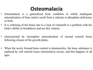
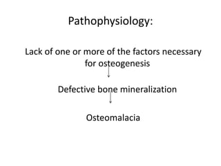
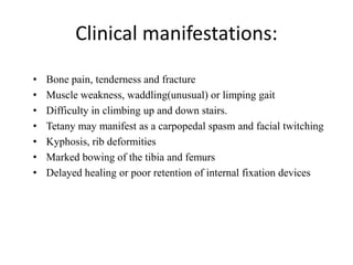
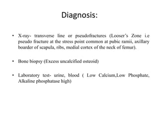
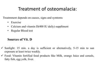
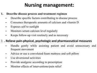
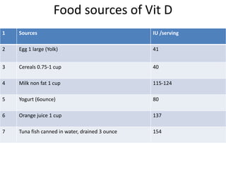
Osteomalacia
Prepare by: RN Arpana Bhusal
BNS
Osteomalacia
• Osteomalacia is a generalized bone condition in which inadequate
mineralization of bone matrix result from a calcium or phosphate deficiency
or both.
• It is softening of the bones due to a lack of vitaminD or a problem with the
body's ability to breakdown and use this vitamin.
• characterized by incomplete mineralization of normal osteoid tissue
following closure of the growth plates.
• When the newly formed bone content is demineralize, the bony substance is
replaced by soft osteoid tissue osteomalacia occurs, and this happens in all
ages.
Osteoids
• Bone matrix primarily composed of type I collagen.
• It does not mineralize properly when there insufficient minerals or
osteoblast dysfunction.
• When the newly formed bone of the growth plate does not
mineralize, the growth plate becomes thick, wide and irregular and
cause rickets in children.
• When the remodeled bone does not mineralize, osteomalacia occurs.
Causes:
• Insufficient calcium absorption from the intestine—lack of
calcium or resistance to the action of vitammin D
• Increased renal phosphorus level
• Under nutrition during pregnancy
• Malabsorption Syndrome
• Partial Gastrectomy
Risk factors:
• Dietary deficiency (dietary, sunlight)
• Sunlight exposure inadequate
• Malabsorption- celiac disease, chronic biliary tract obstruction,
small bowel resection
• Severe renal insufficiency, liver disease
• Prolonged anticonvulsant therapy
Pathogenic Mechanism
• Deficiency of activated vitamin D- promotes calcium absorption from
GIT and facilitate demineralization of bone.
• Low extracellular supply of calcium and phosphate and doesnot
move calcification sites I bone.
• Failed calcium absorption or from excessissive loss of calcium from
the body.
• Chronic billiary tract obstruction, pancreatitis lead to fat inadequately
absorbed and lead to loss of vitamin D and calcium excreted with
fatty acid.
• Liver & Kidney disease lead to lack of Vitamin D and doesnot covert
In activated form.
• Hyperparathyroidism leads to skeletal decalcification and thus to
osteomalacia by increasing excretion of phosphate.
Pathophysiology:
Lack of one or more of the factors necessary
for osteogenesis
Defective bone mineralization
Osteomalacia
Clinical manifestations:
• Bone pain, tenderness and fracture
• Muscle weakness, waddling(unusual) or limping gait
• Difficulty in climbing up and down stairs.
• Tetany may manifest as a carpopedal spasm and facial twitching
• Kyphosis, rib deformities
• Marked bowing of the tibia and femurs
• Delayed healing or poor retention of internal fixation devices
Diagnosis:
• X-ray- transverse line or pseudofractures (Looser’s Zone i.e
pseudo fracture at the stress point common at pubic ramii, axillary
boarder of scapula, ribs, medial cortex of the neck of femur).
• Bone biopsy (Excess uncalcified osteoid)
• Laboratory test- urine, blood ( Low Calcium,Low Phosphate,
Alkaline phosphatase high)
Treatment of osteomalacia:
Treatmment depends on causes, signs and symtoms
• Exercise
• Calcium and vitamin D(400 IU daily) suppliment
• Regular Blood test
Sources of Vit. D
 Sunlight: 15 min. a day is sufficient or alternatively, 5-15 min to sun
exposure at least twice weekly.
 Food: Vitamin fortified food products like Milk, orange Juice and cereals,
fatty fish, egg yolk, liver.
Recommended Vit.D
Age Male Female
0-12 months 400 IU (10mcg) 400 IU (10mcg)
1-13 years 600 IU (15mcg) 600 IU (15mcg)
14-18 years 600 IU (15mcg) 600 IU (15mcg)
19-50 years 600 IU (15mcg) 600 IU (15mcg)
51-70 years 600 IU (15mcg) 600 IU (15mcg)
More than 70 years 800 IU (20mcg) 800 IU (20mcg)
Prognosis
• Improvement can be seen within a few weeks in some people with
vitamin deficiency disorders.
• Complete healing with treatment takes place in 6 months.
Prevention
• Diet rich in Vitamin D
• Get sunlight
Nursing management:
1. Describe disease process and treatment regimen
– Describe specific factors contributing to disease process
– Consumes therapeutic amounts of calcium and vitamin D
– Exposes self to sunlight
– Monitors serum calcium level regularly
– Keeps follow-up visit routinely and as necessary
2. . Relieve pain-physical, psychologic and pharmaceutical measures
– Handle gently while assisting patient and avoid unnecessary and
frequent movement
– Advice or use a convoluted foam mattress and soft pillow
– Use diversional activities
– Provide analgesic according to prescription
– Monitor effects of interventions/pain relief
Food sources of Vit D
1 Sources IU /serving
2 Egg 1 large (Yolk) 41
3 Cereals 0.75-1 cup 40
4 Milk non fat 1 cup 115-124
5 Yogurt (6ounce) 80
6 Orange juice 1 cup 137
7 Tuna fish canned in water, drained 3 ounce 154
osteomalacia

osteomalacia

  • 1.
    Osteomalacia Prepare by: RNArpana Bhusal BNS
  • 2.
    Osteomalacia • Osteomalacia isa generalized bone condition in which inadequate mineralization of bone matrix result from a calcium or phosphate deficiency or both. • It is softening of the bones due to a lack of vitaminD or a problem with the body's ability to breakdown and use this vitamin. • characterized by incomplete mineralization of normal osteoid tissue following closure of the growth plates. • When the newly formed bone content is demineralize, the bony substance is replaced by soft osteoid tissue osteomalacia occurs, and this happens in all ages.
  • 3.
    Osteoids • Bone matrixprimarily composed of type I collagen. • It does not mineralize properly when there insufficient minerals or osteoblast dysfunction. • When the newly formed bone of the growth plate does not mineralize, the growth plate becomes thick, wide and irregular and cause rickets in children. • When the remodeled bone does not mineralize, osteomalacia occurs.
  • 4.
    Causes: • Insufficient calciumabsorption from the intestine—lack of calcium or resistance to the action of vitammin D • Increased renal phosphorus level • Under nutrition during pregnancy • Malabsorption Syndrome • Partial Gastrectomy
  • 5.
    Risk factors: • Dietarydeficiency (dietary, sunlight) • Sunlight exposure inadequate • Malabsorption- celiac disease, chronic biliary tract obstruction, small bowel resection • Severe renal insufficiency, liver disease • Prolonged anticonvulsant therapy
  • 6.
    Pathogenic Mechanism • Deficiencyof activated vitamin D- promotes calcium absorption from GIT and facilitate demineralization of bone. • Low extracellular supply of calcium and phosphate and doesnot move calcification sites I bone. • Failed calcium absorption or from excessissive loss of calcium from the body. • Chronic billiary tract obstruction, pancreatitis lead to fat inadequately absorbed and lead to loss of vitamin D and calcium excreted with fatty acid. • Liver & Kidney disease lead to lack of Vitamin D and doesnot covert In activated form. • Hyperparathyroidism leads to skeletal decalcification and thus to osteomalacia by increasing excretion of phosphate.
  • 7.
    Pathophysiology: Lack of oneor more of the factors necessary for osteogenesis Defective bone mineralization Osteomalacia
  • 8.
    Clinical manifestations: • Bonepain, tenderness and fracture • Muscle weakness, waddling(unusual) or limping gait • Difficulty in climbing up and down stairs. • Tetany may manifest as a carpopedal spasm and facial twitching • Kyphosis, rib deformities • Marked bowing of the tibia and femurs • Delayed healing or poor retention of internal fixation devices
  • 9.
    Diagnosis: • X-ray- transverseline or pseudofractures (Looser’s Zone i.e pseudo fracture at the stress point common at pubic ramii, axillary boarder of scapula, ribs, medial cortex of the neck of femur). • Bone biopsy (Excess uncalcified osteoid) • Laboratory test- urine, blood ( Low Calcium,Low Phosphate, Alkaline phosphatase high)
  • 10.
    Treatment of osteomalacia: Treatmmentdepends on causes, signs and symtoms • Exercise • Calcium and vitamin D(400 IU daily) suppliment • Regular Blood test Sources of Vit. D  Sunlight: 15 min. a day is sufficient or alternatively, 5-15 min to sun exposure at least twice weekly.  Food: Vitamin fortified food products like Milk, orange Juice and cereals, fatty fish, egg yolk, liver.
  • 11.
    Recommended Vit.D Age MaleFemale 0-12 months 400 IU (10mcg) 400 IU (10mcg) 1-13 years 600 IU (15mcg) 600 IU (15mcg) 14-18 years 600 IU (15mcg) 600 IU (15mcg) 19-50 years 600 IU (15mcg) 600 IU (15mcg) 51-70 years 600 IU (15mcg) 600 IU (15mcg) More than 70 years 800 IU (20mcg) 800 IU (20mcg)
  • 12.
    Prognosis • Improvement canbe seen within a few weeks in some people with vitamin deficiency disorders. • Complete healing with treatment takes place in 6 months. Prevention • Diet rich in Vitamin D • Get sunlight
  • 13.
    Nursing management: 1. Describedisease process and treatment regimen – Describe specific factors contributing to disease process – Consumes therapeutic amounts of calcium and vitamin D – Exposes self to sunlight – Monitors serum calcium level regularly – Keeps follow-up visit routinely and as necessary 2. . Relieve pain-physical, psychologic and pharmaceutical measures – Handle gently while assisting patient and avoid unnecessary and frequent movement – Advice or use a convoluted foam mattress and soft pillow – Use diversional activities – Provide analgesic according to prescription – Monitor effects of interventions/pain relief
  • 14.
    Food sources ofVit D 1 Sources IU /serving 2 Egg 1 large (Yolk) 41 3 Cereals 0.75-1 cup 40 4 Milk non fat 1 cup 115-124 5 Yogurt (6ounce) 80 6 Orange juice 1 cup 137 7 Tuna fish canned in water, drained 3 ounce 154