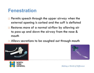
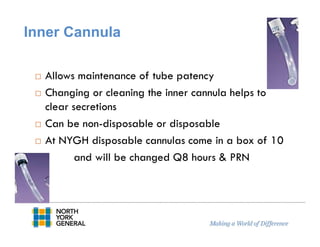
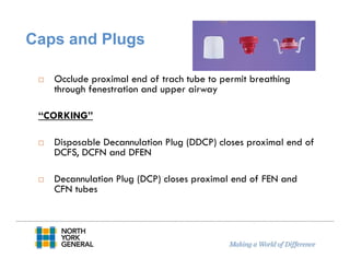
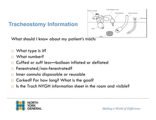
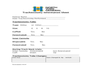
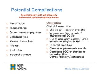
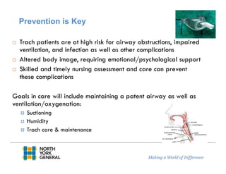
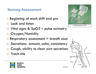
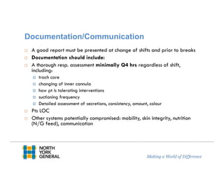
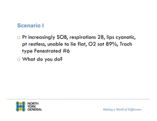
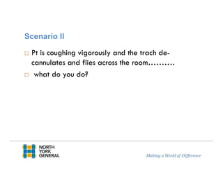
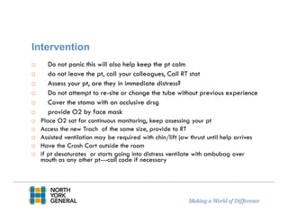
Tracheostomy Care
&
Management
Maria Monteiro, CNE
Revised: January 2015
Objectives
Review of Evidenced-Based Guidelines in the Care & Maintenance
Review Definition, Types of Tracheostomies & their uses
Potential Complications
Nursing Care Guidelines at NYGH:
Assessment,
Suctioning,
Dressing changes,
Inner cannula changes,
Other nursing considerations
Documentation in powerchart
Emergency Scenarios
Definitions
Tracheotomy: Incision made below the cricoid
cartilage through the 2nd-4th tracheal rings
Tracheostomy: the opening or stoma made by this
incision
Tracheostomy Tube: Artificial airway inserted into
the trachea
Why does your patient have a
tracheostomy?
To maintain a patent airway when the ability to do
this is temporarily or permanently compromised
Bypass Obstructed airway
Tumor
Laryngeal edema
Foreign body obstruction
Facilitate removal of secretions
Permit long-term ventilation/prevent aspiration with prolonged
coma
Decrease work of breathing---severe COPD
Anatomy
Types
Cuffed or Un-cuffed
Fenestrated or Non-fenestrated
Disposable or Non-disposable inner cannula
NYGH uses ‘Shiley’ tubes with disposable inner
cannulas
Parts of a Trach
1. Flange- secured with trach ties, stabilizes the trach
2. Outer Cannula-tube connected to flange
3. Inner Cannula- removable for cleaning
4. Obturator-a plastic guide with a smooth rounded tip
that is used to guide the outer cannula during insertion
5. Cuff-Soft balloon around the end of the
trach that can be inflated to allow for
mechanical ventilation
3
1
5
4
Cuffed
Purpose:
Increase or improve ventilation/oxygenation
Prevent aspiration with feeding tubes, decreased
gag reflex, gastro-esophageal reflux
Identification:
DCT- disposable cannula
DFEN- disposable cannula fenestrated
Cuff Complications
Pressure from the cuff can cause damage the trachea
Necrosis
Low pressure cuffs are used
RT will inflate/deflate and monitor pressure
Un-cuffed
Plastic or metal
Allows air to flow freely around the tracheostomy
tube through the larynx
Reduces the risk of tracheal damage
Identification:
DCFS- disposable cannula cuffless
DCFN- disposable cannula fenestrated cuffless
Fenestration
Permits speech through the upper airway when the
external opening is corked and the cuff is deflated
Restores more of a normal airflow by allowing air
to pass up and down the airway from the nose &
mouth
Allows secretions to be coughed out through mouth
Inner Cannula
Allows maintenance of tube patency
Changing or cleaning the inner cannula helps to
clear secretions
Can be non-disposable or disposable
At NYGH disposable cannulas come in a box of 10
and will be changed Q8 hours & PRN
Caps and Plugs
Occlude proximal end of trach tube to permit breathing
through fenestration and upper airway
“CORKING”
Disposable Decannulation Plug (DDCP) closes proximal end of
DCFS, DCFN and DFEN
Decannulation Plug (DCP) closes proximal end of FEN and
CFN tubes
Tracheostomy Information
What should I know about my patient’s trach:
What type is it?
What number?
Cuffed or cuff less—balloon inflated or deflated
Fenestrated/non-fenestrated?
Inner cannula disposable or reusable
Corked? For how long? What is the goal?
Is the Trach NYGH information sheet in the room and visible?
Potential Complications
Hemorrhage
Pneumothorax
Subcutaneous emphysema
Dislodged tube
Airway obstructions
Infection
Aspiration
Tracheal damage
Obstruction
Clinical Presentation:
Skin colour—pallour, cyanotic
Increase respiratory rate, P,
BP,Decreased O2 Sat
Use of accessory muscles, flared
nostrils, inability to lie flat
Labored breathing
Clammy appearance/cyanosis
Decreased LOC or changes to
behaviour (i.e.)
Distress/anxiety/restlessness
Recognizing early S & S will allow early
intervention & prevent negative outcomes
Prevention is Key
Trach patients are at high risk for airway obstructions, impaired
ventilation, and infection as well as other complications
Altered body image, requiring emotional/psychological support
Skilled and timely nursing assessment and care can prevent
these complications
Goals in care will include maintaining a patent airway as well as
ventilation/oxygenation:
Suctioning
Humidity
Trach care & maintenance
Nursing Assessment
Beginning of each shift and prn
Look and listen
Vital signs & SpO2 – pulse oximetry
Oxygen/Humidity
Respiratory assessment = breath sounds
Secretions- amount, color, consistency
Cough, ability to clear own secretions
Trach site
Equipment in Room
**emergency equipment**
Each Shift the nurse responsible for the pt must check and document that
equipment is present and in working order
Functioning Suction & O2/Air for humidity
Ambubag with trach adaptor/connector
Trach set of same size and type—VISIBLE TO ALL
Trach Info sheet with pt info.
Obturator
Box of disposable Inner Cannulas or trach tray for cleaning non-disposable
Suction catheters
Normal saline bottle
Hydrogen peroxide (for non-disposable inner cannulas cleaning)
Small ampules of normal saline for suctioning
Mouth swabs
Occlusive drsg—tegaderm
Trach 4X4 sponges for dressing changes
Trach care to be entered in powerchart by nurse
Q 8 hours ( 0600-1400-2200) & PRN
Suction pt prior to trach care
Assess skin around trach for redness, drainage, secretions,
bleeding, maceration or excoriation and skin breakdown
caused by flange pressure
Clean around stoma & flange with Q-tips/ 2X2 moistened
with NS
dry stoma area if needed and apply 4X4 gauze
With newly established trach- are sutures still in place? Can
they be removed?
trach ties secure?? Should be able to fit one finger under the
tie
Trach Care Guidelines
Changing/Cleaning Inner Cannula
NYGH uses Shiley trach tubes with disposable inner cannulas
If a pt does have a trach from home or other institution, it is crucial
to know if the inner cannula is disposable or reusable
Non-disposable inner cannulas are cleaned with Normal Saline
diluted hydrogen peroxide, rinsed off with N/S remove excess
fluid before re-inserting
Disposable inner cannulas are replaced with trach care Q8 hours
& PRN( come in a box of 10)
Trach ties-are changed only when wet or soiled and 2 people should
assist with this procedure---Leave one finger between ties and
neck--Velcro hooks attach easily to tracheostomy tube flange
Decision to Suction
Frequency of suction will vary and must be
individually assessed & not done on a schedule
Factors to Consider:
Is the pt able to cough &/or clear secretions?
Increased work to breath?
Changes to respiratory rate
Amount and consistency of secretions
Decreased O2 saturation
Secretions are audible
Pt request
Other Respiratory S & S (i.e. SOB, cyanosis, restless,
anxiety)
Complications with Suctioning
Hypoxemia—dysrhythmia
Atelectasis or lung collapse
Mucosal trauma/damage---bleeding
Broncho spasm
Dysrhythmias
Nosocomial pulmonary tract infection
Sepsis
Cardiac arrest
Procedure Considerations
Suctioning removes secretions, & also O2
Suction pressure too high (>120mmHg) can cause
mucosa damage & bleeding
Suction pressure too low may not clear secretions & be
ineffective
Suction mouth with a (yankauer) not the same suction
catheter as trachea to avoid cross contamination
Do not apply suction while inserting the catheter
May be necessary to pre-oxygenate the patient prior to
and after suctioning
Use personal protective equipment (i.e. goggles, mask,
face shield)
Suctioning
Insert catheter until you meet resistance &/or pt coughs forcibly then pull back slightly &
start suctioning
Carina
Suctioning
Test suction pressure before instilling catheter 60-120 mm Hg
Suction catheter: ≤ ½ diameter of tube
Prepare clean cup with NS to lubricate and clear secretions from
suction catheter
Dominant hand remains sterile with clean glove, and will be
inserting the catheter, while the non-dominant gloved hand grasps
the suction port
Apply suction only on removal of catheter no during insertion
Suction efficiently and quickly depending on secretion amount,
consistency
Do not exceed 3 attempts and allow 20 to 30 seconds between
each, oxygenate pt between PRN
Inducing Cough
If you suction and don’t obtain secretions you can instill
normal saline (sterile ampule) to loosen secretions
( induce cough reflex) for suctioning
Note: this should not be done as a routine
(may flush particles into resp system, increase infection, aspiration pneumonia)
Secretions may build up within the inner cannula, and
narrow the passage----pull out cannula and observe if
copious secretions discard (if disposable) and place new
one (if not disposable) clean and reapply
Additional Nursing Considerations
Good mouth care
Brush teeth
Yankeur suction
Prevent pneumonia
Communication
Alteration in
communication
Lip reading
Communication
board
Corking for
speech
Hygiene
Dysphagia
May not be able to eat
orally
May have difficulty
swallowing, require puree
or thickened fluids
May have N/G or G-
tube
Risk for aspiration
Risk of
pneumonia/respiratory
infections increased
Humidity applications
Clean equipment
Change suction
catheters/tubes
Clean trach
Hand washing
Cough etiquette
Infection Control
Documentation/Communication
A good report must be presented at change of shifts and prior to breaks
Documentation should include:
A thorough resp. assessment minimally Q4 hrs regardless of shift,
including:
trach care
changing of inner cannula
how pt is tolerating interventions
suctioning frequency
Detailed assessment of secretions, consistency, amount, colour
Pts LOC
Other systems potentially compromised: mobility, skin integrity, nutrition
(N/G feed), communication
Scenario I
Pt increasingly SOB, respirations 28, lips cyanotic,
pt restless, unable to lie flat, O2 sat 89%, Trach
type Fenestrated #6
What do you do?
Interventions
Reassure patient
suction pt quickly and efficiently monitor O2 Sat continuously– if no
improvement
apply N/S 1cc & suction again
Evaluate, anxiety,O2 Sat, colour of skin
If pt has a Fenestrated trach tube
keep a non fenestrated inner cannula of the same size close at hand
Remove fenestrated inner cannula, Replace with non-fenestrated one and
bag pt with 100% O2
Call RT stat
Scenario II
Pt is coughing vigorously and the trach de-
cannulates and flies across the room……….
what do you do?
Intervention
Do not panic this will also help keep the pt calm
do not leave the pt, call your colleagues, Call RT stat
Assess your pt, are they in immediate distress?
Do not attempt to re-site or change the tube without previous experience
Cover the stoma with an occlusive drsg
provide O2 by face mask
Place O2 sat for continuous monitoring, keep assessing your pt
Access the new Trach of the same size, provide to RT
Assisted ventilation may be required with chin/lift jaw thrust until help arrives
Have the Crash Cart outside the room
if pt desaturates or starts going into distress ventilate with ambubag over
mouth as any other pt---call code if necessary
References
Nance-Floyd, B. Tracheostomy care:An evidence-based guide to suctioning
and dressing changes. American Nurse Today. 2011;6(7):14-16.
Higgens, D. (2009). Basic nursing principles of caring for patients with a
trachestomy, Nursing Times. 105 (3), 14-15
Reference Perry & Potter (2006), Clinical Nursing Skills & Techniques 6th
Edition, Elsevier Mosby, Missouri, USA.
Tyco-Healthcare (2004) Tracheostomy Tube: Adult Home Care Guide
(Shiley tracheostomy tubes)
SIMS Portex Inc. (1998). Tracheostomy Care Handbook: Guide for the
health care provider
These guidelines were developed based on current research & are subject to updates &
change
THANK YOU
QUESTIONS??

Tracheostomy care & management

  • 1.
  • 2.
    Objectives Review of Evidenced-BasedGuidelines in the Care & Maintenance Review Definition, Types of Tracheostomies & their uses Potential Complications Nursing Care Guidelines at NYGH: Assessment, Suctioning, Dressing changes, Inner cannula changes, Other nursing considerations Documentation in powerchart Emergency Scenarios
  • 3.
    Definitions Tracheotomy: Incision madebelow the cricoid cartilage through the 2nd-4th tracheal rings Tracheostomy: the opening or stoma made by this incision Tracheostomy Tube: Artificial airway inserted into the trachea
  • 4.
    Why does yourpatient have a tracheostomy? To maintain a patent airway when the ability to do this is temporarily or permanently compromised Bypass Obstructed airway Tumor Laryngeal edema Foreign body obstruction Facilitate removal of secretions Permit long-term ventilation/prevent aspiration with prolonged coma Decrease work of breathing---severe COPD
  • 5.
  • 6.
    Types Cuffed or Un-cuffed Fenestratedor Non-fenestrated Disposable or Non-disposable inner cannula NYGH uses ‘Shiley’ tubes with disposable inner cannulas
  • 7.
    Parts of aTrach 1. Flange- secured with trach ties, stabilizes the trach 2. Outer Cannula-tube connected to flange 3. Inner Cannula- removable for cleaning 4. Obturator-a plastic guide with a smooth rounded tip that is used to guide the outer cannula during insertion 5. Cuff-Soft balloon around the end of the trach that can be inflated to allow for mechanical ventilation 3 1 5 4
  • 8.
    Cuffed Purpose: Increase or improveventilation/oxygenation Prevent aspiration with feeding tubes, decreased gag reflex, gastro-esophageal reflux Identification: DCT- disposable cannula DFEN- disposable cannula fenestrated
  • 9.
    Cuff Complications Pressure fromthe cuff can cause damage the trachea Necrosis Low pressure cuffs are used RT will inflate/deflate and monitor pressure
  • 10.
    Un-cuffed Plastic or metal Allowsair to flow freely around the tracheostomy tube through the larynx Reduces the risk of tracheal damage Identification: DCFS- disposable cannula cuffless DCFN- disposable cannula fenestrated cuffless
  • 11.
    Fenestration Permits speech throughthe upper airway when the external opening is corked and the cuff is deflated Restores more of a normal airflow by allowing air to pass up and down the airway from the nose & mouth Allows secretions to be coughed out through mouth
  • 12.
    Inner Cannula Allows maintenanceof tube patency Changing or cleaning the inner cannula helps to clear secretions Can be non-disposable or disposable At NYGH disposable cannulas come in a box of 10 and will be changed Q8 hours & PRN
  • 13.
    Caps and Plugs Occludeproximal end of trach tube to permit breathing through fenestration and upper airway “CORKING” Disposable Decannulation Plug (DDCP) closes proximal end of DCFS, DCFN and DFEN Decannulation Plug (DCP) closes proximal end of FEN and CFN tubes
  • 14.
    Tracheostomy Information What shouldI know about my patient’s trach: What type is it? What number? Cuffed or cuff less—balloon inflated or deflated Fenestrated/non-fenestrated? Inner cannula disposable or reusable Corked? For how long? What is the goal? Is the Trach NYGH information sheet in the room and visible?
  • 16.
    Potential Complications Hemorrhage Pneumothorax Subcutaneous emphysema Dislodgedtube Airway obstructions Infection Aspiration Tracheal damage Obstruction Clinical Presentation: Skin colour—pallour, cyanotic Increase respiratory rate, P, BP,Decreased O2 Sat Use of accessory muscles, flared nostrils, inability to lie flat Labored breathing Clammy appearance/cyanosis Decreased LOC or changes to behaviour (i.e.) Distress/anxiety/restlessness Recognizing early S & S will allow early intervention & prevent negative outcomes
  • 17.
    Prevention is Key Trachpatients are at high risk for airway obstructions, impaired ventilation, and infection as well as other complications Altered body image, requiring emotional/psychological support Skilled and timely nursing assessment and care can prevent these complications Goals in care will include maintaining a patent airway as well as ventilation/oxygenation: Suctioning Humidity Trach care & maintenance
  • 18.
    Nursing Assessment Beginning ofeach shift and prn Look and listen Vital signs & SpO2 – pulse oximetry Oxygen/Humidity Respiratory assessment = breath sounds Secretions- amount, color, consistency Cough, ability to clear own secretions Trach site
  • 19.
    Equipment in Room **emergencyequipment** Each Shift the nurse responsible for the pt must check and document that equipment is present and in working order Functioning Suction & O2/Air for humidity Ambubag with trach adaptor/connector Trach set of same size and type—VISIBLE TO ALL Trach Info sheet with pt info. Obturator Box of disposable Inner Cannulas or trach tray for cleaning non-disposable Suction catheters Normal saline bottle Hydrogen peroxide (for non-disposable inner cannulas cleaning) Small ampules of normal saline for suctioning Mouth swabs Occlusive drsg—tegaderm Trach 4X4 sponges for dressing changes
  • 20.
    Trach care tobe entered in powerchart by nurse Q 8 hours ( 0600-1400-2200) & PRN Suction pt prior to trach care Assess skin around trach for redness, drainage, secretions, bleeding, maceration or excoriation and skin breakdown caused by flange pressure Clean around stoma & flange with Q-tips/ 2X2 moistened with NS dry stoma area if needed and apply 4X4 gauze With newly established trach- are sutures still in place? Can they be removed? trach ties secure?? Should be able to fit one finger under the tie Trach Care Guidelines
  • 21.
    Changing/Cleaning Inner Cannula NYGHuses Shiley trach tubes with disposable inner cannulas If a pt does have a trach from home or other institution, it is crucial to know if the inner cannula is disposable or reusable Non-disposable inner cannulas are cleaned with Normal Saline diluted hydrogen peroxide, rinsed off with N/S remove excess fluid before re-inserting Disposable inner cannulas are replaced with trach care Q8 hours & PRN( come in a box of 10) Trach ties-are changed only when wet or soiled and 2 people should assist with this procedure---Leave one finger between ties and neck--Velcro hooks attach easily to tracheostomy tube flange
  • 22.
    Decision to Suction Frequencyof suction will vary and must be individually assessed & not done on a schedule Factors to Consider: Is the pt able to cough &/or clear secretions? Increased work to breath? Changes to respiratory rate Amount and consistency of secretions Decreased O2 saturation Secretions are audible Pt request Other Respiratory S & S (i.e. SOB, cyanosis, restless, anxiety)
  • 23.
    Complications with Suctioning Hypoxemia—dysrhythmia Atelectasisor lung collapse Mucosal trauma/damage---bleeding Broncho spasm Dysrhythmias Nosocomial pulmonary tract infection Sepsis Cardiac arrest
  • 24.
    Procedure Considerations Suctioning removessecretions, & also O2 Suction pressure too high (>120mmHg) can cause mucosa damage & bleeding Suction pressure too low may not clear secretions & be ineffective Suction mouth with a (yankauer) not the same suction catheter as trachea to avoid cross contamination Do not apply suction while inserting the catheter May be necessary to pre-oxygenate the patient prior to and after suctioning Use personal protective equipment (i.e. goggles, mask, face shield)
  • 25.
    Suctioning Insert catheter untilyou meet resistance &/or pt coughs forcibly then pull back slightly & start suctioning Carina
  • 26.
    Suctioning Test suction pressurebefore instilling catheter 60-120 mm Hg Suction catheter: ≤ ½ diameter of tube Prepare clean cup with NS to lubricate and clear secretions from suction catheter Dominant hand remains sterile with clean glove, and will be inserting the catheter, while the non-dominant gloved hand grasps the suction port Apply suction only on removal of catheter no during insertion Suction efficiently and quickly depending on secretion amount, consistency Do not exceed 3 attempts and allow 20 to 30 seconds between each, oxygenate pt between PRN
  • 27.
    Inducing Cough If yousuction and don’t obtain secretions you can instill normal saline (sterile ampule) to loosen secretions ( induce cough reflex) for suctioning Note: this should not be done as a routine (may flush particles into resp system, increase infection, aspiration pneumonia) Secretions may build up within the inner cannula, and narrow the passage----pull out cannula and observe if copious secretions discard (if disposable) and place new one (if not disposable) clean and reapply
  • 28.
    Additional Nursing Considerations Goodmouth care Brush teeth Yankeur suction Prevent pneumonia Communication Alteration in communication Lip reading Communication board Corking for speech Hygiene
  • 29.
    Dysphagia May not beable to eat orally May have difficulty swallowing, require puree or thickened fluids May have N/G or G- tube Risk for aspiration Risk of pneumonia/respiratory infections increased Humidity applications Clean equipment Change suction catheters/tubes Clean trach Hand washing Cough etiquette Infection Control
  • 30.
    Documentation/Communication A good reportmust be presented at change of shifts and prior to breaks Documentation should include: A thorough resp. assessment minimally Q4 hrs regardless of shift, including: trach care changing of inner cannula how pt is tolerating interventions suctioning frequency Detailed assessment of secretions, consistency, amount, colour Pts LOC Other systems potentially compromised: mobility, skin integrity, nutrition (N/G feed), communication
  • 33.
    Scenario I Pt increasinglySOB, respirations 28, lips cyanotic, pt restless, unable to lie flat, O2 sat 89%, Trach type Fenestrated #6 What do you do?
  • 34.
    Interventions Reassure patient suction ptquickly and efficiently monitor O2 Sat continuously– if no improvement apply N/S 1cc & suction again Evaluate, anxiety,O2 Sat, colour of skin If pt has a Fenestrated trach tube keep a non fenestrated inner cannula of the same size close at hand Remove fenestrated inner cannula, Replace with non-fenestrated one and bag pt with 100% O2 Call RT stat
  • 35.
    Scenario II Pt iscoughing vigorously and the trach de- cannulates and flies across the room………. what do you do?
  • 36.
    Intervention Do not panicthis will also help keep the pt calm do not leave the pt, call your colleagues, Call RT stat Assess your pt, are they in immediate distress? Do not attempt to re-site or change the tube without previous experience Cover the stoma with an occlusive drsg provide O2 by face mask Place O2 sat for continuous monitoring, keep assessing your pt Access the new Trach of the same size, provide to RT Assisted ventilation may be required with chin/lift jaw thrust until help arrives Have the Crash Cart outside the room if pt desaturates or starts going into distress ventilate with ambubag over mouth as any other pt---call code if necessary
  • 37.
    References Nance-Floyd, B. Tracheostomycare:An evidence-based guide to suctioning and dressing changes. American Nurse Today. 2011;6(7):14-16. Higgens, D. (2009). Basic nursing principles of caring for patients with a trachestomy, Nursing Times. 105 (3), 14-15 Reference Perry & Potter (2006), Clinical Nursing Skills & Techniques 6th Edition, Elsevier Mosby, Missouri, USA. Tyco-Healthcare (2004) Tracheostomy Tube: Adult Home Care Guide (Shiley tracheostomy tubes) SIMS Portex Inc. (1998). Tracheostomy Care Handbook: Guide for the health care provider These guidelines were developed based on current research & are subject to updates & change
  • 38.