Downloaded 945 times


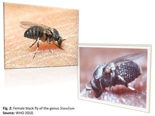


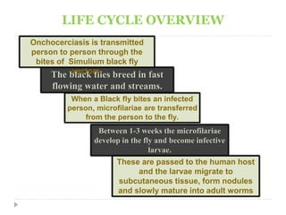
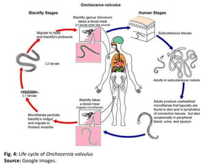


Onchocerciasis, caused by the parasite Onchocerca volvulus, is a chronic disease resulting in blindness and severe skin conditions, with Nigeria being the most endemic country. An estimated 7-10 million Nigerians are infected, primarily affecting individuals aged 20-30 years, and the disease is transmitted through bites from the black fly, which breeds in fast-flowing water. While ivermectin can kill microfilariae, there is no cure for blindness caused by the disease, necessitating increased awareness and community treatment efforts.


