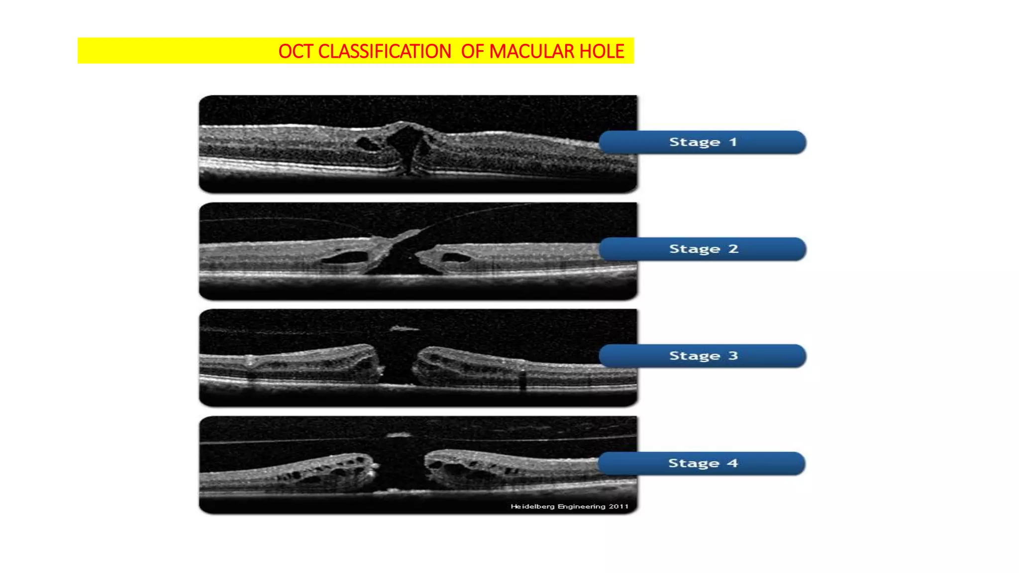
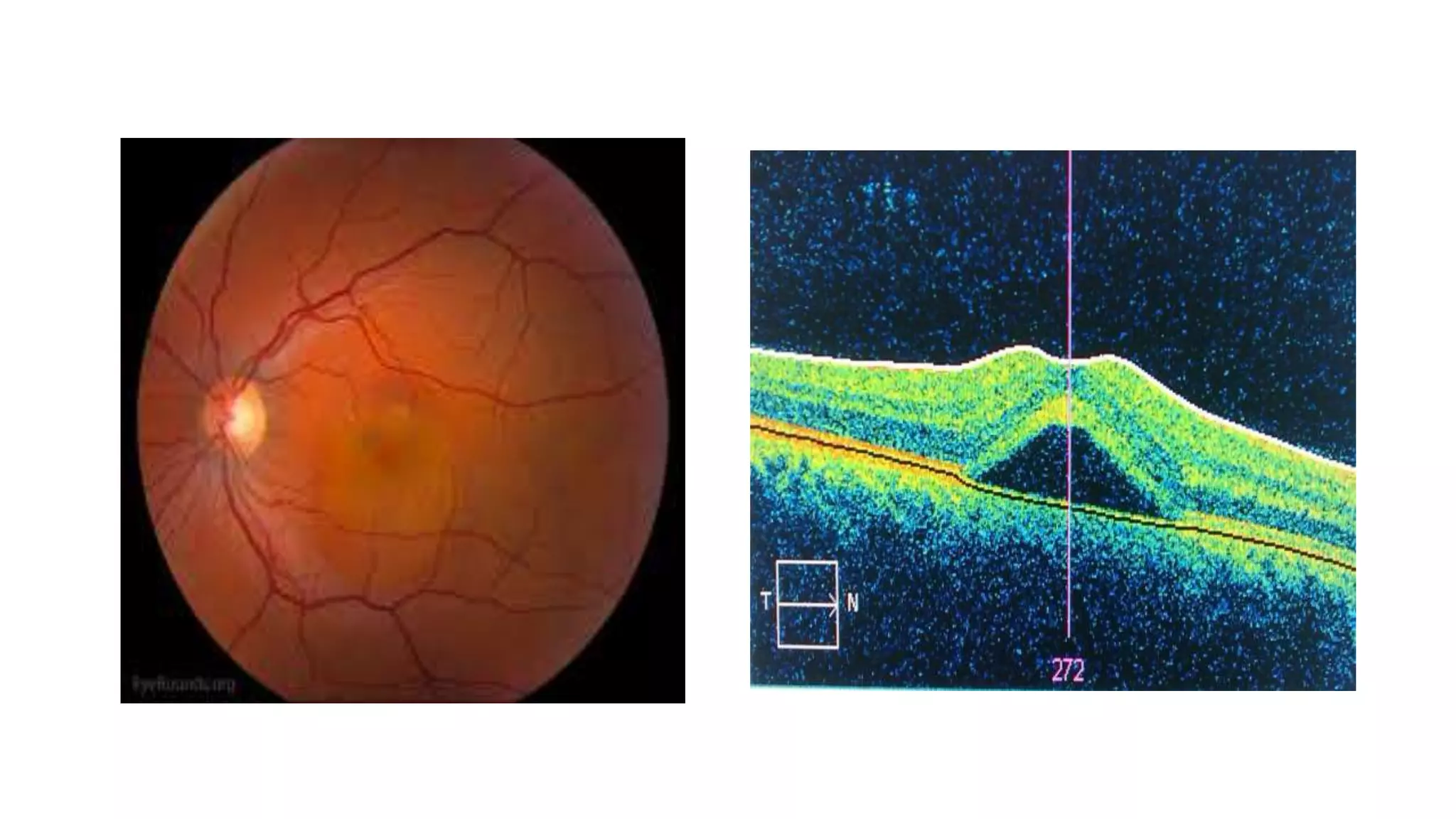
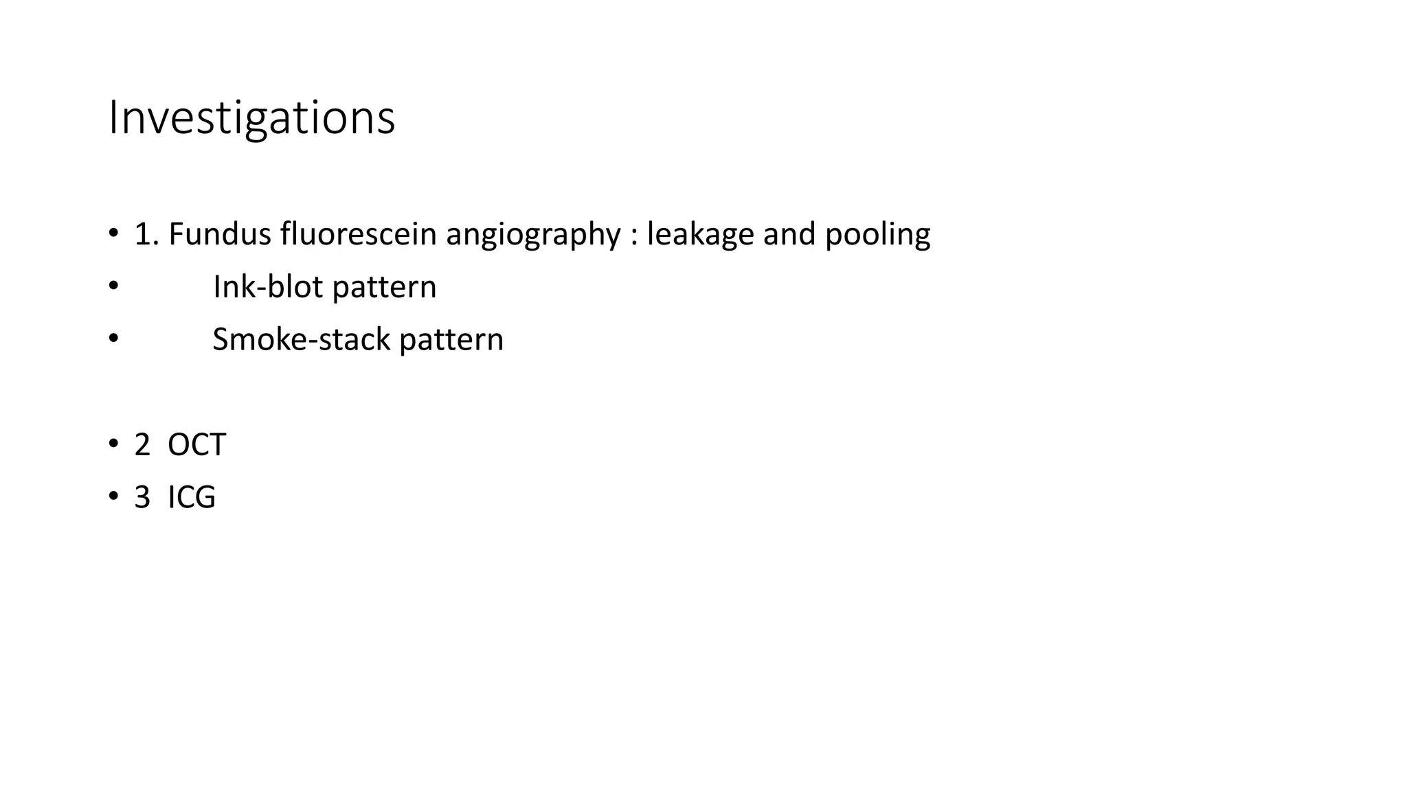
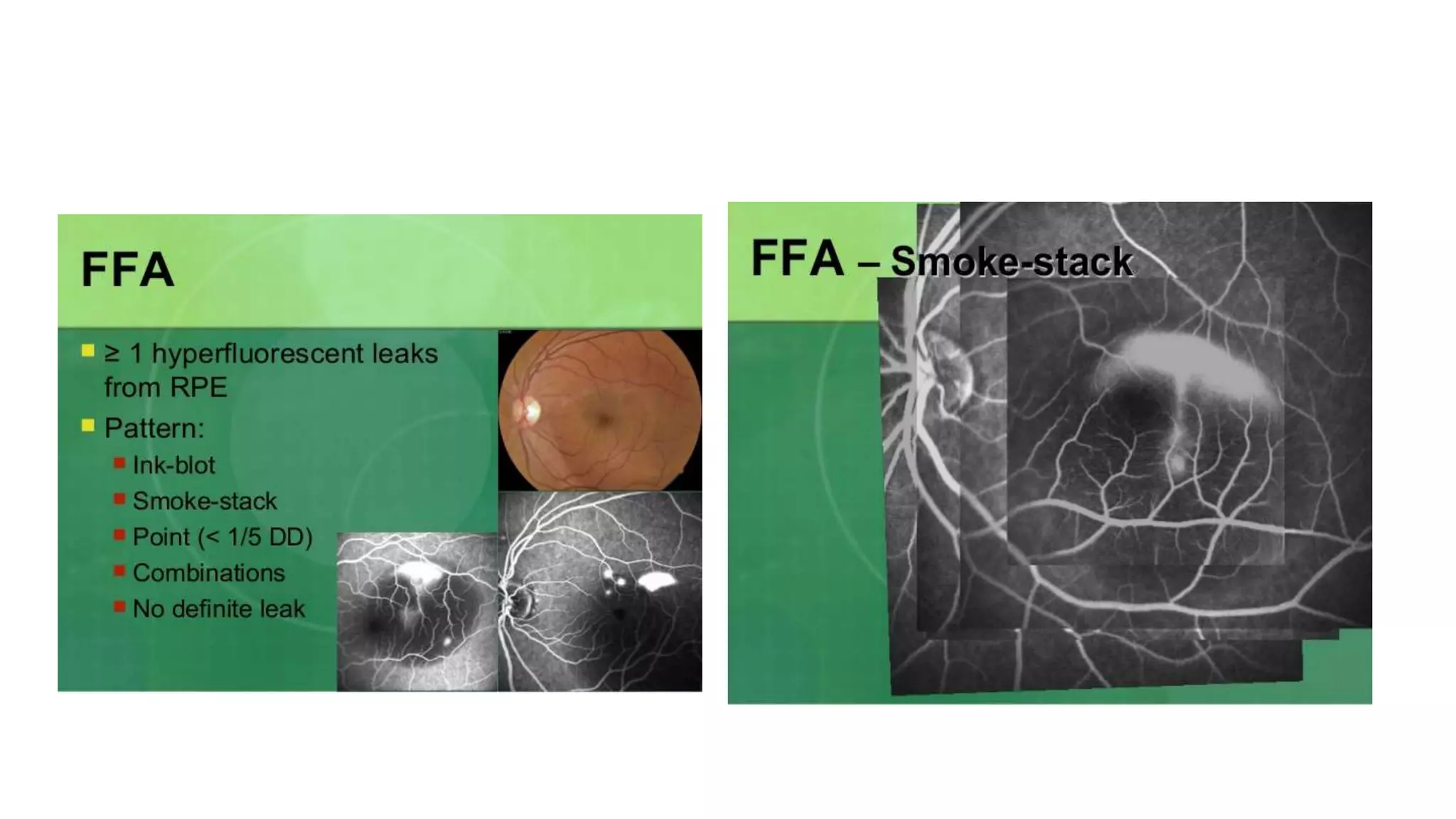
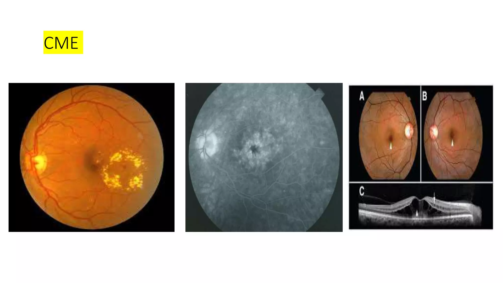
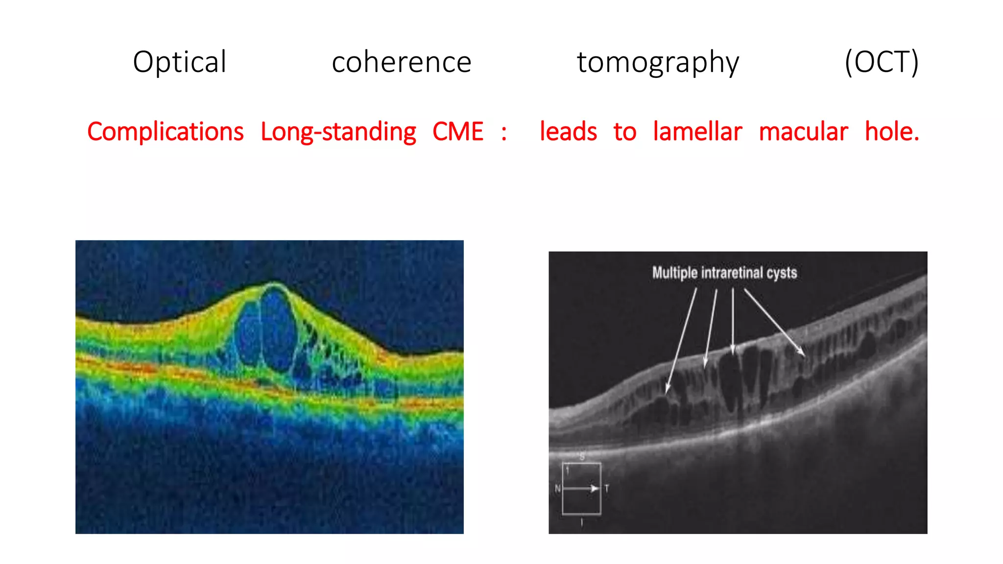
Central serous chorioretinopathy (CSCR) is characterized by spontaneous serous detachment of the neurosensory retina in the macular region. It typically affects young to middle-aged males and is associated with type A personality and steroid use. The leading pathogenesis theory is choroidal vascular hyperpermeability causing a breach in the outer blood-retinal barrier and leakage of fluid. On examination, CSCR presents as mild macular elevation with or without retinal pigment epithelium detachment. It usually resolves spontaneously but can recur in 30-50% of cases.
Cystoid macular edema (CME) refers to fluid collection in the macula and is a complication of ocular surgery,
LIMBUS… • The limbus forms the border between the transparent cornea and opaque sclera, contains the pathways of aqueous humour outflow, and is the site of surgical incisions for cataract and glaucoma
2. Anatomical Limbus: Circumcorneal transitional zone of the conjunctivocorneal & corneoscleral junction Conjunctivo-corneal junction: • Bulbar conjunctiva is firmly adherent to underlying structures • Substantia propria of the conjunctiva stops here but its epithelium continues with that of the cornea. Sclero-corneal junction: • Transparent corneal lamellae become continuous • With the oblique, circular and opaque fibres of sclera
3. CONTINUE…. • In the area near limbus, the conjunctiva, tenon’s capsule & the episcleral tissue are fused into a dense tissue which is strongly adherent to corneo scleral junction.It is preferred site for obtaining a firm hold of the eyeball during ocular surgery. • The limbus is a common site for the occurrence of corneal epithelial neoplasm. • The Limbus contains radially oriented fibrovascular ridge known as the palisades of Vogt that may harbour a stem cell population. The palisades of Vogt are more common in the superior and inferior quadrants around the eye
Update knowledge about Muntifocal IOL made by Asaduzzaman
Working as Associate Optometrist in Ispahani Islamia Eye Institute &Hospita, Dhaka 1215
Email:asad.optom92@yaho. com
Update knowledge about Muntifocal IOL made by Asaduzzaman
Working as Associate Optometrist in Ispahani Islamia Eye Institute &Hospita, Dhaka 1215
Email:asad.optom92@yaho. com
MANAGEMENT OF MACULAR HOLE, Ophthalmology presentation, eye care in the elderly , macular hole as a consequence of trauma, Vitreoretinal surgical cases, ,
This presentation introduces myopia, high myopia, and in more details, pathologic myopia (aka malignant myopia). It is intended for training ophthalmologists, ophthalmology residents, medical students in ophthalmology rotations.
strabismus , gaze , ocular movements , classification etc
presented by senior optometrist & orthoptician at Sagarmatha Choudhary Eye Hospital, SCEH, LAHAN (NEPAL )
He explain details about the binocular gaze , EOMs, etc & work up of a patient of squint etc.
visual acuity testing in children is challenging
VEP, OKN,PLT etc
CARDIFF, BOEK CANDY, WORTH IVORY BAAL, STYCAR
HOTV , MINIACTURE TOY TEST
SHEREDN GARED
SNELLEN CHART
ETDRS CHART
LOGMAR CHART
these are charts used in ophthalmology in pediatric age group
cover test
uncover test
alternate cover
hirschburg corneal light reflex test
10 D verticle prism bar test
Couples presenting to the infertility clinic- Do they really have infertility...Sujoy Dasgupta
Dr Sujoy Dasgupta presented the study on "Couples presenting to the infertility clinic- Do they really have infertility? – The unexplored stories of non-consummation" in the 13th Congress of the Asia Pacific Initiative on Reproduction (ASPIRE 2024) at Manila on 24 May, 2024.
Pulmonary Thromboembolism - etilogy, types, medical- Surgical and nursing man...VarunMahajani
Disruption of blood supply to lung alveoli due to blockage of one or more pulmonary blood vessels is called as Pulmonary thromboembolism. In this presentation we will discuss its causes, types and its management in depth.
These lecture slides, by Dr Sidra Arshad, offer a quick overview of physiological basis of a normal electrocardiogram.
Learning objectives:
1. Define an electrocardiogram (ECG) and electrocardiography
2. Describe how dipoles generated by the heart produce the waveforms of the ECG
3. Describe the components of a normal electrocardiogram of a typical bipolar leads (limb II)
4. Differentiate between intervals and segments
5. Enlist some common indications for obtaining an ECG
Study Resources:
1. Chapter 11, Guyton and Hall Textbook of Medical Physiology, 14th edition
2. Chapter 9, Human Physiology - From Cells to Systems, Lauralee Sherwood, 9th edition
3. Chapter 29, Ganong’s Review of Medical Physiology, 26th edition
4. Electrocardiogram, StatPearls - https://www.ncbi.nlm.nih.gov/books/NBK549803/
5. ECG in Medical Practice by ABM Abdullah, 4th edition
6. ECG Basics, http://www.nataliescasebook.com/tag/e-c-g-basics
Flu Vaccine Alert in Bangalore Karnatakaaddon Scans
As flu season approaches, health officials in Bangalore, Karnataka, are urging residents to get their flu vaccinations. The seasonal flu, while common, can lead to severe health complications, particularly for vulnerable populations such as young children, the elderly, and those with underlying health conditions.
Dr. Vidisha Kumari, a leading epidemiologist in Bangalore, emphasizes the importance of getting vaccinated. "The flu vaccine is our best defense against the influenza virus. It not only protects individuals but also helps prevent the spread of the virus in our communities," he says.
This year, the flu season is expected to coincide with a potential increase in other respiratory illnesses. The Karnataka Health Department has launched an awareness campaign highlighting the significance of flu vaccinations. They have set up multiple vaccination centers across Bangalore, making it convenient for residents to receive their shots.
To encourage widespread vaccination, the government is also collaborating with local schools, workplaces, and community centers to facilitate vaccination drives. Special attention is being given to ensuring that the vaccine is accessible to all, including marginalized communities who may have limited access to healthcare.
Residents are reminded that the flu vaccine is safe and effective. Common side effects are mild and may include soreness at the injection site, mild fever, or muscle aches. These side effects are generally short-lived and far less severe than the flu itself.
Healthcare providers are also stressing the importance of continuing COVID-19 precautions. Wearing masks, practicing good hand hygiene, and maintaining social distancing are still crucial, especially in crowded places.
Protect yourself and your loved ones by getting vaccinated. Together, we can help keep Bangalore healthy and safe this flu season. For more information on vaccination centers and schedules, residents can visit the Karnataka Health Department’s official website or follow their social media pages.
Stay informed, stay safe, and get your flu shot today!
MANAGEMENT OF ATRIOVENTRICULAR CONDUCTION BLOCK.pdfJim Jacob Roy
Cardiac conduction defects can occur due to various causes.
Atrioventricular conduction blocks ( AV blocks ) are classified into 3 types.
This document describes the acute management of AV block.
Report Back from SGO 2024: What’s the Latest in Cervical Cancer?bkling
Are you curious about what’s new in cervical cancer research or unsure what the findings mean? Join Dr. Emily Ko, a gynecologic oncologist at Penn Medicine, to learn about the latest updates from the Society of Gynecologic Oncology (SGO) 2024 Annual Meeting on Women’s Cancer. Dr. Ko will discuss what the research presented at the conference means for you and answer your questions about the new developments.
Prix Galien International 2024 Forum ProgramLevi Shapiro
June 20, 2024, Prix Galien International and Jerusalem Ethics Forum in ROME. Detailed agenda including panels:
- ADVANCES IN CARDIOLOGY: A NEW PARADIGM IS COMING
- WOMEN’S HEALTH: FERTILITY PRESERVATION
- WHAT’S NEW IN THE TREATMENT OF INFECTIOUS,
ONCOLOGICAL AND INFLAMMATORY SKIN DISEASES?
- ARTIFICIAL INTELLIGENCE AND ETHICS
- GENE THERAPY
- BEYOND BORDERS: GLOBAL INITIATIVES FOR DEMOCRATIZING LIFE SCIENCE TECHNOLOGIES AND PROMOTING ACCESS TO HEALTHCARE
- ETHICAL CHALLENGES IN LIFE SCIENCES
- Prix Galien International Awards Ceremony
TEST BANK for Operations Management, 14th Edition by William J. Stevenson, Ve...kevinkariuki227
TEST BANK for Operations Management, 14th Edition by William J. Stevenson, Verified Chapters 1 - 19, Complete Newest Version.pdf
Anti ulcer drugs and their Advance pharmacology ||
Anti-ulcer drugs are medications used to prevent and treat ulcers in the stomach and upper part of the small intestine (duodenal ulcers). These ulcers are often caused by an imbalance between stomach acid and the mucosal lining, which protects the stomach lining.
||Scope: Overview of various classes of anti-ulcer drugs, their mechanisms of action, indications, side effects, and clinical considerations.
1. CSCR ( CENTRAL SEROUS CHORIORETINOPATHY )
Macular oedema & Macular hole
Dr. ANITA KUMARI
SCEH, LAHAN
O.A 2nd YEAR
2. • Central serous chorioretinopathy (CSCR) is characterised by spontaneous serous
detachment of neurosensory retina in the macular region, with or without retinal
pigment epithelium detachment .
3.
4. Etiopathogenesis & Risk factors
• Age and sex. The disease affects typically young adult (20–50 years), males
more than females
• • Personality. Type A personality individuals are more prone
• • Steroid intake is an important risk factor
• • Emotional stress ( thyroid & Cushings syndrome )
• Hypertension • Pregnancy (usually 3rd trimester)
5. Pathogenesis of CSCR
• Pathogenesis is not known exactly. Various theories have been postulated.
Presently, the most accepted theory is of ‘choroidal vascular hyperpermeability’
• sympathetic drive, sympathomimetics and corticosteroids alter the choroidal
vascular permeability either directly or indirectly by affecting its autoregulation.
6. • This, in turn, increases the tissue hydrostatic pressure in the choroid causing
pigment epithelial defect (PED) resulting in a breach in the outer blood retinal
barrier .
• Leakage of fluid across this area results in development of localized serous
detachment of neurosensory retina .
7. • Clinical features Symptoms. Patient may present with:
• • Sudden painless loss of vision (6/9–6/24) associated with
• • Relative positive scotoma, micropsia & metamorphopsia.
8. Signs. fundus examination shows
• : • Mild elevation of macular area, demarcated by a circular ring reflex.
• • Small yellow grey elevations may be seen due to RPE /PED detachment.
• • Foveal reflex is absent or distorted
• • Subretinal deposits & pigmentary changes
9. Clinical course CSR is usually self-limiting but often recurrent
• Acute classic CSCR : spontaneous resolution within 3–6 months . Recurrences are
known in 30–50% cases
• Chronic CSCR . It is characterized by a chronic course lasting more than 12
months, typically affecting individuals above 50 years of age.
• Bullous CSCR is rare presentation characterized by larger and more numerous
areas of serous retinal and RPE detachments
12. Treatment
• 1. Conservative measures. Reassurance mostly in 80% cases .
• CSR undergoes spontaneous resolution in 80 to 90% cases. Visual acuity returns to
normal or near normal within 3 to 6 months .
• Discontinuation of steroids
• Life style changes to reduces stress in life
• 2. Anti-VEGF can be considered if CNV
• 3. Laser photocoagulation : Long-standing cases (more than 6 months).
• • Patients having recurrent CSR with visual loss.
• • Patients having permanent loss of vision in the other eye due to this condition
• 4. Photodynamic therapy (PDT)
14. CYSTOID MACULAR OEDEMA (CME )
• DEFINE : It refers to collection of fluid in the outer plexiform and inner nuclear
layer of the retina, centred around the foveola.
• Etiology :
• 1. Complication of ocular treatment such as: • Ocular surgery, e.g., cataract
extraction (ICCE>ECCE) (Irvine Gass syndrome), keratoplasty, glaucoma surgery &
RD surgery.
• Topical ocular therapy with eye drops like epinephrine and prostaglandin
etc.
15. • 2. Retinal vascular disorders : such as CRVO, BRVO , Coats’ disease, Eales disease,
and hypertensive retinopathy
• 3. Intraocular inflammations & intraocular tumours e.g. posterior uveitis and
anterior uveitis
• 4. Retinal dystrophies, e.g., retinitis pigmentosa & VMR ( Vitreomacular traction
syndrome )
• 5. Systemic diseases such as leukaemia, chronic renal failure and multiple
myeloma
16. Pathogenesis CME
• It develops due to leakage of fluid following breakdown of inner blood-retinal
barrier (i.e., leakage from the retinal capillaries) & accumulating fluid in the outer
plexiform and inner nuclear layer of retina .
17. Clinical features
• 1. Visual loss.
• 2. Direct Ophthalmoscopy or slit lamp biomicroscopy with 90 D lens : : shows loss
of foveal reflex, retinal thickening, a yellow spot at the center of fovea .
• Typical ‘Honeycomb appearance’ of macula (due to multiple cystoid oval spaces )
• 3. Fundus fluorescein angiography : ‘flower petal appearance’
• 4. Optical coherence tomography (OCT) reveals loss of foveal depression, intrareti
with round optically clear (cystoid spaces) and retinal thicknening
20. Treatment
• 1. Treatment of the causative factor, e.g., photocoagulation for diabetic CSME, STOP
use of topical 2% adrenaline eye drops etc.
• 2. Topical antiprostaglandin (NSAID) drops like ketorolac or nepafenac when used
pre- and postoperatively, prevent the occurrence of CME associated with intraocular
surgery .
• 3. Topical and systemic steroid for inflammation
• 4. Systemic carbonic anhydrase inhibitors (CAIs) Tab. Diamox may be beneficial in some
cases of CME, e.g., in retinitis pigmentosa
22. • Macular hole refers to a partial thickness of full thickness hole in the
neurosensory retina in the foveal region .
• Etiology : 1. Senile or idiopathic (83%), more common in females aged 60–80
years than males (F:M, 3:1).
• 2. Traumatic macular hole account for (5%) cases
• 3. Other causes of macular hole include: CME, vitreomacular traction,
postsurgery, myopia, post-laser treatment and post-inflammatory.
23.
24. Clinical features : Symptoms
• : • Decreased vision, typically around 6/60 level for a full thickness hole and
better for a partial hole.
• • Metamorphopsia or distortion of vision may be there.
• • Central scotoma
• Pathogenesis: Senile macular holes are caused by tractional forces associated with
early PVD .
25. Clinical features & Symptoms include
• Decreased vision, typically around 6/60
• Metamorphopsia or distortion of vision
• Central scotoma
• Signs.
• Based on the fundus appearance (best examined with 78/90D slit-lamp
examination)
• the macular hole can be classified into four stages ( Gass Classification)
26. • Stage 1 or impending hole : it shows absent foveal reflex & a yellow spot or
yellow ring in the foveal region .
• Stage 2. A small full thickness hole ( either centre or marginal ) full thickness hole
less than 400 um in size
• Stage 3. A full thickness hole is seen as round reddish spot surrounded by a grey
halo [cuff of subretinal fluid (SRF)]& no PVD ,more than 400 um in size
• Stage 4. Full thickness hole with with posterior vitreous detached from the disc
and macula .
29. Treatment
• ■Stage 1. Treatment is not recommended as spontaneous hole closure can occur.
But close follow-up and observation is required as 50% cases .
• Stage 2 to 4 holes of recent onset (<1 year) with reduced visual acuity (<6/24)
should be treated with pars plana vitrectomy with posterior hyaloid removal,
internal limiting membrane (ILM) peeling and gas or silicon oil tamponade with strict
postoperative face down position for 7–14 days
30. • Prognosis. Of M.H = Anatomical closure is reported in 60–85% of cases. Visual
improvement is reported in 70% cases with recent onset hole .
• complications include:
• retinal breaks, retinal detachment, late reopening of the hole, RPE loss under the
hole, phototoxicity and endophthalmitis.
31. HOME WORK ?
1. What is CSCR ? What are types & etiology of CSCR?
2. What is cystoid macular odema ? Etiology & managemt of macular odema?
3. what are clinical features & signs of CSCR on slit lamp biomicroscopy ?
4. What is macular hole ? Etiology ?
5. Explain in brief OCT classification / Gass classification of macular hole ?
6. Management of macular hole explain briefly ?
7. What are the different types of macular odema ? What is pathogenesis ?
8. Explain in short management of cystoid macular odema ?