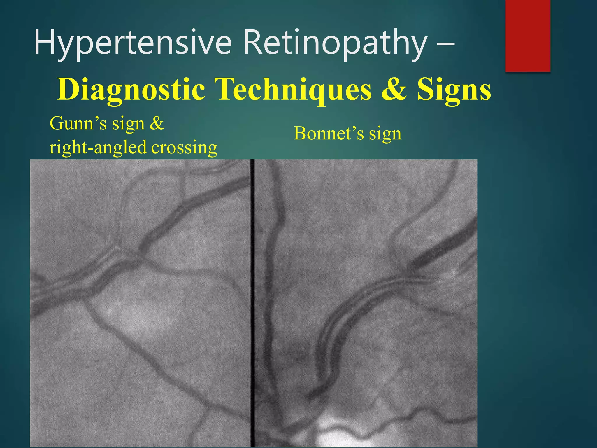
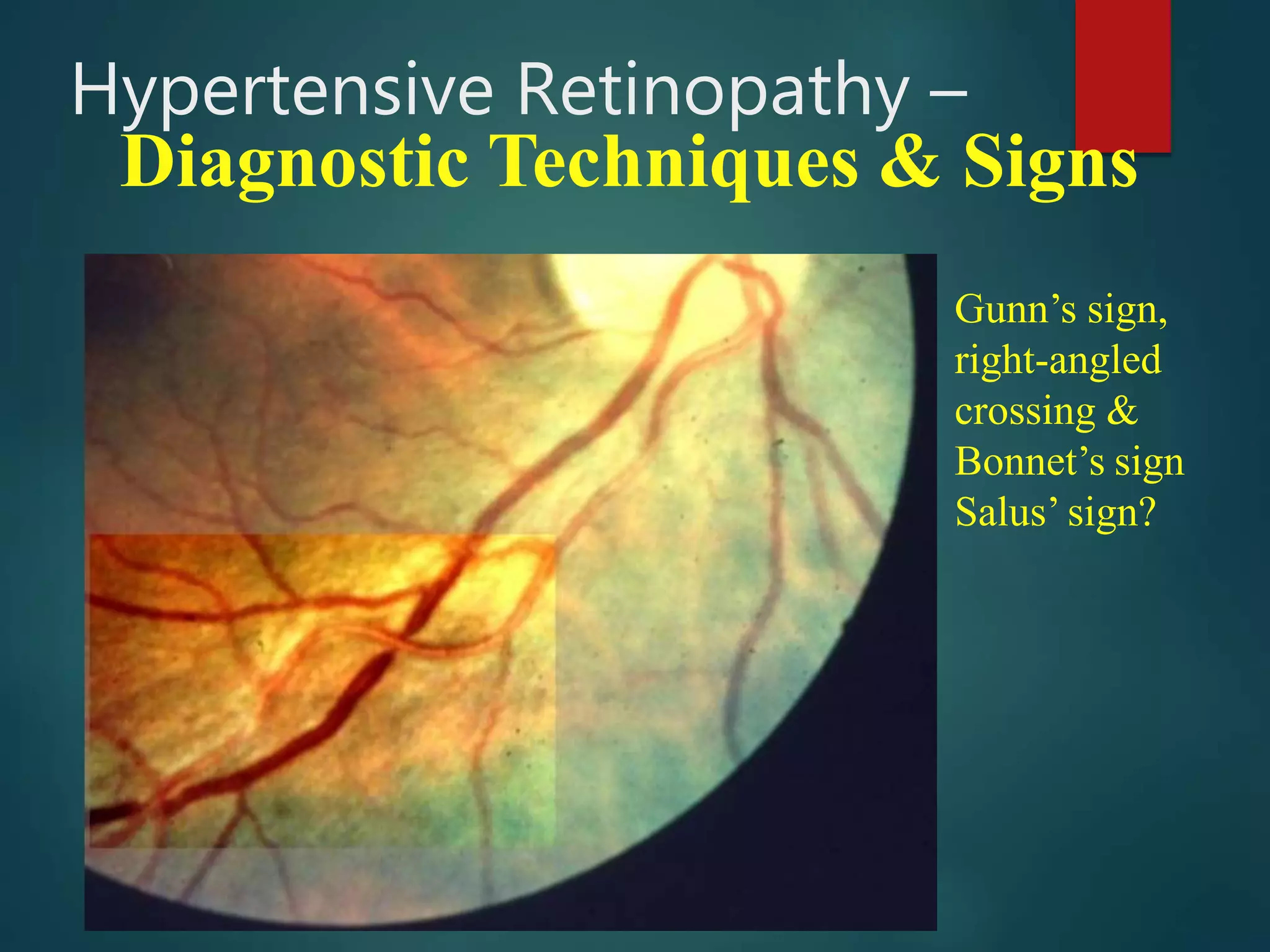
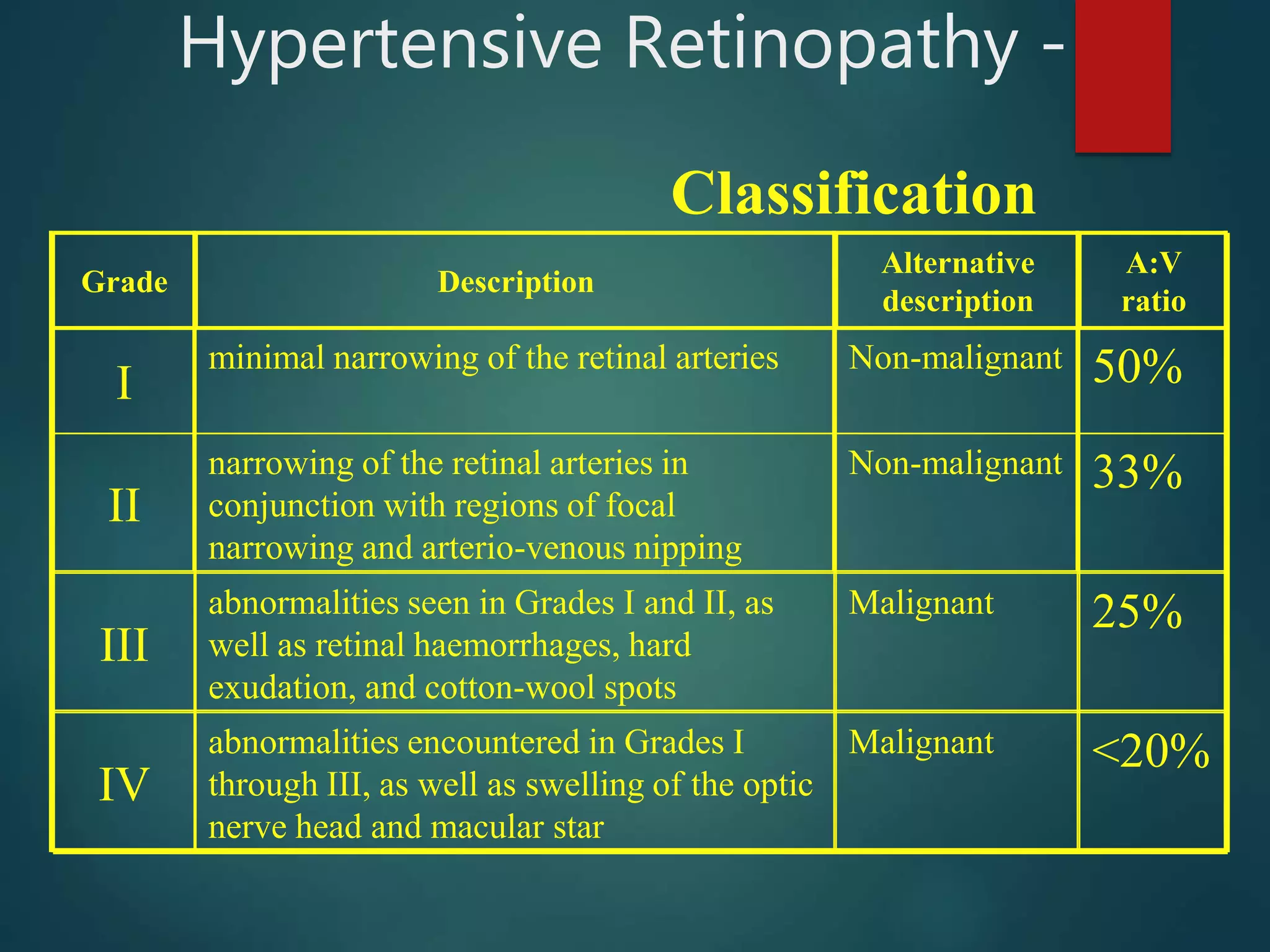
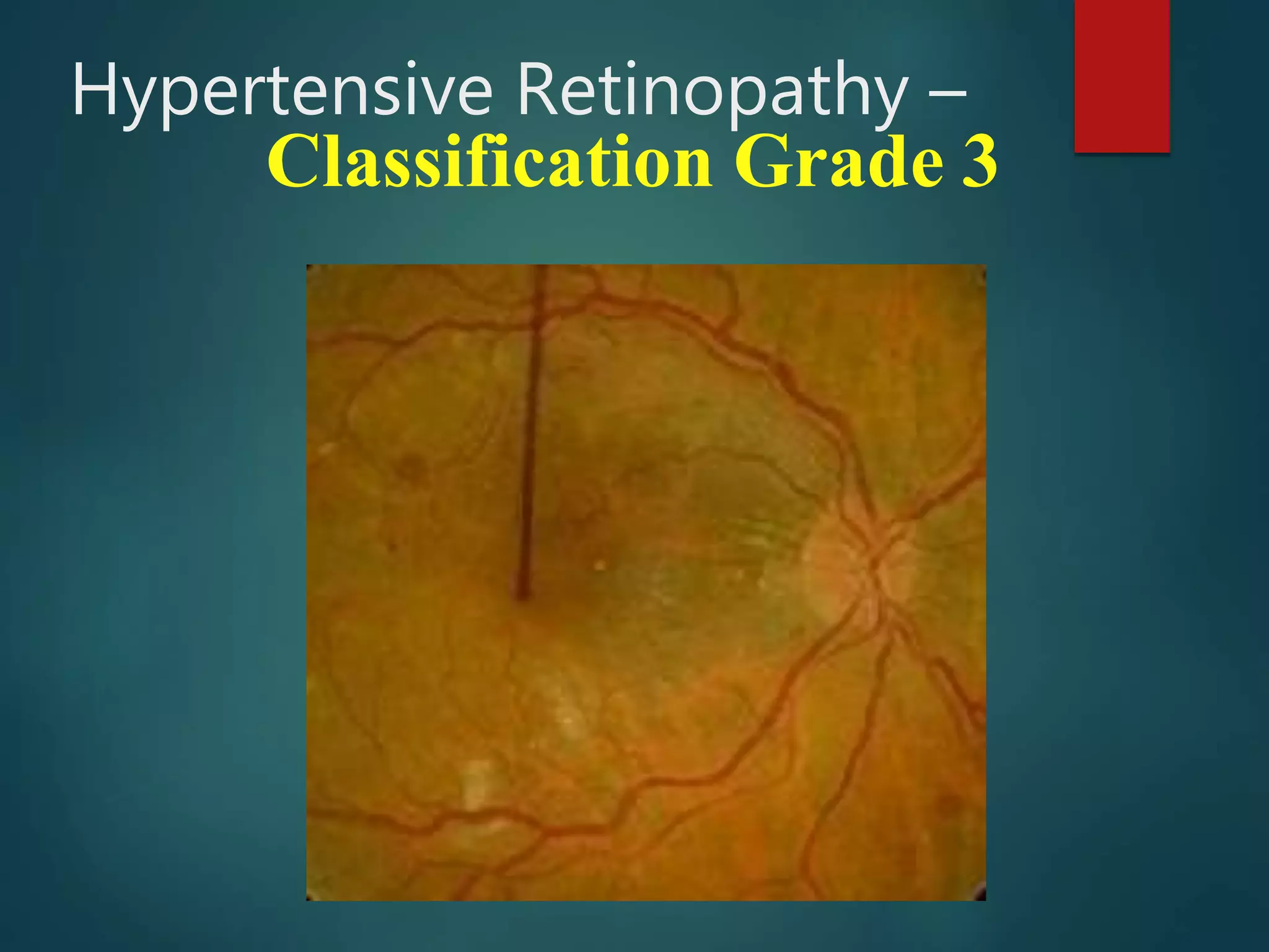
This document discusses hypertensive retinopathy, which is caused by high blood pressure damaging the small blood vessels in the retina. It begins by covering the prevalence of hypertensive retinopathy and associated risk factors. Next, it describes the diagnostic techniques and signs seen during an eye examination, such as arteriolar narrowing, tortuosity, and arteriovenous nicking. The classification of hypertensive retinopathy severity from Grade I to IV is then explained. The document concludes by discussing the pathophysiology, management through blood pressure control, and associated ocular conditions.
Hypertensive retinopathy is a very important topic for PG examinations of all types. Especially, the fundal changes are important; Keith and Wegner Grading is also a repeated topic in PG. This slide represents all information in a compressed fashion. Have fun!
This lecture is based on medical students those are preparing for postgraduate degree namely FCPS/MS/MD/ any any subject coz hypertension is a systemic disease and by seeing the ocular fundus we can asses the general condition of blood vessels in major organ.
Hypertensive Retinopathy (HTN-R) for undergraduate MBBS Students.
Covers the basics of Aetiology, pathophysiology, clinical features, Classification and management of HTN-R.
Also encompasses salient points for PGMEE
Hypertensive Retinopathy (HTN-R) for undergraduate MBBS Students.
Covers the basics of Aetiology, pathophysiology, clinical features, Classification and management of HTN-R.
Also encompasses salient points for PGMEE
KAWASAKI DISEASE (MUCOCUTANEOUS LYMPH NODE SYNDROME) 10-15 per 100,000 children < 5 years in USA
150 per 100,000 children of Japanese descent
ACUTE RHEUMATIC FEVER / RHEUMATIC HEART DISEASE
0.5-3 per 100,000 population in developed countries 200-300 per 100,000 in developing countries
MYOCARDITIS / PERICARDITIS
BACTERIAL ENDOCARDITIS
CARADIOMYOPATHY
CARDIAC TUMOR
Similar to Hypertensive retinopathy converted (20)
strabismus , gaze , ocular movements , classification etc
presented by senior optometrist & orthoptician at Sagarmatha Choudhary Eye Hospital, SCEH, LAHAN (NEPAL )
He explain details about the binocular gaze , EOMs, etc & work up of a patient of squint etc.
visual acuity testing in children is challenging
VEP, OKN,PLT etc
CARDIFF, BOEK CANDY, WORTH IVORY BAAL, STYCAR
HOTV , MINIACTURE TOY TEST
SHEREDN GARED
SNELLEN CHART
ETDRS CHART
LOGMAR CHART
these are charts used in ophthalmology in pediatric age group
cover test
uncover test
alternate cover
hirschburg corneal light reflex test
10 D verticle prism bar test
Cscr ( central serous chorioretinopathy )Vinitkumar MJ
What is the difference between disc odema & papillodema ?
Enumerate causes of papillodema ? & management of that ?
what is macular hole
what is CSCR
WHAT IS macular odema ?
These simplified slides by Dr. Sidra Arshad present an overview of the non-respiratory functions of the respiratory tract.
Learning objectives:
1. Enlist the non-respiratory functions of the respiratory tract
2. Briefly explain how these functions are carried out
3. Discuss the significance of dead space
4. Differentiate between minute ventilation and alveolar ventilation
5. Describe the cough and sneeze reflexes
Study Resources:
1. Chapter 39, Guyton and Hall Textbook of Medical Physiology, 14th edition
2. Chapter 34, Ganong’s Review of Medical Physiology, 26th edition
3. Chapter 17, Human Physiology by Lauralee Sherwood, 9th edition
4. Non-respiratory functions of the lungs https://academic.oup.com/bjaed/article/13/3/98/278874
Title: Sense of Taste
Presenter: Dr. Faiza, Assistant Professor of Physiology
Qualifications:
MBBS (Best Graduate, AIMC Lahore)
FCPS Physiology
ICMT, CHPE, DHPE (STMU)
MPH (GC University, Faisalabad)
MBA (Virtual University of Pakistan)
Learning Objectives:
Describe the structure and function of taste buds.
Describe the relationship between the taste threshold and taste index of common substances.
Explain the chemical basis and signal transduction of taste perception for each type of primary taste sensation.
Recognize different abnormalities of taste perception and their causes.
Key Topics:
Significance of Taste Sensation:
Differentiation between pleasant and harmful food
Influence on behavior
Selection of food based on metabolic needs
Receptors of Taste:
Taste buds on the tongue
Influence of sense of smell, texture of food, and pain stimulation (e.g., by pepper)
Primary and Secondary Taste Sensations:
Primary taste sensations: Sweet, Sour, Salty, Bitter, Umami
Chemical basis and signal transduction mechanisms for each taste
Taste Threshold and Index:
Taste threshold values for Sweet (sucrose), Salty (NaCl), Sour (HCl), and Bitter (Quinine)
Taste index relationship: Inversely proportional to taste threshold
Taste Blindness:
Inability to taste certain substances, particularly thiourea compounds
Example: Phenylthiocarbamide
Structure and Function of Taste Buds:
Composition: Epithelial cells, Sustentacular/Supporting cells, Taste cells, Basal cells
Features: Taste pores, Taste hairs/microvilli, and Taste nerve fibers
Location of Taste Buds:
Found in papillae of the tongue (Fungiform, Circumvallate, Foliate)
Also present on the palate, tonsillar pillars, epiglottis, and proximal esophagus
Mechanism of Taste Stimulation:
Interaction of taste substances with receptors on microvilli
Signal transduction pathways for Umami, Sweet, Bitter, Sour, and Salty tastes
Taste Sensitivity and Adaptation:
Decrease in sensitivity with age
Rapid adaptation of taste sensation
Role of Saliva in Taste:
Dissolution of tastants to reach receptors
Washing away the stimulus
Taste Preferences and Aversions:
Mechanisms behind taste preference and aversion
Influence of receptors and neural pathways
Impact of Sensory Nerve Damage:
Degeneration of taste buds if the sensory nerve fiber is cut
Abnormalities of Taste Detection:
Conditions: Ageusia, Hypogeusia, Dysgeusia (parageusia)
Causes: Nerve damage, neurological disorders, infections, poor oral hygiene, adverse drug effects, deficiencies, aging, tobacco use, altered neurotransmitter levels
Neurotransmitters and Taste Threshold:
Effects of serotonin (5-HT) and norepinephrine (NE) on taste sensitivity
Supertasters:
25% of the population with heightened sensitivity to taste, especially bitterness
Increased number of fungiform papillae
- Video recording of this lecture in English language: https://youtu.be/kqbnxVAZs-0
- Video recording of this lecture in Arabic language: https://youtu.be/SINlygW1Mpc
- Link to download the book free: https://nephrotube.blogspot.com/p/nephrotube-nephrology-books.html
- Link to NephroTube website: www.NephroTube.com
- Link to NephroTube social media accounts: https://nephrotube.blogspot.com/p/join-nephrotube-on-social-media.html
Local Advanced Lung Cancer: Artificial Intelligence, Synergetics, Complex Sys...Oleg Kshivets
Overall life span (LS) was 1671.7±1721.6 days and cumulative 5YS reached 62.4%, 10 years – 50.4%, 20 years – 44.6%. 94 LCP lived more than 5 years without cancer (LS=2958.6±1723.6 days), 22 – more than 10 years (LS=5571±1841.8 days). 67 LCP died because of LC (LS=471.9±344 days). AT significantly improved 5YS (68% vs. 53.7%) (P=0.028 by log-rank test). Cox modeling displayed that 5YS of LCP significantly depended on: N0-N12, T3-4, blood cell circuit, cell ratio factors (ratio between cancer cells-CC and blood cells subpopulations), LC cell dynamics, recalcification time, heparin tolerance, prothrombin index, protein, AT, procedure type (P=0.000-0.031). Neural networks, genetic algorithm selection and bootstrap simulation revealed relationships between 5YS and N0-12 (rank=1), thrombocytes/CC (rank=2), segmented neutrophils/CC (3), eosinophils/CC (4), erythrocytes/CC (5), healthy cells/CC (6), lymphocytes/CC (7), stick neutrophils/CC (8), leucocytes/CC (9), monocytes/CC (10). Correct prediction of 5YS was 100% by neural networks computing (error=0.000; area under ROC curve=1.0).
NVBDCP.pptx Nation vector borne disease control programSapna Thakur
NVBDCP was launched in 2003-2004 . Vector-Borne Disease: Disease that results from an infection transmitted to humans and other animals by blood-feeding arthropods, such as mosquitoes, ticks, and fleas. Examples of vector-borne diseases include Dengue fever, West Nile Virus, Lyme disease, and malaria.
CDSCO and Phamacovigilance {Regulatory body in India}NEHA GUPTA
The Central Drugs Standard Control Organization (CDSCO) is India's national regulatory body for pharmaceuticals and medical devices. Operating under the Directorate General of Health Services, Ministry of Health & Family Welfare, Government of India, the CDSCO is responsible for approving new drugs, conducting clinical trials, setting standards for drugs, controlling the quality of imported drugs, and coordinating the activities of State Drug Control Organizations by providing expert advice.
Pharmacovigilance, on the other hand, is the science and activities related to the detection, assessment, understanding, and prevention of adverse effects or any other drug-related problems. The primary aim of pharmacovigilance is to ensure the safety and efficacy of medicines, thereby protecting public health.
In India, pharmacovigilance activities are monitored by the Pharmacovigilance Programme of India (PvPI), which works closely with CDSCO to collect, analyze, and act upon data regarding adverse drug reactions (ADRs). Together, they play a critical role in ensuring that the benefits of drugs outweigh their risks, maintaining high standards of patient safety, and promoting the rational use of medicines.
New Drug Discovery and Development .....NEHA GUPTA
The "New Drug Discovery and Development" process involves the identification, design, testing, and manufacturing of novel pharmaceutical compounds with the aim of introducing new and improved treatments for various medical conditions. This comprehensive endeavor encompasses various stages, including target identification, preclinical studies, clinical trials, regulatory approval, and post-market surveillance. It involves multidisciplinary collaboration among scientists, researchers, clinicians, regulatory experts, and pharmaceutical companies to bring innovative therapies to market and address unmet medical needs.
Title: Sense of Smell
Presenter: Dr. Faiza, Assistant Professor of Physiology
Qualifications:
MBBS (Best Graduate, AIMC Lahore)
FCPS Physiology
ICMT, CHPE, DHPE (STMU)
MPH (GC University, Faisalabad)
MBA (Virtual University of Pakistan)
Learning Objectives:
Describe the primary categories of smells and the concept of odor blindness.
Explain the structure and location of the olfactory membrane and mucosa, including the types and roles of cells involved in olfaction.
Describe the pathway and mechanisms of olfactory signal transmission from the olfactory receptors to the brain.
Illustrate the biochemical cascade triggered by odorant binding to olfactory receptors, including the role of G-proteins and second messengers in generating an action potential.
Identify different types of olfactory disorders such as anosmia, hyposmia, hyperosmia, and dysosmia, including their potential causes.
Key Topics:
Olfactory Genes:
3% of the human genome accounts for olfactory genes.
400 genes for odorant receptors.
Olfactory Membrane:
Located in the superior part of the nasal cavity.
Medially: Folds downward along the superior septum.
Laterally: Folds over the superior turbinate and upper surface of the middle turbinate.
Total surface area: 5-10 square centimeters.
Olfactory Mucosa:
Olfactory Cells: Bipolar nerve cells derived from the CNS (100 million), with 4-25 olfactory cilia per cell.
Sustentacular Cells: Produce mucus and maintain ionic and molecular environment.
Basal Cells: Replace worn-out olfactory cells with an average lifespan of 1-2 months.
Bowman’s Gland: Secretes mucus.
Stimulation of Olfactory Cells:
Odorant dissolves in mucus and attaches to receptors on olfactory cilia.
Involves a cascade effect through G-proteins and second messengers, leading to depolarization and action potential generation in the olfactory nerve.
Quality of a Good Odorant:
Small (3-20 Carbon atoms), volatile, water-soluble, and lipid-soluble.
Facilitated by odorant-binding proteins in mucus.
Membrane Potential and Action Potential:
Resting membrane potential: -55mV.
Action potential frequency in the olfactory nerve increases with odorant strength.
Adaptation Towards the Sense of Smell:
Rapid adaptation within the first second, with further slow adaptation.
Psychological adaptation greater than receptor adaptation, involving feedback inhibition from the central nervous system.
Primary Sensations of Smell:
Camphoraceous, Musky, Floral, Pepperminty, Ethereal, Pungent, Putrid.
Odor Detection Threshold:
Examples: Hydrogen sulfide (0.0005 ppm), Methyl-mercaptan (0.002 ppm).
Some toxic substances are odorless at lethal concentrations.
Characteristics of Smell:
Odor blindness for single substances due to lack of appropriate receptor protein.
Behavioral and emotional influences of smell.
Transmission of Olfactory Signals:
From olfactory cells to glomeruli in the olfactory bulb, involving lateral inhibition.
Primitive, less old, and new olfactory systems with different path
micro teaching on communication m.sc nursing.pdfAnurag Sharma
Microteaching is a unique model of practice teaching. It is a viable instrument for the. desired change in the teaching behavior or the behavior potential which, in specified types of real. classroom situations, tends to facilitate the achievement of specified types of objectives.
2. Blood pressure (BP) is the pressure of circulating blood on the
walls of blood vessels. Most of this pressure is due to work done
by the heart by pumping blood through the circulatory system.
8. Hypertensive Retinopathy -
Introduction
• Bilateral
• Symmetrical
• Small blood vessel disease
• Caused by systemic hypertension
– Acute or chronic
– Systolic or diastolic
• End organ disease manifestation
9. Hypertensive Retinopathy -
Prevalence
• The second most common retinal vascular
disease
• Systemic hypertension (>160/90mmHg) 10-
15% in the UK >40 age group
• Malignant hypertension (240/140mmhg) 0.5-
0.75%
• Hypertensive retinopathy 4-10%
10. Hypertensive Retinopathy –
Prevalence, Risk factors
• Afro-Caribbeans = relative risk factor 2x
• Age
• Family history
• Medications
• Obesity
• Smoking
• Stress
• Alcohol consumption
• Lack of exercise
12. Hypertensive Retinopathy –
History & Symptoms
• Possible history of systemic hypertension
• Systemic hypertension largely
asymptomatic
• Hypertensive retinopathy largely
asymptomatic
• The eye examination will often give the
first clue of systemic hypertension
13. Hypertensive Retinopathy –
Diagnostic Techniques & Signs
• Ophthalmoscopy (non-malignant retinopathy)
– Arteriosclerosis from chronic disease
• focal arteriolar narrowing
• arterio-venous crossing changes
– venous constriction and deflection
– distal banking
• arteriolar colour changes
• vessel sclerosis
– Similar signs with ageing
• Sphygmomanometry
– blood pressure measurement is required to make a
positive diagnosis in the absence of malignant
retinopathy changes
14. Hypertensive Retinopathy –
Diagnostic Techniques & Signs
• Arteriolar Narrowing
– Young patients, autoregulation causes uniform
narrowing of retinal arterioles
– Older patients, arteriosclerosis and autoregulation cause
focal arteriolar narrowing
– Assess the arterio-venous calibre ratio as a percentage
• adjacent arteries and veins
• equivalent numbers of bifurcations
• between 1 and 3 DD from optic disc
17. Hypertensive Retinopathy –
Diagnostic Techniques & Signs
• Tortuosity of the retinal arterioles
– not, in itself, a sign of hypertensive retinopathy
– segmental arteriolar tortuosity is such a sign
• commonly found in the nasal retina
– Almost 80% of patients with hypertension do
not show tortuosity
– A standard 5 point grading scale can be used
– Record tortuosity type, severity and location
18. Hypertensive Retinopathy –
Diagnostic Techniques & Signs
• Arteriosclerosis
– Thickening of the arteriolar wall
– Assess using the arteriolar reflex
• brightness
• thickness ratio
– Assess using arterio-venous crossing changes
• venous deflection (Salus’ sign)
• localised venous narrowing (nipping; Gunn’s sign)
• right-angled crossing caused by venous deflection
• venous distal banking (Bonnet’s sign)
21. Hypertensive Retinopathy –
Diagnostic Techniques & Signs
• Malignant Hypertensive Retinopathy
– A:V ratio of 25% & arterial reflex ratio of 60%
• “copper wiring”
– A:V ratio of <20% & arterial reflex ratio of 100%
• “silver wiring”
– cotton wool spots
– hard exudates
– dot and flame shaped haemorrhages
– if advanced – retinal or macula oedema or
papilloedema
– all non-advanced changes due to focal hypoperfusion
– note presence, number, size, position (photograph!)
22. Hypertensive Retinopathy –
Diagnostic Techniques & Signs
Early malignant
Dot and blot haemorrhages
Hard and soft exudates
Diffuse arteriolar
narrowing
Arterio-venous crossing
defects
24. Hypertensive Retinopathy -
Classification
<20%Malignantabnormalities encountered in Grades I
through III, as well as swelling of the optic
nerve head and macular star
IV
25%Malignantabnormalities seen in Grades I and II, as
well as retinal haemorrhages, hard
exudation, and cotton-wool spots
III
33%Non-malignantnarrowing of the retinal arteries in
conjunction with regions of focal
narrowing and arterio-venous nipping
II
50%Non-malignantminimal narrowing of the retinal arteries
I
A:V
ratio
Alternative
description
DescriptionGrade
28. Hypertensive Retinopathy –
Classification
• HR grades I and II are typically chronic
• HR grades III and IV are typically acute
– diastolic blood pressure >= 110 correlates with
grade III
– diastolic blood pressure >= 130 correlates with
grade IV
29. Hypertensive Retinopathy –
Choroidopathy
• Hypertensive choroidopathy frequently occurs
with grade IV Hypertensive Retinopathy
– yellow spots (Elshnig Nodules) are visible at the level
of the retinal pigment epithelium
– hyperfluorescent on fluorescein angiography
– secondary to arteriosclerosis within the choriocapillaris
– in severe cases they cause serous retinal detachment
– resolve to become pigmented or depigmented
– linear groups of spots occur they are referred to as
Siegrist's streaks
30. Hypertensive Retinopathy –
Pathophysiology
• A disease of the retinal microvasculature
• Cholesterol deposition in the tunica intima of
medium and large arteries
– reduction in the lumen size of these vessels
• Arteriolosclerosis causes a breakdown in
autoregulation
– the high pressures in the arterioles are transmitted to
the retinal capillaries
– capillary closure or haemorrhage occurs
31. Hypertensive Retinopathy –
Pathophysiology
• Dot haemorrhages are ruptures of the deep
capillary bed
– leakage of blood into the outer plexiform layer
– their depth leads to a round, small area of blood
– Phagocytosis of the red and white blood cells leaves
hard exudates
– the hard exudates are at a similar depth and have a
similar size (slightly larger) and shape to the dot
haemorrhages
– hard exudates will last for more than 12 months, even
following successful treatment.
32. Hypertensive Retinopathy –
Pathophysiology
• Flame shaped haemorrhages are ruptures of the
superficial capillary bed
– the blood disperses within the retinal nerve fibre layer
• Either capillary rupture or capillary closure gives:
– RGC oxygen starvation
– RGC waste removal failure
– Axoplasmic transport failure
• accumulation of waste material at the boundary between
perfused and non-perfused retina
• clinically visible as cotton wool spots (CWS)
• In extreme cases, disc oedema
– a hypertension-related increase in intracranial pressure
33. Hypertensive Retinopathy –
Pathophysiology
• Arteriosclerotic changes persist after Tx
• Hypertensive retinopathy changes resolve over time
following Tx
– Cotton wool spots develop in 24 to 48 hours with the elevation
of blood pressure, and resolve in 2 to 10 weeks
– A macular star develops within several weeks of the
development of elevated blood pressure and resolves within
months to years
– Papilloedema develops within days to weeks of increased blood
pressure and resolves within weeks to months
– Visual recovery is limited if the macula or optic nerve have been
affected
34. Hypertensive Retinopathy –
Management
• Appropriate treatment of the underlying
hypertension
• If the patient is previously undiagnosed the patient
needs referral to their general practitioner for
assessment
• A grade I or grade II hypertensive retinopathy
– non-urgent referral
• A grade III hypertensive retinopathy
– more urgent referral to the GP
• A grade IV hypertensive retinopathy
– Px is in medical crisis. This patient needs immediate
referral to a hospital eye casualty department
36. Hypertensive Retinopathy –
Clinical Pearls
• If CWS are present, autoregulation has
failed: diastolic BP >110mmHg
• Papilloedema means malignant
hypertension
BP > 250/150mmHg
• Fluorescein angiography is not indicated as
it provides no diagnostic information