Downloaded 285 times


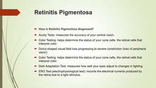


Retinitis pigmentosa is a genetic disorder that causes the rods and cones in the retina to deteriorate over time. This leads to progressive vision loss starting with night blindness and loss of peripheral vision, which can eventually cause total blindness. There is no cure, but vitamin A supplementation and an omega-3 rich diet have been shown to potentially slow the progression of the disease. It is diagnosed through visual field tests and ERG testing, and patients are referred to low vision specialists who can prescribe aids to help maximize remaining vision.


![Hypothalamus short ppt by Dr. Neha [PT].pptx](https://cdn.slidesharecdn.com/ss_thumbnails/hypothalamusbydr-260124145759-b9f94a93-thumbnail.jpg?width=640&height=640&fit=bounds)