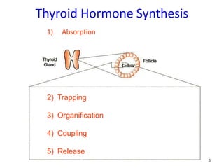
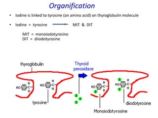
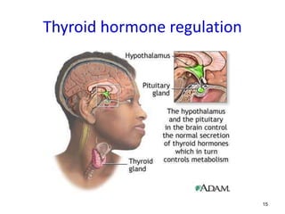
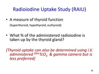
Thyroid Anatomy/Physiology/
Thyroid scintigraphy principles
NMT631
2
Thyroid - position
• The thyroid is a butterfly shaped gland that sits in front of the
trachea, midway between the thyroid cartilage (“Adam’s
apple”) and the top of the sternum
Supra Sternal Notch
SSN
3
Thyroid - Anatomy
A. Normal Adult Thyroid weighs 20-
25g
B. Components
1) Two Lateral lobes
2) Isthmus centrally connects the
lobes
3) A Pyramidal lobe in 50% of the
population
4
Thyroid –
Cellular Anatomy
• Epithelial cells arranged in spheres called thyroid
follicles.
• Follicles are filled with colloid
- Contains two active hormones, T3 and T4
- bound to thyroglobulin
• Parafollicular or C cells secrete hypercalcemia
hormone calcitonin.
Microscopic structure of Thyroid follicles
5
Thyroid Hormone Synthesis
1) Absorption
2) Trapping
3) Organification
4) Coupling
5) Release
6
Absorption
• Typical diet contains 300 -1000 μg of
Iodine/day
• Iodine ingested in food is reduced to iodide,
then absorbed into the blood
7
Trapping –
Iodide pump/trap
• Iodine Pump:
 30-50% of the
circulating iodine
taken up (rest lost in
urine)
 “Iodine trapping”
achieved by active
transport (iodide
pump).
 Iodide pump works
against both
electrical and
concentration
gradients.
8
Trapping
• Iodide then trapped by
follicular cells
• Trapped iodide ions are
oxidized by peroxidase
enzyme.
IP – Iodide Pump
9
Organification –
Thyroglobulin’s (TG) role
• TG is a large protein in
the colloid
• It has 140 molecules of
aminoacid tyrosin used
for thyroid hormone
synthesis
• Synthesis takes place
within the TG molecules
in the colloid.
• The trapped iodine is
now capable of
combining with tyrosine
10
Organification
• Iodine is linked to tyrosine (an amino acid) on thyroglobulin molecule
• Iodine + tyrosine MIT & DIT
MIT = monoiodotyrosine
DIT = diiodotyrosine
11
Coupling
• With the help of an enzyme, MIT and DIT combine to
form T3 and T4 (thyroxine)
MIT + DIT  T3
DIT + DIT  T4
• Both hormones are stored in thyroid follicular cells and
bound to thyroglobulin
12
Release
• In response to TSH (thyroid stimulating hormone),
T3 and T4 split from thyroglobulin to be sent into the
circulation
Steps:
1) TG taken up by thyroid cells by
pinocytosis
2) Lysosomes digest thyroglobluin,
thereby liberating T3, T4, MIT & DIT.
3) Fee thyroid hormones (T3 & T4) diffuse
out through the cell membrane into
blood
4) In the circulation, T3 and T4 are bound
to TBG (thyroxine-binding globulin)
5) MIT & DIT are deiodinated by an
enzyme and is recycled.
13 TSH stimulates iodide trap, thyroid hormone synthesis & release
14
Thyroid Hormone synthesis - Summary
• A normal thyroid produces about 10x more T4 than T3
Concise Human Physiology M. Y. Sukkar, H. a. El-Munshid, M. S. M. Ardawi
15
Thyroid hormone regulation
16
Thyroid Hormone Regulation
17
Hypothalamus – Pituitary – Thyroid
feedback system
• The activity of the thyroid
gland regulated by
neuroendocrine negative
feedback loop
• maintains a stable
amount of thyroid
hormones in the
circulation.
18
THYROID IMAGING
19
Thyroid Imaging
Clinical Indications
Radiopharmaceutical
used
Dosage &
Administration
Technique
The Normal Scan
Artifacts & Pitfalls
20
1) Relate structure to function
2) Evaluation of thyroid nodules
3) Location of ectopic tissue
4) Follow-up exams (post therapy, post surgery)
Clinical Indications
21
1) Tc-99m pertechnetate
2) I-123 sodium iodide
3) (I-131 sodium iodide)
Radiopharmaceuticals used
22
1) Tc-99m Pertechnetate (99mTcO4
-)
• Can substitute for iodide in the iodide transport
channel in follicular cells
• Trapped but not organified
• Released from cells over time
Radiopharmaceuticals used
• Only 1– 5% of injected dose trapped (↑ Bkg)
• Ideal physical characteristics (140keV; T1/2 - 6hrs)
• Readily available & cheap
• Low dose to thyroid
• Preferred over iodine when:
- patient on thyroid blocking agents
- patient unable to take meds orally
- study must be completed in < 2hrs
Dosage & Administration
23
Drugs - hyperthyroidism:
• Propylthiouracil
• Tapazole® (methimazole)
Drugs - hypothyroidism:
• LEVOTHROID®/ SYNTHROID ®
(synthetic T4)
• Cytomel® (synthetic T3)
Thyroid drug abstinence before imaging
(blocks thyroperoxidase)
24
123I Sodium iodide (Na123I)
• Trapped & organified
• 159keV gamma emission and
excellent trapping makes it ideal
imaging agent with low background
• Cyclotron produced (↑ cost,
problems with availability &
delivery)
• 200-600 μCi capsules
• T1/2 - 13 hrs; maximum uptake at 24hrs
Radiopharmaceuticals used
Dosage & Administration
25
131I Sodium Iodide (Na131I)
• Same uptake principles as Na123I
• Used for imaging (50 – 200 μCi) & therapy
• β emission & T1/2 of 8 days - ↑ radiation dose to
thyroid (disadvantages for imaging/ advantages for
therapy followed by delayed imaging)
• 364keV gamma emission used for imaging
• High energy collimator required
• ↓ cost & readily available
medic.usm.my
Administered as solution (Tx)
or capsules (Dx/Tx)
Radiopharmaceuticals used
Dosage & Administration
26
Tc-99m
pertechnetate
I-123 sodium
iodide
I-131 sodium
iodide
Activity 2-10 mCi 200-600 μCi 50-200 μCi
Route IV Oral Oral
Localization Trapped but not
organified
Trapped and
organified
Trapped and
organified
Time to Imaging 20 mins – 1 hr 4 – 24 hrs 4 – 24 hrs
Half-life 6 hrs 13 hrs 8 days
Gamma energy 140 keV 159 keV 364 keV
Radiation dose 0.13 rad/mCi 0.007 rad/μCi 1-3 rads/μCi
Thyroid Imaging Radiopharmaceuticals Properties
Radiopharmaceuticals used
27
1) ID patient; verify physician’s order; review clinical
indication for thyroid imaging
2) Explain procedure to patient; obtain relevant
medical history
Technique
Clinical Procedure
28
Relevant Medical History
• Physical findings (neck
palpation, vital signs)
• Symptoms of
hyper/hypothyroidism
• Medications/dietary
supplements/birth
control pills
• Surgery, esp neck,
upper chest
• Malignancies
• Changes in ability to
swallow/voice
• Previous medical
imaging
• Lab values: thyroid
hormone levels
• Pregnancy/lactation
– TcO4-: Stop for 12h
– I-123: Stop for 3d
– I-131: Stop for 3m
Technique
29
Symptoms of Hyper/Hypothyroidism
Hyperthyroidism
• Nervousness
• Palpitations
• Diarrhea
• Sweating
• Increased appetite
• Heat intolerance
Either
• Fatigue
• Dyspnea
• Weight change up or
down
Hypothyroidism
• Coarse hair
• Puffy eyelids
• Dry skin
• Myxedema
• Constipation
• Paresthesia
• Decreased appetite
• Cold intolerance
Technique
30
Clinical Procedure (cont’d)
3) Prepare patient
- ensure that female patients are not pregnant or breast
feeding (if breast feeding provide instructions)
- rule out substances that may affect radioiodine uptake
into thyroid gland
Technique
31
Factors Influencing
Thyroid Uptake of Iodine
Decreased Uptake
• Iodine-rich foods (shellfish)
• Radiographic contrast containing iodine
• Iodine-containing medications (vitamin/mineral
supplements, cough medicines, certain skin ointments)
• Non-iodine containing medications (penicillin, steroids,
antithyroid drugs)
• Thyroid hormones
Technique
Steroid Tyrosine
32
Factors Influencing
Thyroid Uptake of Iodine
Increased Uptake
• Iodine deficiency
• Pregnancy
• Renal failure
Technique
33
Clinical Procedure (cont’d)
4) Administer radiopharmaceutical
5) Image patient
Imaging time post tracer administration:
Tc-99m pertechnetate: 15-30 min
I-123 sodium iodide: 4-6 hrs/16-24 hrs
I-131 sodium iodide: 16-24 hrs
Patient positioning: supine with neck hyperextended
Views: anterior, obliques
Pinhole collimator
Mark anatomical landmarks
Technique
Pinhole collimator
34
Marking anatomical landmarks
• supra sternal notch
• thyroid cartilage
• chin (approx.)
Technique
One or more landmarks and
palpable nodules may be
marked with the help of a
radioactive/ radioopaque
source:
35
The Normal Scan
Right Lobe Left Lobe
Isthmus
Normal:
Butterfly-shaped gland with uniform,
symmetrical tracer distribution
Normal radioiodine Image
Normal pertechnetate image
Chin
SSN
Anterior w/ markers
36
THYROID
Non-Imaging Procedures
37
Thyroid Uptake Study
38
Radioiodine Uptake Study (RAIU)
• A measure of thyroid function
(hyperthyroid, hypothyroid, euthyroid)
• What % of the administered radioiodine is
taken up by the thyroid gland?
(Thyroid uptake can also be determined using i.V.
administered 99mTcO4
- & gamma camera but is
less preferred)
39
Clinical Procedure
1) At 4-6 hrs and/or 24 hrs following radioiodine
administration, collect counts over the following
areas using an uptake probe:
patient’s neck
patient’s thigh
standard (neck phantom)
room background
neck phantom
Standard Capsule Method
40
41
Thyroid Neck Phantom
• designed to simulate a patient’s
neck
• made of lucite
• Has two part insert that allows
counting from a bottle, vial or
capsule
• capsule holder enables counting
capsules directly
• phantom’s cylinder and carrier
have scribelines for accurate
alignment
• flat surface on the cylinder allows
either vertical or horizontal
positioning
42
Clinical Procedure (cont’d)
2) Place all counts in
counts/minute
(cpm). Calculate
net patient and
standard counts by
subtracting the
appropriate
background
counts.
Counts
C1
(cpm)
Counts
C2
(cpm)
Average
(C1+C2)/2
(cpm)
Net
(Avg – Bkg)
(cpm)
Capsule
counts 90 110 100 100 – 10 =
90
Room Bkg* 9 11 10
Neck
counts @
4 hrs
40 50 45 45 – 25 =
20
Thigh bkg*
counts @
4 hrs
30 20 25
Neck
counts @
24 hrs
60 70 65 65 – 20 =
45
Thigh bkg*
counts @
24 hrs
15 25 20
* Bkg - Background
Sample uptake work sheet
Standard Capsule Method
43
Uptake calculation
3) Calculate the % radioiodine uptake using the
following formula:
% uptake = neck cpm - thigh cpm x100
standard cpm - room bkg cpm
= Net neck cpm x 100
Net capsule cpm
Standard Capsule Method
44
Example
The following data were collected 24 hrs after
administration of two 100 μCi I-123 capsules.
Avg Counts Time (min) cpm
Neck 15380 5 3076
Thigh 860 5 172
Standard 12600 2 6300
(1 capsule)
Room bkg 350 5 70
Standard Capsule Method
45
% uptake = neck cpm - thigh cpm . x 100
standard cpm - room bkg cpm
= 3076 cpm - 172 cpm . x 100
2(6300 cpm – 70 cpm)
= 23%
Standard Capsule Method
46
Avg. counts CPM Net
counts
Capsule
(Std.1+Std.2)
12600 +
12600
25200 / 2 min
12600 12530
Room
BKG
350 / 5min 70
Neck 15380 / 5min 3076 2904
Thigh 860 / 5min 172
Net Neck cpm X 100
Net Capsule cpm
% Thyroid Uptake =
at 24hrs
2904 X 100
12530
= 23%
Standard Capsule Method
47
Normal Ranges
(may vary from dept to dept)
4 hr uptake 6 - 18%
24 hr uptake 10 - 35%
48
Using same capsule for both patient and
standard
• Count capsule with uptake probe before
administering to patient; note counts and time
• When patient counts are collected, decay correct
initial capsule counts to be used in calculation
Net neck cpm x 100
Net capsule(s) cpm x Decay Factor
Capsule Decay Method
49
Sources of Error
• Patient contains residual radioactivity from
previous test/therapy
• Counting geometry must remain constant for
patient and standard
• Elevated room background
50
Perchlorate Discharge Test
51
Why is it done?
• Used to evaluate any organification defect –
thyroid traps iodide but it does not combine
with tyrosine
52
Clinical Procedure
1) Patient receives radioiodine capsule
2) Two hrs following radioiodine administration, a
baseline uptake is performed. Then potassium
perchlorate (KClO4) is administered orally.
3) Sixty to ninety minutes later, another thyroid
uptake is performed.
53
Interpretation of Results
• If an organification defect is present, the perchlorate
ion (ClO4
-) displaces the iodide ion (I-) that has not
been organified and the post-perchlorate uptake will
be lower (10-15%) than the initial value.

Thyroid anatomy,physiology,thyroid scintigraphy principles

  • 1.
  • 2.
    2 Thyroid - position •The thyroid is a butterfly shaped gland that sits in front of the trachea, midway between the thyroid cartilage (“Adam’s apple”) and the top of the sternum Supra Sternal Notch SSN
  • 3.
    3 Thyroid - Anatomy A.Normal Adult Thyroid weighs 20- 25g B. Components 1) Two Lateral lobes 2) Isthmus centrally connects the lobes 3) A Pyramidal lobe in 50% of the population
  • 4.
    4 Thyroid – Cellular Anatomy •Epithelial cells arranged in spheres called thyroid follicles. • Follicles are filled with colloid - Contains two active hormones, T3 and T4 - bound to thyroglobulin • Parafollicular or C cells secrete hypercalcemia hormone calcitonin. Microscopic structure of Thyroid follicles
  • 5.
    5 Thyroid Hormone Synthesis 1)Absorption 2) Trapping 3) Organification 4) Coupling 5) Release
  • 6.
    6 Absorption • Typical dietcontains 300 -1000 μg of Iodine/day • Iodine ingested in food is reduced to iodide, then absorbed into the blood
  • 7.
    7 Trapping – Iodide pump/trap •Iodine Pump:  30-50% of the circulating iodine taken up (rest lost in urine)  “Iodine trapping” achieved by active transport (iodide pump).  Iodide pump works against both electrical and concentration gradients.
  • 8.
    8 Trapping • Iodide thentrapped by follicular cells • Trapped iodide ions are oxidized by peroxidase enzyme. IP – Iodide Pump
  • 9.
    9 Organification – Thyroglobulin’s (TG)role • TG is a large protein in the colloid • It has 140 molecules of aminoacid tyrosin used for thyroid hormone synthesis • Synthesis takes place within the TG molecules in the colloid. • The trapped iodine is now capable of combining with tyrosine
  • 10.
    10 Organification • Iodine islinked to tyrosine (an amino acid) on thyroglobulin molecule • Iodine + tyrosine MIT & DIT MIT = monoiodotyrosine DIT = diiodotyrosine
  • 11.
    11 Coupling • With thehelp of an enzyme, MIT and DIT combine to form T3 and T4 (thyroxine) MIT + DIT  T3 DIT + DIT  T4 • Both hormones are stored in thyroid follicular cells and bound to thyroglobulin
  • 12.
    12 Release • In responseto TSH (thyroid stimulating hormone), T3 and T4 split from thyroglobulin to be sent into the circulation Steps: 1) TG taken up by thyroid cells by pinocytosis 2) Lysosomes digest thyroglobluin, thereby liberating T3, T4, MIT & DIT. 3) Fee thyroid hormones (T3 & T4) diffuse out through the cell membrane into blood 4) In the circulation, T3 and T4 are bound to TBG (thyroxine-binding globulin) 5) MIT & DIT are deiodinated by an enzyme and is recycled.
  • 13.
    13 TSH stimulatesiodide trap, thyroid hormone synthesis & release
  • 14.
    14 Thyroid Hormone synthesis- Summary • A normal thyroid produces about 10x more T4 than T3 Concise Human Physiology M. Y. Sukkar, H. a. El-Munshid, M. S. M. Ardawi
  • 15.
  • 16.
  • 17.
    17 Hypothalamus – Pituitary– Thyroid feedback system • The activity of the thyroid gland regulated by neuroendocrine negative feedback loop • maintains a stable amount of thyroid hormones in the circulation.
  • 18.
  • 19.
    19 Thyroid Imaging Clinical Indications Radiopharmaceutical used Dosage& Administration Technique The Normal Scan Artifacts & Pitfalls
  • 20.
    20 1) Relate structureto function 2) Evaluation of thyroid nodules 3) Location of ectopic tissue 4) Follow-up exams (post therapy, post surgery) Clinical Indications
  • 21.
    21 1) Tc-99m pertechnetate 2)I-123 sodium iodide 3) (I-131 sodium iodide) Radiopharmaceuticals used
  • 22.
    22 1) Tc-99m Pertechnetate(99mTcO4 -) • Can substitute for iodide in the iodide transport channel in follicular cells • Trapped but not organified • Released from cells over time Radiopharmaceuticals used • Only 1– 5% of injected dose trapped (↑ Bkg) • Ideal physical characteristics (140keV; T1/2 - 6hrs) • Readily available & cheap • Low dose to thyroid • Preferred over iodine when: - patient on thyroid blocking agents - patient unable to take meds orally - study must be completed in < 2hrs Dosage & Administration
  • 23.
    23 Drugs - hyperthyroidism: •Propylthiouracil • Tapazole® (methimazole) Drugs - hypothyroidism: • LEVOTHROID®/ SYNTHROID ® (synthetic T4) • Cytomel® (synthetic T3) Thyroid drug abstinence before imaging (blocks thyroperoxidase)
  • 24.
    24 123I Sodium iodide(Na123I) • Trapped & organified • 159keV gamma emission and excellent trapping makes it ideal imaging agent with low background • Cyclotron produced (↑ cost, problems with availability & delivery) • 200-600 μCi capsules • T1/2 - 13 hrs; maximum uptake at 24hrs Radiopharmaceuticals used Dosage & Administration
  • 25.
    25 131I Sodium Iodide(Na131I) • Same uptake principles as Na123I • Used for imaging (50 – 200 μCi) & therapy • β emission & T1/2 of 8 days - ↑ radiation dose to thyroid (disadvantages for imaging/ advantages for therapy followed by delayed imaging) • 364keV gamma emission used for imaging • High energy collimator required • ↓ cost & readily available medic.usm.my Administered as solution (Tx) or capsules (Dx/Tx) Radiopharmaceuticals used Dosage & Administration
  • 26.
    26 Tc-99m pertechnetate I-123 sodium iodide I-131 sodium iodide Activity2-10 mCi 200-600 μCi 50-200 μCi Route IV Oral Oral Localization Trapped but not organified Trapped and organified Trapped and organified Time to Imaging 20 mins – 1 hr 4 – 24 hrs 4 – 24 hrs Half-life 6 hrs 13 hrs 8 days Gamma energy 140 keV 159 keV 364 keV Radiation dose 0.13 rad/mCi 0.007 rad/μCi 1-3 rads/μCi Thyroid Imaging Radiopharmaceuticals Properties Radiopharmaceuticals used
  • 27.
    27 1) ID patient;verify physician’s order; review clinical indication for thyroid imaging 2) Explain procedure to patient; obtain relevant medical history Technique Clinical Procedure
  • 28.
    28 Relevant Medical History •Physical findings (neck palpation, vital signs) • Symptoms of hyper/hypothyroidism • Medications/dietary supplements/birth control pills • Surgery, esp neck, upper chest • Malignancies • Changes in ability to swallow/voice • Previous medical imaging • Lab values: thyroid hormone levels • Pregnancy/lactation – TcO4-: Stop for 12h – I-123: Stop for 3d – I-131: Stop for 3m Technique
  • 29.
    29 Symptoms of Hyper/Hypothyroidism Hyperthyroidism •Nervousness • Palpitations • Diarrhea • Sweating • Increased appetite • Heat intolerance Either • Fatigue • Dyspnea • Weight change up or down Hypothyroidism • Coarse hair • Puffy eyelids • Dry skin • Myxedema • Constipation • Paresthesia • Decreased appetite • Cold intolerance Technique
  • 30.
    30 Clinical Procedure (cont’d) 3)Prepare patient - ensure that female patients are not pregnant or breast feeding (if breast feeding provide instructions) - rule out substances that may affect radioiodine uptake into thyroid gland Technique
  • 31.
    31 Factors Influencing Thyroid Uptakeof Iodine Decreased Uptake • Iodine-rich foods (shellfish) • Radiographic contrast containing iodine • Iodine-containing medications (vitamin/mineral supplements, cough medicines, certain skin ointments) • Non-iodine containing medications (penicillin, steroids, antithyroid drugs) • Thyroid hormones Technique Steroid Tyrosine
  • 32.
    32 Factors Influencing Thyroid Uptakeof Iodine Increased Uptake • Iodine deficiency • Pregnancy • Renal failure Technique
  • 33.
    33 Clinical Procedure (cont’d) 4)Administer radiopharmaceutical 5) Image patient Imaging time post tracer administration: Tc-99m pertechnetate: 15-30 min I-123 sodium iodide: 4-6 hrs/16-24 hrs I-131 sodium iodide: 16-24 hrs Patient positioning: supine with neck hyperextended Views: anterior, obliques Pinhole collimator Mark anatomical landmarks Technique Pinhole collimator
  • 34.
    34 Marking anatomical landmarks •supra sternal notch • thyroid cartilage • chin (approx.) Technique One or more landmarks and palpable nodules may be marked with the help of a radioactive/ radioopaque source:
  • 35.
    35 The Normal Scan RightLobe Left Lobe Isthmus Normal: Butterfly-shaped gland with uniform, symmetrical tracer distribution Normal radioiodine Image Normal pertechnetate image Chin SSN Anterior w/ markers
  • 36.
  • 37.
  • 38.
    38 Radioiodine Uptake Study(RAIU) • A measure of thyroid function (hyperthyroid, hypothyroid, euthyroid) • What % of the administered radioiodine is taken up by the thyroid gland? (Thyroid uptake can also be determined using i.V. administered 99mTcO4 - & gamma camera but is less preferred)
  • 39.
    39 Clinical Procedure 1) At4-6 hrs and/or 24 hrs following radioiodine administration, collect counts over the following areas using an uptake probe: patient’s neck patient’s thigh standard (neck phantom) room background neck phantom Standard Capsule Method
  • 40.
  • 41.
    41 Thyroid Neck Phantom •designed to simulate a patient’s neck • made of lucite • Has two part insert that allows counting from a bottle, vial or capsule • capsule holder enables counting capsules directly • phantom’s cylinder and carrier have scribelines for accurate alignment • flat surface on the cylinder allows either vertical or horizontal positioning
  • 42.
    42 Clinical Procedure (cont’d) 2)Place all counts in counts/minute (cpm). Calculate net patient and standard counts by subtracting the appropriate background counts. Counts C1 (cpm) Counts C2 (cpm) Average (C1+C2)/2 (cpm) Net (Avg – Bkg) (cpm) Capsule counts 90 110 100 100 – 10 = 90 Room Bkg* 9 11 10 Neck counts @ 4 hrs 40 50 45 45 – 25 = 20 Thigh bkg* counts @ 4 hrs 30 20 25 Neck counts @ 24 hrs 60 70 65 65 – 20 = 45 Thigh bkg* counts @ 24 hrs 15 25 20 * Bkg - Background Sample uptake work sheet Standard Capsule Method
  • 43.
    43 Uptake calculation 3) Calculatethe % radioiodine uptake using the following formula: % uptake = neck cpm - thigh cpm x100 standard cpm - room bkg cpm = Net neck cpm x 100 Net capsule cpm Standard Capsule Method
  • 44.
    44 Example The following datawere collected 24 hrs after administration of two 100 μCi I-123 capsules. Avg Counts Time (min) cpm Neck 15380 5 3076 Thigh 860 5 172 Standard 12600 2 6300 (1 capsule) Room bkg 350 5 70 Standard Capsule Method
  • 45.
    45 % uptake =neck cpm - thigh cpm . x 100 standard cpm - room bkg cpm = 3076 cpm - 172 cpm . x 100 2(6300 cpm – 70 cpm) = 23% Standard Capsule Method
  • 46.
    46 Avg. counts CPMNet counts Capsule (Std.1+Std.2) 12600 + 12600 25200 / 2 min 12600 12530 Room BKG 350 / 5min 70 Neck 15380 / 5min 3076 2904 Thigh 860 / 5min 172 Net Neck cpm X 100 Net Capsule cpm % Thyroid Uptake = at 24hrs 2904 X 100 12530 = 23% Standard Capsule Method
  • 47.
    47 Normal Ranges (may varyfrom dept to dept) 4 hr uptake 6 - 18% 24 hr uptake 10 - 35%
  • 48.
    48 Using same capsulefor both patient and standard • Count capsule with uptake probe before administering to patient; note counts and time • When patient counts are collected, decay correct initial capsule counts to be used in calculation Net neck cpm x 100 Net capsule(s) cpm x Decay Factor Capsule Decay Method
  • 49.
    49 Sources of Error •Patient contains residual radioactivity from previous test/therapy • Counting geometry must remain constant for patient and standard • Elevated room background
  • 50.
  • 51.
    51 Why is itdone? • Used to evaluate any organification defect – thyroid traps iodide but it does not combine with tyrosine
  • 52.
    52 Clinical Procedure 1) Patientreceives radioiodine capsule 2) Two hrs following radioiodine administration, a baseline uptake is performed. Then potassium perchlorate (KClO4) is administered orally. 3) Sixty to ninety minutes later, another thyroid uptake is performed.
  • 53.
    53 Interpretation of Results •If an organification defect is present, the perchlorate ion (ClO4 -) displaces the iodide ion (I-) that has not been organified and the post-perchlorate uptake will be lower (10-15%) than the initial value.