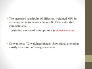
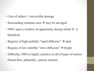
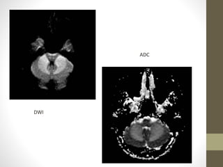
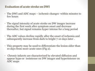
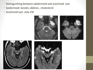
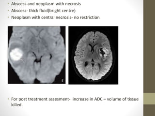
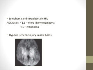
This document discusses diffusion weighted imaging (DWI) and its application in evaluating brain pathologies. It provides details on how DWI works using diffusion gradients and endogenous contrast from water motion. Areas of restricted diffusion like cytotoxic edema appear brighter on DWI. DWI is highly sensitive for detecting acute ischemia within minutes. It is useful for distinguishing acute from subacute lesions based on apparent diffusion coefficient (ADC) maps. DWI also has applications in evaluating other conditions like abscesses, tumors, infections and injuries.
1. Diffusion weighted
imaging – principles and its
application in brain pathologies
Moderator- DR. JEEVIKA.M.U
Sharath
2. Diffusion-weighted MRI
• is a example of endogenous contrast, using the motion of protons to
produce signal changes.
• DWI images is obtained by applying pairs of opposing and balanced
magnetic field gradients (but of differing durations and amplitudes)
around a spin-echo refocusing pulse of a T2 weighted sequence.
• Stationary water molecules - unaffected by the paired gradients-
retain their signal.
• Nonstationary water molecules - overall loss of the MR signal
3. • The normal motion of water molecules within living tissues is random
(brownian motion).
• Any pathology that affects this normal diffusion of water molecules provides
basis of contrast.
• Areas of cytotoxic edema- motion of water molecules is restricted- appear
brighter on DWI - lesser signal losses
• Tissues with a higher rate of diffusion undergo a greater loss of signal in a
given period of time than do tissues with a lower diffusion rate
• Classical diffusion weighted images are based on isotropic diffusion
Anisotropic diffusion- diffusion tensor imaging{tractography}
4. • DW images usually performed with echo-planar sequences which
1.markedly decrease imaging time, motion artifacts
2. increase sensitivity to signal changes due to molecular motion.
-Acquired by SJETSKAL- TANNER pulsed gradient spin echo sequence.
• The primary application of DW imaging has been in brain imaging, mainly
because of its exquisite sensitivity to early detection of ischemic stroke
5. • The increased sensitivity of diffusion-weighted MRI in
detecting acute ischemia - the result of the water shift
intracellularly,
restricting motion of water protons (cytotoxic edema),
• Conventional T2 weighted images show signal alteration
mostly as a result of vasogenic edema
6. • Core of infarct = irreversible damage
• Surrounding ischemic area may be salvaged
• DWI: open a window of opportunity during which Tt is
beneficial
• Regions of high mobility “rapid diffusion” dark
• Regions of low mobility “slow diffusion” bright
• Difficulty: DWI is highly sensitive to all of types of motion
(blood flow, pulsatility, patient motion).
7.
8.
9. B- value
• B – value - magnitude of the diffusion weighting provided by diffusion gradients.
Expressed in sec/mm2.
• Typically b values used in clinical practice ,range from 0-1000 s/mm2, however b values
upto 4000 s/mm2 are available on modern MRI scanners.
• Useful rule of thumb is to pick b value such that b x ADC is approximately equal to 1.
• Brain- 0, 500, 1000
• Neck- 0 and 800
• Breast- 0 ,500, 1000
• Prostate- 0,750 , 1500
• Liver – 50, 500,1000
10. • B- value- amplitude, separation and duration of
diffusion gradient. Increases with gradient strength ,
duration of application and time between application
of two diffusion gradients
11. Apparent Diffusion Coefficient
It is a measure of diffusion .
ADC value is Calculated for each voxel by acquiring two or more images
with different b-values .(usually b value zero and higher b value images).
ADC= - Ln{A(b)/A(o)} A(b)- measured echo magnitude
b A(0)- echo magnitude without diffusion gradient
Images of the ADC are called as ADC map.
An ADC map shows parametric images containing the apparent diffusion
coefficients of diffusion weighted images. Also called diffusion map
12. Apparent Diffusion Coefficient(contd)
Differentiate T2 shine through effects or artifacts from real
ischemic lesions.
T 2 shine through
• Signal intensity on DWI – not only on ADC but also on tissue T2. this
can cause paradoxical decrease in signals of restricted diffusion or when
diffusion is normal, can be mistaken for diffusion restriction.
14. • The ADC may be useful for distinguishing acute from subacute DWI
lesions.
• Acute ischemic lesions can be divided into hyperacute lesions (low
ADC and DWI-positive) and subacute lesions (normalized ADC).
• Chronic lesions can be differentiated from acute lesions by
normalization of ADC and DWI.
• a tumour would exhibit more restricted apparent diffusion
compared with a cyst because intact cellular membranes in a tumour
would hinder the free movement of water molecules
15. Nonischemic causes for decreased ADC
• Abscess
• Lymphoma and other tumors
• Multiple sclerosis
• Seizures
• Metabolic (Canavans )
17. EvaluationofacutestrokeonDWI
• The DWI and ADC maps – ischemic changes -within minutes to
few hours
• The signal intensity of acute stroke on DW images increase
during the first week after symptom onset and decrease
thereafter, but signal remains hyper intense for a long period
• The ADC values decline rapidly after the onset of ischemia and
subsequently increase from dark to bright 7-10 days later .
• This property may be used to differentiate the lesion older than
10 days from more acute ones (Fig 2).
• Chronic infarcts are characterized by elevated diffusion and
appear hypo or isointense on DW images and hyperintense on
ADC maps
21. • Distinguishing between epidermoid and arachnoid cyst
• Epidermoid- keratin, debries , cholesterol
• Arachnoid cyst- only CSF
22. • Abscess and neoplasm with necrosis
• Abscess- thick fluid(bright centre)
• Neoplasm with central necrosis- no restriction
• For post treatment assesment- increase in ADC – volume of tissue
killed.
23. • Lymphoma and toxoplasma in HIV
ADC ratio : > 1.6 – more likely toxoplasma
< 1 – lymphoma
• Hypoxic ischemic injury in new borns
24. Thank you
References
• Mri made easy- govind chauhan
• Christensens physics of diagnostic radiology
• Osborns textbook of neuroimaging
• Diagnostic imaging series- Brain
Editor's Notes
4.acquire phase information from the first gradient, but are not rephased by the second gradient, leading to an