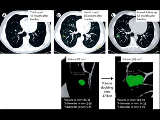
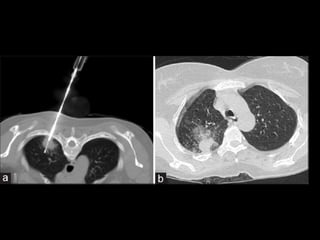
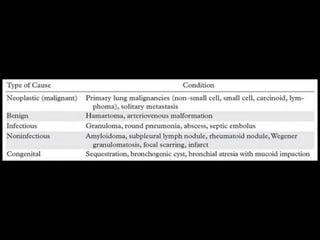
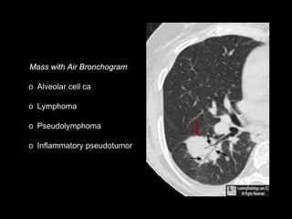
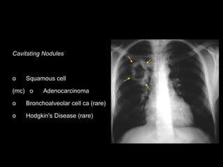
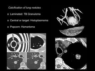
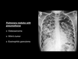
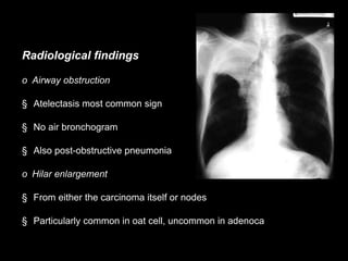
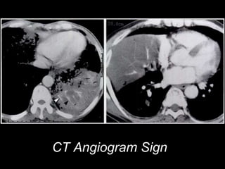
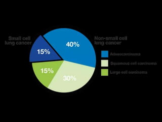
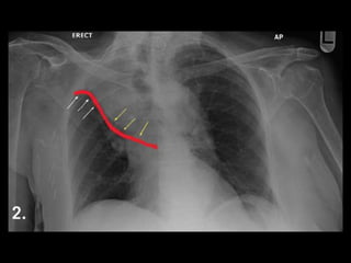
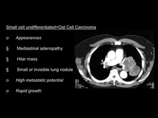
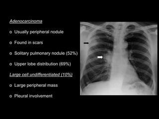
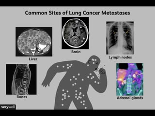
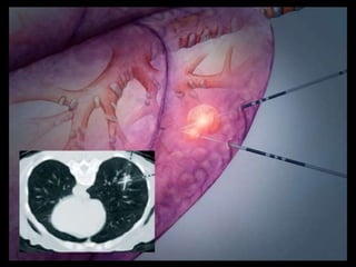
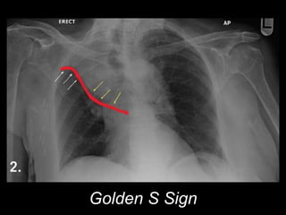
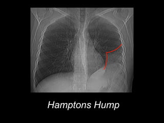
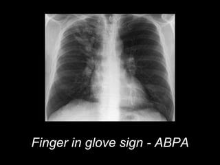
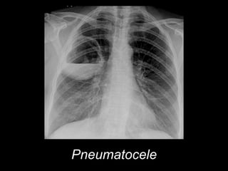
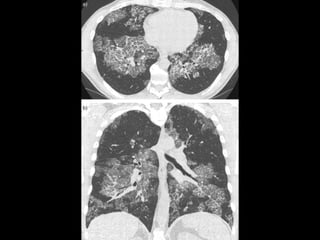
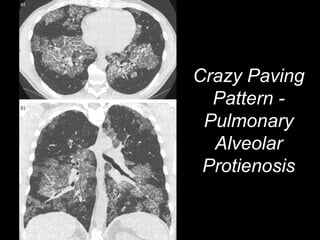
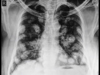
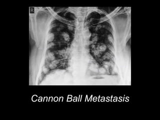
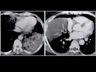
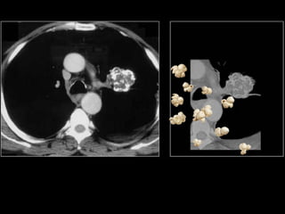
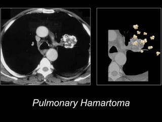
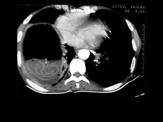
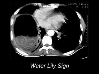
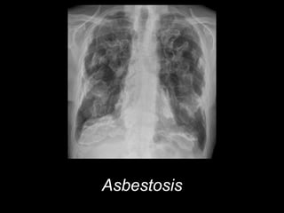
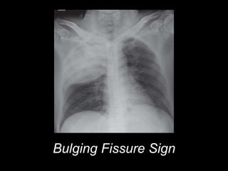
Lung Masses
Dr. Girendra Shankar
PG Resident
Dr. R N Dash
Associate Professor
Department of Radiodiagnosis and Imaging,KIMS
Doubling time
If a lesion doubles in volume
>6 weeks and <16 months,
usually malignant
Incidence
o On routine survey, <5% are malignant nodules
o At Biopsy/Surgery, 40% of nodules are Ca, 40% Granulomas
o Alveolar cell ca
o Lymphoma
o Pseudolymphoma
o Inflammatory pseudotumor
Cavitating Nodules
o Squamous cell
(mc) o Adenocarcinoma
o Bronchoalveolar cell ca (rare)
o Hodgkin's Disease (rare)
Calcification of lung nodules
o Laminated: TB Granuloma
o Central or target: Histoplasmoma
o Popcorn: Hamartoma
Pulmonary nodules with
pneumothorax
o Osteosarcoma
o Wilm's tumor
o Eosinophilic granuloma
Radiological findings
o Airway obstruction
§ Atelectasis most common sign
§ No air bronchogram
§ Also post-obstructive pneumonia
o Hilar enlargement
§ From either the carcinoma itself or nodes
§ Particularly common in oat cell, uncommon in adenoca
CT Angiogram Sign
o Mediastinal node enlargement
§ Particularly anaplastic ca
o Cavitation
§ Common (2-16%)
§ Especially in squamous cell, mostly in upper lobes
§ Cavity is usually thick-walled with nodular inner margin
o Pleural involvement
§ Common: 10%
§ Hemorrhagic effusion denotes direct tumor invasion
§ Effusion carries a poor prognosis even if no malignant cells are found
Squamous cell ca
Central Location (2/3)
Atelectasis
"Reverse S sign of Golden”
Post-obstructive pneumonia
May cavitate
Most closely associated with smoking
Small cell undifferentiated=Oat Cell Carcinoma
o Appearances
§ Mediastinal adenopathy
§ Hilar mass
§ Small or invisible lung nodule
o High metastatic potential
o Rapid growth
Adenocarcinoma
o Usually peripheral nodule
o Found in scars
o Solitary pulmonary nodule (52%)
o Upper lobe distribution (69%)
Large cell undifferentiated (10%)
o Large peripheral mass
o Pleural involvement
SPOTTERS
Thymic Sail Sign
Golden S Sign
Hamptons Hump
Finger in glove sign - ABPA
Pneumatocele
Crazy Paving
Pattern -
Pulmonary
Alveolar
Protienosis
Cannon Ball Metastasis
CT Angiogram Sign
Pulmonary Hamartoma
Water Lily Sign
Asbestosis
Bulging Fissure Sign

Imaging in lung masses - UG

Editor's Notes

  • #21 May be associated with §       Hypoglycemia §       Cushing's syndrome §       Inappropriate secretion of ADH §       Excessive gonadotropin secretion
  • #23 o      §       Osteolytic mets (3/4) Kidney, GI tract, liver, contralateral lung
  • #31  Hamptons Hump dome-shaped, pleural-based opacification in the lung most commonly due to pulmonary embolism and lung infarction
  • #32  Hamptons Hump dome-shaped, pleural-based opacification in the lung most commonly due to pulmonary embolism and lung infarction
  • #33 allergic bronchopulmonary aspergillosis (ABPA) cystic fibrosis to the characteristic sign of a bronchocoele.
  • #34 allergic bronchopulmonary aspergillosis (ABPA) cystic fibrosis to the characteristic sign of a bronchocoele.
  • #45 Water Lily Sign
  • #46 Water Lily Sign
  • #47 Asbestosis
  • #48 Asbestosis
  • #49 Bulging Fissure Sign
  • #50 Bulging Fissure Sign