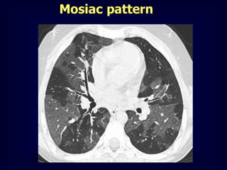
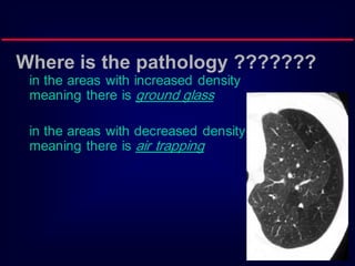
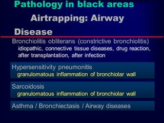
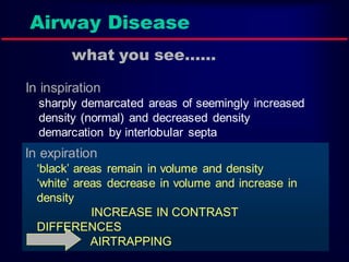
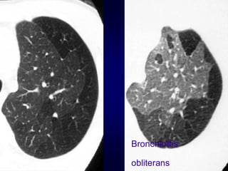
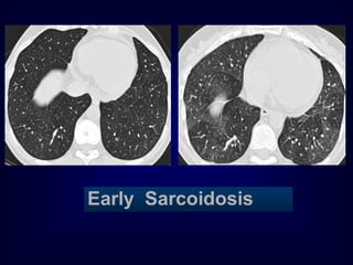
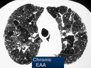
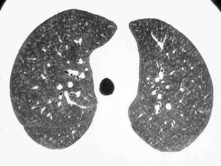
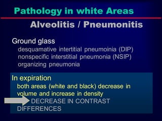
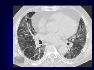
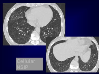
This document discusses the mosaic pattern seen on CT scans of the chest. It describes how the pattern shows areas of increased and decreased density that correspond to areas of ground glassing and air trapping, respectively. The black areas represent air trapping and can be seen in conditions like bronchiolitis obliterans, sarcoidosis, and asthma. The white areas represent ground glassing and alveolitis/pneumonitis, seen in diseases like desquamative interstitial pneumonia and nonspecific interstitial pneumonia. The mosaic pattern results from airway disease causing air trapping or alveolitis/pneumonitis causing ground glassing.
Role of hrct in interstitial lung diseases pk uploadDr pradeep Kumar
Role of hrct in interstitial lung diseases pk , This is best powerpoint slides presentation including Latest American thoracic society and fleishners society guidelines . this includes radiographic images a well HRCT chest findings of various ILD. This will help alot for md pg radiology resident and radiologist. Thanks
Describes parts of the mediastinum and anatomical landmarks and common mediastinal pathologies and there radiological features and differentiation in a simple educational way with multiple CT examples of different cases .
HRCT in Diffuse Lung Diseases - II (Honeycombing, UIP pattern, IPF)Bhavin Jankharia
This is the second part of this series on HRCT in diffuse lung diseases, focussing on the diagnosis of honeycombing, UIP pattern and IPF and the associated complications and differential diagnoses
Imaging plays an important role in diagnosis and formulating differential diagnosis in case of Solitary pulmonary nodule. It helps in differentiating and predicting benign and malignant nodules.
Describes cross sectional anatomy of the mediastinum , and lobar and segmental anatomy of the lung with teaching points and radiological guidelines and multiple examples of lobar and segmental pathologies and how we localize these pathologies .Also the types of chest CT images and indications of chest CT.
Bronchiectasis ( Bronchos- airways ; ectasia- dilatation) is a morphological term used to describe abnormal irreversibly dilated and often thick walled bronchi.
Bronchiectasis represents the end stage of variety of pathological precesses that cause destruction of bronchial wall and its surrounding tissues.
Pneumonia is characterized by the emergence of new lung infiltrates, accompanied by clinical signs such as fever, purulent sputum, leukocytosis, and decreased oxygenation and Nosocomial Pneumonia is a non-incubating lower respiratory infection that presents clinically two or more days after hospitalization. In this presentation "Nosocomial Pneumonias" has been described including their causes, therapy, Principles, diagnosis, symptoms, management, etc. For more information, please contact us: 9779030507.
Oxygen Therapy is not Beneficial in COPD Patients with Moderate HypoxaemiaGamal Agmy
A Randomized Trial of Long-Term Oxygen for COPD with Moderate Desaturation
The Long-Term Oxygen Treatment Trial Research Group*
N Engl J Med. 2016 October 27; 375(17): 1617–1627
4. Where is the pathology ???????
in the areas with increased density
meaning there is ground glass
in the areas with decreased density
meaning there is air trapping
5. Pathology in black areas
Airtrapping: Airway
Disease
Bronchiolitis obliterans (constrictive bronchiolitis)
idiopathic, connective tissue diseases, drug reaction,
after transplantation, after infection
Hypersensitivity pneumonitis
granulomatous inflammation of bronchiolar wall
Sarcoidosis
granulomatous inflammation of bronchiolar wall
Asthma / Bronchiectasis / Airway diseases
6. Airway Disease
what you see……
In inspiration
sharply demarcated areas of seemingly increased
density (normal) and decreased density
demarcation by interlobular septa
In expiration
‘black’ areas remain in volume and density
‘white’ areas decrease in volume and increase in
density
INCREASE IN CONTRAST
DIFFERENCES
AIRTRAPPING
13. Pathology in white Areas
Alveolitis / Pneumonitis
Ground glass
desquamative intertitial pneumoinia (DIP)
nonspecific interstitial pneumonia (NSIP)
organizing pneumonia
In expiration
both areas (white and black) decrease in
volume and increase in density
DECREASE IN CONTRAST
DIFFERENCES