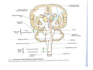
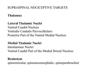
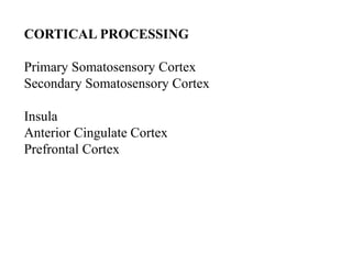
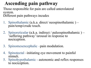
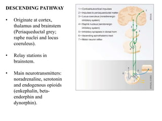
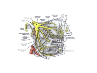
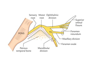
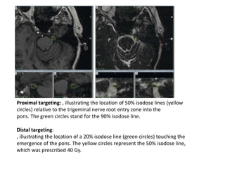
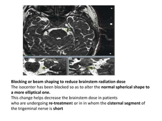
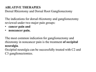
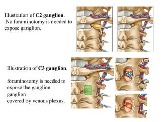
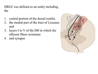
PAIN PATHWAY
DR GHANSHYAM SASAPARDHI
IASP DEFINATION
“Pain is an unpleasant sensory and emotional
experience associated with actual or potential
tissue damage, or described in terms of such
damage.”
ISAP- INTERNATIONAL ASSOCIATION FOR STUDY OF PAIN
.
The pain experience has two dimensions:
1. Sensory/discriminative - allowing us to
locate tissue damage
2. Affective/aversive - ‘unpleasant’ and
‘emotional’ in the IASP definition.
Avoid stimuli that can damage tissue.
Ascending pain pathway consists of-
Peripheral receptor – nerve ending of Aδ and C-
fibres , detect stimulus. Signal carried by:
1st order neuron – from periphery to spinal cord.
2nd order neuron – cell body in dorsal horn.
3rd order neuron – cell body in thalamus, ascend
ipsilaterally to project to somatosensory cortex.
SUPRASPINAL NOCICEPTIVE TARGETS
Thalamus
Lateral Thalamic Nuclei
Ventral Caudal Nucleus
Ventralis Caudalis Parvocellularis
Posterior Part of the Ventral Medial Nucleus
Medial Thalamic Nuclei
Intralaminar Nuclei
Ventral Caudal Part of the Medial Dorsal Nucleus
Brainstem
spinoreticular, spinomesencephalic, spinoparabrachial
CORTICAL PROCESSING
Primary Somatosensory Cortex
Secondary Somatosensory Cortex
Insula
Anterior Cingulate Cortex
Prefrontal Cortex
Ascending pain pathway
Those responsible for pain are called anterolateral
system.
Different pain pathways incudes
1. Spinothalamic (a.k.a. direct/ neospinothalamic ) –
pain/temp/crude touch.
2. Spinoreticular (a.k.a. indirect / paleospinothalamic ) –
’suffering pathway’/arousal in response to
nociception.
3. Spinomesencephalic –pain modulation.
4. Spinotectal –initiating eye movement to painful
stimuli.
5. Spinohypothalamic - autonomic and reflex responses
to nociception.
DESCENDING PATHWAY
• Originate at cortex,
thalamus and brainstem
(Periaqueductal grey;
raphe nuclei and locus
coeruleus).
• Relay stations in
brainstem.
• Main neurotransmitters:
noradrenaline, serotonin
and endogenous opioids
(enkephalin, beta-
endorphin and
dynorphin).
The most important descending control system has
links in the midbrain ( PAG) periaqueductal gray
and to the rostral ventromedial medulla (RVM),
which constitute the PAG/RVM system.
Electrical stimulation at either site can enhance or
inhibit pain, depending on the exact site, current
level, and the behavioral context
Connections with:
1. Nucleus raphe nucleus (serotonergic)  inhibitory
interneurones.
2. Locus coeruleus (Noradrenergic)  dorsal horn.
CRANIOFACIAL PAIN SYNDROMES
Possible pathways for facial pain include:
. ETIOLOGIES
1. CEPHALIC NEURALGIAS
a) trigeminal neuralgia
● vascular compression
● MS:
b) glossopharyngeal neuralgia : pain usually in base of tongue
and adjacent pharynx.- PICA
c) geniculate neuralgia : otalgia – nervus inermedius-
somatosensory branch of facial n.
d) tic convulsif : geniculate neuralgia with hemifacial spasm-
AICA-
e) occipital neuralgia
h) herpes zoster:
Characteristic vesicles and crusting usually follow pain, most
often in distribution of V1
i) Post herpetic neuralgia (Ramsay-Hunt syndrome)
j) supraorbital neuralgia (SON)
k) trigeminal neuropathic pain (AKA trigeminal deafferentation
pain): injuries from sinus or dental surgery, head trauma
2. OPHTHALMIC PAIN
a) Tolosa-Hunt syndrome : painful ophthalmoplegia
b) (Raeder’s) paratrigeminal neuralgia : unilateral Horner’s
syndrome +trigeminal neuralgia
c) orbital pseudotumor : proptosis, pain, and EOM dysfunction
3. otalgia
4. masticatory disorders
a) dental or periodontal disease
b) nerve injury (inferior and/or superior alveolar nerves)
c) temporo-mandibular joint (TMJ) dysfunct ion
d) elongated styloid process
e) temporal & masseter myositis
5. vascular pain syndromes
a) migraine headaches:
b) cluster H/A ; subtypes: episodic, chronic, chronic
paroxysmal hemicrania.
c) giant cell arteritis (temporal arteritis). Tenderness over STA
e) hypertensive H/A
f) aneurysm or AVM (due either to mass effect or hemorrhage)
g) carotidynia: e.g. with carotid dissection
6. sinusitis (maximally, frontal, ethmoidal, sphenoidal)
8. neoplasm: may cause referred pain or fifth nerve
compression
a) extracranial
b) intracranial tumor: primarily posterior fossa lesions,
9. atypical facial pain (AFP) (prosopalgia):
10. primary (nonvascular) H/A: including
a) tension (muscle contract ion) H/A
b) post-traumatic H/A
Trigeminal neuralgia
80–90% of cases -SCA
MS plaque
Surgical options
1. peripheral trigeminal nerve branch procedures to block or ablate
the division involved.
1. blocking the trigger: either via percutaneous rhizotomy or alcohol
block.
2. percutaneous trigeminal rhizotomy (PTR): AKA percutaneous
(stereotactic) rhizotomy (PSR) of trigeminal (Gasserian) ganglion
3. intradural retrogasserian trigeminal nerve section (sensory
portion ± motor root, : if no vascular compression
4. microvascular decompression (MVD):
5. stereotactic radiosurgery:
Proximal targeting: , illustrating the location of 50% isodose lines (yellow
circles) relative to the trigeminal nerve root entry zone into the
pons. The green circles stand for the 90% isodose line.
Distal targeting:
, illustrating the location of a 20% isodose line (green circles) touching the
emergence of the pons. The yellow circles represent the 50% isodose line,
which was prescribed 40 Gy.
Blocking or beam shaping to reduce brainstem radiation dose
The isocenter has been blocked so as to alter the normal spherical shape to
a more elliptical one.
This change helps decrease the brainstem dose in patients
who are undergoing re-treatment or in in whom the cisternal segment of
the trigeminal nerve is short
NEUROSURGICAL THERAPIES FOR INTRACTABLE
PAIN
1.Anatomic;
Correction of structural deformity
2.Augmentative
• Stimulation:
Peripheral nerve
Spinal cord
Deep brain structures
Motor cortex
• Neuraxial /drug infusion:
Intrathecal /epidural
Intraventricular
3.Ablative
Neurectomy
Sympathectomy
Ganglionectomy
Rhizotomy
Spinal DREZ lesion
Cordotomy
Myelotomy
Nucleus caudalis DREZ
lesion
Trigeminal tractotomy
Mesencephalotomy
Thalamotomy
Cingulotomy
Hypophysectomy
Augmentative therapies
INTRACRANIAL NEUROSTIMULATION
Deep Brain Stimulation
performed successfully to treat pain not responsive to
other neuromodulation techniques,
• including cluster headaches,
• failed back surgery syndrome,
• peripheral neuropathic pain, facial
• deafferentation pain,
• pain that is secondary to brachial plexus avulsion.
Stimulation sites included
the periventricular/periaqueductal gray matter (PVG/PAG), the
internal capsule, the sensory thalamus, and the posterior hypothalamus.
DBS has had its best success in treating cluster headaches and nociceptive
syndromes such as chronic low back pain
MOTOR CORTEX STIMULATION
particular promise in the treatment of trigeminal neuropathic
pain and central pain syndromes such as thalamic pain syndrome
OCCIPITAL NERVE STIMULATION
Less invasive and less risky alternative to deep brain intracranial
stimulation.
Useful in some pt with cluster headaches
SPINAL CORD STIMULATION
Spinal cord stimulation modifies the perception of pain by stimulating
the dorsal columns of the spinal cord and may relieve
neuropathic or ischemic pain.
Relief of pain from failed back surgery syndrome (FBSS)
appears to respond best to SCS.
PERIPHERAL NERVE STIMULATION
surgical placement of electrodes directly over a
peripheral nerve or percutaneous placement of the
electrodes sufficiently near the nerve to provide an
effective neuromodulating waveform.
Ulnar nerve stimulation. A, Distribution of the ulnar nerve in the right forearm and hand.
B, Surgical anatomy of the approach and placement of the ulnar nerve stimulator. 1, Skin incision, with
musculature, ligaments, and tendons shown. 2, The ulnar nerve is mobilized in the usual fashion and the lead
placed beneath it. 3, The lead is anchored to prevent its mobilization. C, Subcutaneous pacemaker placed
under the right clavicle and with a cord tunneled to the lead at the elbow. IPG, implantable pulse generator.
ABLATIVE THERAPIES
Dorsal Rhizotomy and Dorsal Root Ganglionectomy
The indications for dorsal rhizotomy and ganglionectomy
reviewed under two major pain groups:
• cancer pain and
• noncancer pain.
The most common indication for ganglionectomy and
rhizotomy in noncancer pain is the treatment of occipital
neuralgia.
Occipital neuralgia can be successfully treated with C2 and
C3 ganglionectomies.
Illustration of C2 ganglion.
No foraminotomy is needed to
expose ganglion.
Illustration of C3 ganglion.
foraminotomy is needed to
expose the ganglion.
ganglion
covered by venous plexus.
DREZ was defined as an entity including
the
1. central portion of the dorsal rootlet,
2. the medial part of the tract of Lissauer,
and
3. layers I to V of the DH in which the
afferent fibers terminate
4. and synapse
INDICATIONS
1. Cancer Pain
The first use of DREZ lesioning for control of pain was
to the benefit of a patient with a neoplastic lesion, namely
a Pancoast- tumor invading the brachial plexus
2.Neuropathic Pain
Root Avulsion Pain (Brachial and Lumbosacral Plexus
3.Spinal Cord Injury Pain
DREZ lesioning has been applied to some other
indications, such as complex regional pain syndrome
(CRPS), peripheral nerve injury, postamputation pain,
occipital neuralgia, post-herpetic neuralgia, and
radiation-induced plexopathy, when pain did not
respond to spinal cord stimulation
Percutaneous Cordotomy and Trigeminal
Tractotomy-Nucleotomy
CORDOTOMY
Interruption lat spinothalamic tract fiber in spinal cord
Indications
procedure that is mostly used for cancer-related pain in
terminally ill pt.
The ideal candidate is a cancer patient with unilateral somatic
Pain below c5 and a life expectancy of approximately 1 year.
TRIGEMINAL TRACTOTOMY-NUCLEOTOMY
Indications
Trigeminal neuropathic pain with varying degrees of
trigeminal nerve damage and neuropathy are the main
candidates for TR-NC.
Other conditions such as traumatic trigeminal neuropathy,
post-herpetic neuralgia, cancer pain of the head or face,
glossopharyngeal or geniculate neuralgia, bilateral
trigeminal neuralgia, and anesthesia dolorosa are the typical
indications for TR-NC.
THANK YOU

Pain pathway

  • 1.
  • 2.
    IASP DEFINATION “Pain isan unpleasant sensory and emotional experience associated with actual or potential tissue damage, or described in terms of such damage.” ISAP- INTERNATIONAL ASSOCIATION FOR STUDY OF PAIN .
  • 3.
    The pain experiencehas two dimensions: 1. Sensory/discriminative - allowing us to locate tissue damage 2. Affective/aversive - ‘unpleasant’ and ‘emotional’ in the IASP definition. Avoid stimuli that can damage tissue.
  • 4.
    Ascending pain pathwayconsists of- Peripheral receptor – nerve ending of Aδ and C- fibres , detect stimulus. Signal carried by: 1st order neuron – from periphery to spinal cord. 2nd order neuron – cell body in dorsal horn. 3rd order neuron – cell body in thalamus, ascend ipsilaterally to project to somatosensory cortex.
  • 6.
    SUPRASPINAL NOCICEPTIVE TARGETS Thalamus LateralThalamic Nuclei Ventral Caudal Nucleus Ventralis Caudalis Parvocellularis Posterior Part of the Ventral Medial Nucleus Medial Thalamic Nuclei Intralaminar Nuclei Ventral Caudal Part of the Medial Dorsal Nucleus Brainstem spinoreticular, spinomesencephalic, spinoparabrachial
  • 7.
    CORTICAL PROCESSING Primary SomatosensoryCortex Secondary Somatosensory Cortex Insula Anterior Cingulate Cortex Prefrontal Cortex
  • 8.
    Ascending pain pathway Thoseresponsible for pain are called anterolateral system. Different pain pathways incudes 1. Spinothalamic (a.k.a. direct/ neospinothalamic ) – pain/temp/crude touch. 2. Spinoreticular (a.k.a. indirect / paleospinothalamic ) – ’suffering pathway’/arousal in response to nociception. 3. Spinomesencephalic –pain modulation. 4. Spinotectal –initiating eye movement to painful stimuli. 5. Spinohypothalamic - autonomic and reflex responses to nociception.
  • 9.
    DESCENDING PATHWAY • Originateat cortex, thalamus and brainstem (Periaqueductal grey; raphe nuclei and locus coeruleus). • Relay stations in brainstem. • Main neurotransmitters: noradrenaline, serotonin and endogenous opioids (enkephalin, beta- endorphin and dynorphin).
  • 10.
    The most importantdescending control system has links in the midbrain ( PAG) periaqueductal gray and to the rostral ventromedial medulla (RVM), which constitute the PAG/RVM system. Electrical stimulation at either site can enhance or inhibit pain, depending on the exact site, current level, and the behavioral context Connections with: 1. Nucleus raphe nucleus (serotonergic)  inhibitory interneurones. 2. Locus coeruleus (Noradrenergic)  dorsal horn.
  • 11.
    CRANIOFACIAL PAIN SYNDROMES Possiblepathways for facial pain include: . ETIOLOGIES 1. CEPHALIC NEURALGIAS a) trigeminal neuralgia ● vascular compression ● MS: b) glossopharyngeal neuralgia : pain usually in base of tongue and adjacent pharynx.- PICA c) geniculate neuralgia : otalgia – nervus inermedius- somatosensory branch of facial n. d) tic convulsif : geniculate neuralgia with hemifacial spasm- AICA- e) occipital neuralgia
  • 12.
    h) herpes zoster: Characteristicvesicles and crusting usually follow pain, most often in distribution of V1 i) Post herpetic neuralgia (Ramsay-Hunt syndrome) j) supraorbital neuralgia (SON) k) trigeminal neuropathic pain (AKA trigeminal deafferentation pain): injuries from sinus or dental surgery, head trauma
  • 13.
    2. OPHTHALMIC PAIN a)Tolosa-Hunt syndrome : painful ophthalmoplegia b) (Raeder’s) paratrigeminal neuralgia : unilateral Horner’s syndrome +trigeminal neuralgia c) orbital pseudotumor : proptosis, pain, and EOM dysfunction 3. otalgia 4. masticatory disorders a) dental or periodontal disease b) nerve injury (inferior and/or superior alveolar nerves) c) temporo-mandibular joint (TMJ) dysfunct ion d) elongated styloid process e) temporal & masseter myositis
  • 14.
    5. vascular painsyndromes a) migraine headaches: b) cluster H/A ; subtypes: episodic, chronic, chronic paroxysmal hemicrania. c) giant cell arteritis (temporal arteritis). Tenderness over STA e) hypertensive H/A f) aneurysm or AVM (due either to mass effect or hemorrhage) g) carotidynia: e.g. with carotid dissection
  • 15.
    6. sinusitis (maximally,frontal, ethmoidal, sphenoidal) 8. neoplasm: may cause referred pain or fifth nerve compression a) extracranial b) intracranial tumor: primarily posterior fossa lesions, 9. atypical facial pain (AFP) (prosopalgia): 10. primary (nonvascular) H/A: including a) tension (muscle contract ion) H/A b) post-traumatic H/A
  • 16.
    Trigeminal neuralgia 80–90% ofcases -SCA MS plaque Surgical options 1. peripheral trigeminal nerve branch procedures to block or ablate the division involved. 1. blocking the trigger: either via percutaneous rhizotomy or alcohol block. 2. percutaneous trigeminal rhizotomy (PTR): AKA percutaneous (stereotactic) rhizotomy (PSR) of trigeminal (Gasserian) ganglion 3. intradural retrogasserian trigeminal nerve section (sensory portion ± motor root, : if no vascular compression 4. microvascular decompression (MVD): 5. stereotactic radiosurgery:
  • 19.
    Proximal targeting: ,illustrating the location of 50% isodose lines (yellow circles) relative to the trigeminal nerve root entry zone into the pons. The green circles stand for the 90% isodose line. Distal targeting: , illustrating the location of a 20% isodose line (green circles) touching the emergence of the pons. The yellow circles represent the 50% isodose line, which was prescribed 40 Gy.
  • 20.
    Blocking or beamshaping to reduce brainstem radiation dose The isocenter has been blocked so as to alter the normal spherical shape to a more elliptical one. This change helps decrease the brainstem dose in patients who are undergoing re-treatment or in in whom the cisternal segment of the trigeminal nerve is short
  • 21.
    NEUROSURGICAL THERAPIES FORINTRACTABLE PAIN 1.Anatomic; Correction of structural deformity 2.Augmentative • Stimulation: Peripheral nerve Spinal cord Deep brain structures Motor cortex • Neuraxial /drug infusion: Intrathecal /epidural Intraventricular 3.Ablative Neurectomy Sympathectomy Ganglionectomy Rhizotomy Spinal DREZ lesion Cordotomy Myelotomy Nucleus caudalis DREZ lesion Trigeminal tractotomy Mesencephalotomy Thalamotomy Cingulotomy Hypophysectomy
  • 22.
    Augmentative therapies INTRACRANIAL NEUROSTIMULATION DeepBrain Stimulation performed successfully to treat pain not responsive to other neuromodulation techniques, • including cluster headaches, • failed back surgery syndrome, • peripheral neuropathic pain, facial • deafferentation pain, • pain that is secondary to brachial plexus avulsion. Stimulation sites included the periventricular/periaqueductal gray matter (PVG/PAG), the internal capsule, the sensory thalamus, and the posterior hypothalamus. DBS has had its best success in treating cluster headaches and nociceptive syndromes such as chronic low back pain
  • 23.
    MOTOR CORTEX STIMULATION particularpromise in the treatment of trigeminal neuropathic pain and central pain syndromes such as thalamic pain syndrome OCCIPITAL NERVE STIMULATION Less invasive and less risky alternative to deep brain intracranial stimulation. Useful in some pt with cluster headaches SPINAL CORD STIMULATION Spinal cord stimulation modifies the perception of pain by stimulating the dorsal columns of the spinal cord and may relieve neuropathic or ischemic pain. Relief of pain from failed back surgery syndrome (FBSS) appears to respond best to SCS.
  • 24.
    PERIPHERAL NERVE STIMULATION surgicalplacement of electrodes directly over a peripheral nerve or percutaneous placement of the electrodes sufficiently near the nerve to provide an effective neuromodulating waveform. Ulnar nerve stimulation. A, Distribution of the ulnar nerve in the right forearm and hand. B, Surgical anatomy of the approach and placement of the ulnar nerve stimulator. 1, Skin incision, with musculature, ligaments, and tendons shown. 2, The ulnar nerve is mobilized in the usual fashion and the lead placed beneath it. 3, The lead is anchored to prevent its mobilization. C, Subcutaneous pacemaker placed under the right clavicle and with a cord tunneled to the lead at the elbow. IPG, implantable pulse generator.
  • 25.
    ABLATIVE THERAPIES Dorsal Rhizotomyand Dorsal Root Ganglionectomy The indications for dorsal rhizotomy and ganglionectomy reviewed under two major pain groups: • cancer pain and • noncancer pain. The most common indication for ganglionectomy and rhizotomy in noncancer pain is the treatment of occipital neuralgia. Occipital neuralgia can be successfully treated with C2 and C3 ganglionectomies.
  • 26.
    Illustration of C2ganglion. No foraminotomy is needed to expose ganglion. Illustration of C3 ganglion. foraminotomy is needed to expose the ganglion. ganglion covered by venous plexus.
  • 27.
    DREZ was definedas an entity including the 1. central portion of the dorsal rootlet, 2. the medial part of the tract of Lissauer, and 3. layers I to V of the DH in which the afferent fibers terminate 4. and synapse
  • 28.
    INDICATIONS 1. Cancer Pain Thefirst use of DREZ lesioning for control of pain was to the benefit of a patient with a neoplastic lesion, namely a Pancoast- tumor invading the brachial plexus 2.Neuropathic Pain Root Avulsion Pain (Brachial and Lumbosacral Plexus 3.Spinal Cord Injury Pain DREZ lesioning has been applied to some other indications, such as complex regional pain syndrome (CRPS), peripheral nerve injury, postamputation pain, occipital neuralgia, post-herpetic neuralgia, and radiation-induced plexopathy, when pain did not respond to spinal cord stimulation
  • 29.
    Percutaneous Cordotomy andTrigeminal Tractotomy-Nucleotomy CORDOTOMY Interruption lat spinothalamic tract fiber in spinal cord Indications procedure that is mostly used for cancer-related pain in terminally ill pt. The ideal candidate is a cancer patient with unilateral somatic Pain below c5 and a life expectancy of approximately 1 year.
  • 30.
    TRIGEMINAL TRACTOTOMY-NUCLEOTOMY Indications Trigeminal neuropathicpain with varying degrees of trigeminal nerve damage and neuropathy are the main candidates for TR-NC. Other conditions such as traumatic trigeminal neuropathy, post-herpetic neuralgia, cancer pain of the head or face, glossopharyngeal or geniculate neuralgia, bilateral trigeminal neuralgia, and anesthesia dolorosa are the typical indications for TR-NC.
  • 31.