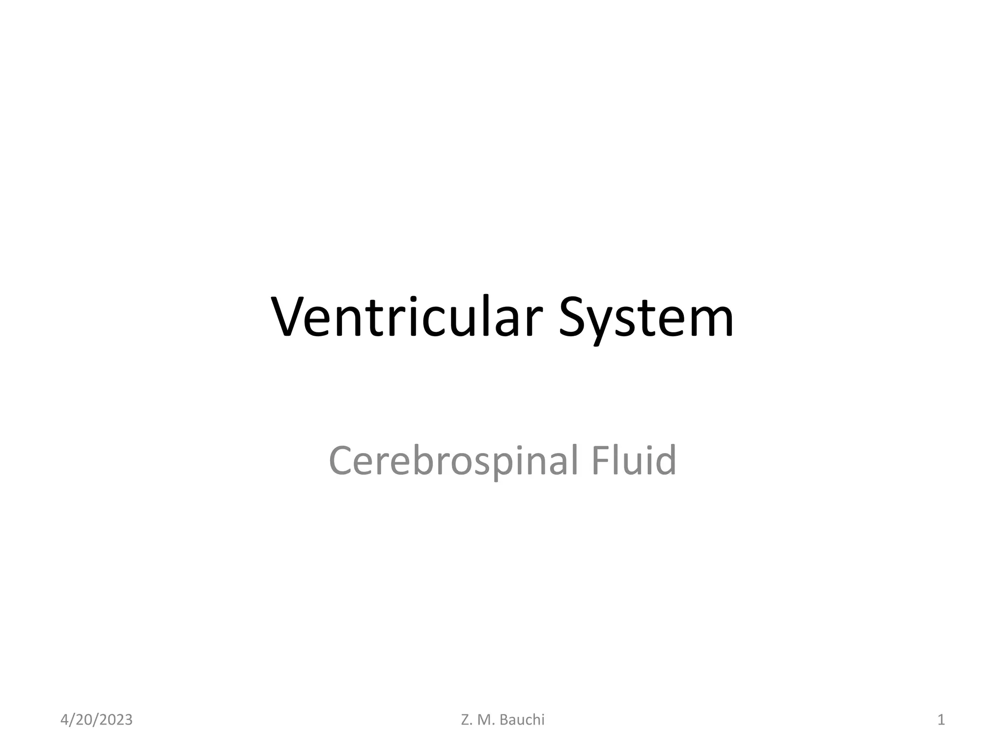
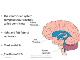
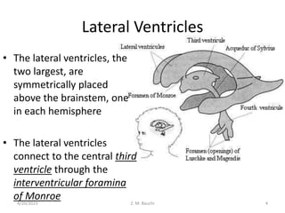
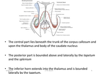
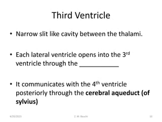
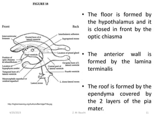
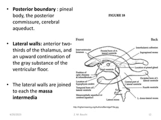
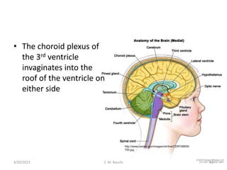
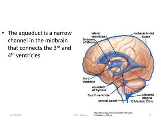
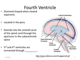
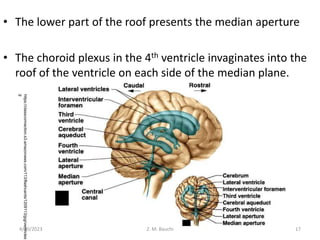
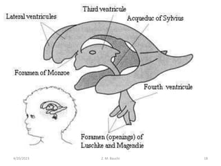
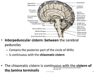
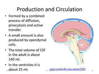
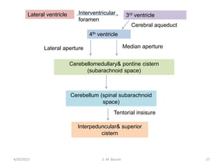
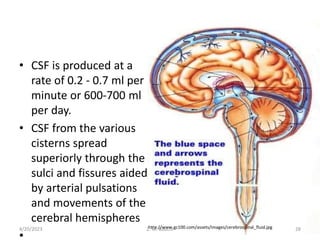
Ventricular System
Cerebrospinal Fluid
4/20/2023 1
Z. M. Bauchi
• The ventricular system
comprises four cavities
called ventricles:
– right and left lateral
ventricles
– third ventricle
– fourth ventricle
4/20/2023 2
http://www.chicagonow.com/nancys-
world/files/2012/12/brain-ventricles.gif
• Comparatively large
spaces filled with
cerebrospinal fluid.
• Of the four ventricles,
three are located in the
forebrain and one in the
brainstem.
4/20/2023 3
Z. M. Bauchi
http://www.buzzle.com/images/diagrams/labeled-brain-diagrams/brain-
ventricles.jpg
Lateral Ventricles
• The lateral ventricles, the
two largest, are
symmetrically placed
above the brainstem, one
in each hemisphere
• The lateral ventricles
connect to the central third
ventricle through the
interventricular foramina
of Monroe
4/20/2023 Z. M. Bauchi 4
• Each lateral ventricle has three horns:
– the anterior or frontal horn extends into the
frontal lobe
– the posterior or occipital horn into the occipital
lobe
– the inferior or temporal horn into the temporal
lobe
4/20/2023 5
Z. M. Bauchi
1. Anterior horn of lateral
ventricle
2. Body of lateral
ventricle
3. Posterior horn of
lateral ventricle
4. Inferior horn of lateral
ventricle
5. Third ventricle
6. Fourth ventricle
anatomy.med.umich.edu/atlas/n1a4p1.html
4/20/2023 6
Z. M. Bauchi
Boundries
• It is bounded below by the rostrum
• In front by the genu of corpus callosum
• Above by the corpus callosum.
• Laterally by the caudate nucleus
• Medially it is separated from the 3rd ventricle by the
septum pellucidum
4/20/2023 7
Z. M. Bauchi
• The central part lies beneath the trunk of the corpus callosum and
upon the thalamus and body of the caudate nucleus
• The posterior part is bounded above and laterally by the tapetum
and the splenium
• The inferior horn extends into the thalamus and is bounded
laterally by the tapetum.
4/20/2023 Z. M. Bauchi 8
• The choroid plexus of each lateral ventricle is
invaginated along a curved line called the
choroid fissure
4/20/2023 9
Z. M. Bauchi
Third Ventricle
• Narrow slit like cavity between the thalami.
• Each lateral ventricle opens into the 3rd
ventricle through the ___________
• It communicates with the 4th ventricle
posteriorly through the cerebral aqueduct (of
sylvius)
4/20/2023 10
Z. M. Bauchi
• The floor is formed by
the hypothalamus and it
is closed in front by the
optic chiasma
• The anterior wall is
formed by the lamina
terminalis
• The roof is formed by the
ependyma covered by
the 2 layers of the pia
mater.
http://highermeaning.org/Authors/Berridge/f18a.jpg
4/20/2023 11
Z. M. Bauchi
• Posterior boundary : pineal
body, the posterior
commissure, cerebral
aqueduct.
• Lateral walls: anterior two-
thirds of the thalamus, and
an upward continuation of
the gray substance of the
ventricular floor.
• The lateral walls are joined
to each the massa
intermedia
4/20/2023 Z. M. Bauchi 12
http://highermeaning.org/Authors/Berridge/f18a.jpg
• The choroid plexus of
the 3rd ventricle
invaginates into the
roof of the ventricle on
either side
4/20/2023 13
Z. M. Bauchi
http://www.cancer.gov/images/cdr/live/CDR748659-
750.jpg
• The aqueduct is a narrow
channel in the midbrain
that connects the 3rd and
4th ventricles.
4/20/2023 Z. M. Bauchi 14
http://25.media.tumblr.com/tumblr_l84oyjOh
5c1qb6etto1_250.jpg
Fourth Ventricle
• Diamond shaped when viewed
superiorly
• Located in the pons
• Extends into the centrall canal
of the spinal cord through the
apertures in the subarachnoid
space
• 3rd and 4th ventricles are
connected through _________.
http://www.obfocus.com/images/csf.gif
4/20/2023 15
Z. M. Bauchi
• Bound laterally by the superior and inferior
cerebellar peduncles
• Anterior boundary/floor (rhomboid fossa) is formed
by the pons above and the medulla below
• Posterior border/roof is thin and is covered by the
cerebellum.
• Superior and inferior medullary vela
4/20/2023 16
Z. M. Bauchi
• The lower part of the roof presents the median aperture
• The choroid plexus in the 4th ventricle invaginates into the
roof of the ventricle on each side of the median plane.
4/20/2023 Z. M. Bauchi 17
https://classconnection.s3.amazonaws.com/113/flashcards/1209113/jpg/ventricles1
g
4/20/2023 18
Z. M. Bauchi
• The vessels of the plexus are derived from the
cerebellar branches of the vertebral and
basilar arteries
4/20/2023 Z. M. Bauchi 19
Cerebrospinal Fluid
• Colourless, alkaline, extracellular fluid that
flows in and around the brain and spinal cord
• Absorbs shocks, maintains uniform pressure
and transports chemical substances within the
CNS
• Formed in the choroid plexus of the lateral,3rd
and 4th ventricles.
4/20/2023 20
Z. M. Bauchi
• Located in the roofs of the 3rd and 4th
ventricles and on the floors of the bodies and
inferior horns of the lateral ventricles
• Plexus in the lateral ventricles most important
4/20/2023 21
Z. M. Bauchi
Cisterns
• Subarachnoid cistern:
between the arachnoid
and pia. Is continuous with
the subarachnoid space of
the spinal cord
• Cerebellomedullary
cistern: (cisterna magna)
– Between inferior part of
medulla and cerebellum.
– Receives CSF from the
medial aperture of the
fourth ventricle
4/20/2023 Z. M. Bauchi 22
http://www.dartmouth.edu/~humananato
my/figures/chapter_43/43-
16_files/IMAGE001.JPG
• Pontine cistern: large
space along the ventral
and lateral surfaces of
the pons.
• Continuous with the
cerebellomedullary
cistern inferiorly.
• And with the
interpeduncular cistern
superiorly.
4/20/2023 Z. M. Bauchi 23
http://www.dartmouth.edu/~humananato
my/figures/chapter_43/43-
16_files/IMAGE001.JPG
• Interpeduncular cistern: between the cerebral
peduncles
– Contains the posterior part of the circle of Willis
– Is continuous with the chiasmatic cistern
• The chiasmatic cistern is continuous with the cistern of
the lamina terminalis
4/20/2023 Z. M. Bauchi 24
http://sci.rutgers.edu/dynarticles/scischemia/Sagit
tal_brain1.gif
• The cistern of the lamina terminalis
continuous as the shallow cistern of the
corpus callosum
• Cistern of the lateral sulcus: anterior to the
temporal lobe
– Contains the middle cerebellar artery.
• Superior cistern: between the splenium of the
corpus callosum and the superior surface of
the cerebrum
– Contains the great cerebral vein and pineal body
4/20/2023 Z. M. Bauchi 25
Production and Circulation
• Formed by a combined
process of diffusion,
pinocytosis and active
transfer.
• A small amount is also
produced by ependymal
cells.
• The total volume of CSF
in the adult is about
140 ml.
• In the ventricles it is
about 25 ml. www.control.tfe.umu.se/Ian/CSF/
4/20/2023 26
Z. M. Bauchi
Lateral ventricle 3rd ventricle
4th ventricle
Cerebellomedullary& pontine cistern
(subarachnoid space)
Cerebellum (spinal subarachnoid
space)
Interpeduncular& superior
cistern
Cerebral aqueduct
Median aperture
Lateral aperture
Tentorial insisure
Interventricular
foramen
4/20/2023 27
Z. M. Bauchi
• CSF is produced at a
rate of 0.2 - 0.7 ml per
minute or 600-700 ml
per day.
• CSF from the various
cisterns spread
superiorly through the
sulci and fissures aided
by arterial pulsations
and movements of the
cerebral hemispheres
•
http://www.gc100.com/assets/images/cerebrospinal_fluid.jpg
4/20/2023 28
Z. M. Bauchi
Composition
• CSF is composed of H2O, sodium chloride,
potassium, glucose and proteins
• The blood-CSF barrier allows selective passage
of substances from the blood to the CSF but
not in reverse direction
4/20/2023 29
Z. M. Bauchi
Absorption
• Main site of absorption of CSF is into the
venous system through the arachnoid villi.
• The villi act as one way valves.
• The rate of absorption correlates with the CSF
pressure.
4/20/2023 30
Z. M. Bauchi
• When erect the CSF is in the subarachnoid
cisterns and in the sulci on the superior and
lateral parts of the brain.
• Small, rapid recurring changes due to
heartbeat and slow recurring changes from
unknown causes
• Momentary large changes caused by coughing
or straining
4/20/2023 31
Z. M. Bauchi
• Monro-kellie doctrine: the cranial cavity is a
closed box, a change in the quantity of
intracranial blood can occur only through the
displacement or replacement of CSF.
4/20/2023 32
Z. M. Bauchi
Functions
• CSF acts as a cushion that protects the brain
from shocks and supports the venous sinuses.
• It also plays an important role in the
homeostasis and metabolism of the central
nervous system.
4/20/2023 33
Z. M. Bauchi
Clinical Anatomy
• Pineal body
• Cisternal puncture: CSF can be obtained from
the cerebellomedullary cistern.
• Hydrocephalus: An obstruction to the normal
flow the symptoms of which can include gait
imbalance, urinary dysfunction, and dementia.
4/20/2023 34
Z. M. Bauchi
• Contusions of the brain
• Fractures: floor of the middle cranial fossa
leads to leakage of CSF from the ear
• Fractures: floor of the anterior cranial fossa
may involve the cribiform plate of the ethmoid
bone leading to leakage of CSF through the
nose.
4/20/2023 35
Z. M. Bauchi
• Blood: Blood may be spilled into the CSF by
accidental puncture of a leptomeningeal vein
during entry of the LP needle.
• The subarachnoid space may also be
punctured for measuring/maintaining CSF
pressure.
• Antibiotics or contrast media can also be
injected.
4/20/2023 36
Z. M. Bauchi

Ventricular System.ppt

  • 1.
  • 2.
    • The ventricularsystem comprises four cavities called ventricles: – right and left lateral ventricles – third ventricle – fourth ventricle 4/20/2023 2 http://www.chicagonow.com/nancys- world/files/2012/12/brain-ventricles.gif
  • 3.
    • Comparatively large spacesfilled with cerebrospinal fluid. • Of the four ventricles, three are located in the forebrain and one in the brainstem. 4/20/2023 3 Z. M. Bauchi http://www.buzzle.com/images/diagrams/labeled-brain-diagrams/brain- ventricles.jpg
  • 4.
    Lateral Ventricles • Thelateral ventricles, the two largest, are symmetrically placed above the brainstem, one in each hemisphere • The lateral ventricles connect to the central third ventricle through the interventricular foramina of Monroe 4/20/2023 Z. M. Bauchi 4
  • 5.
    • Each lateralventricle has three horns: – the anterior or frontal horn extends into the frontal lobe – the posterior or occipital horn into the occipital lobe – the inferior or temporal horn into the temporal lobe 4/20/2023 5 Z. M. Bauchi
  • 6.
    1. Anterior hornof lateral ventricle 2. Body of lateral ventricle 3. Posterior horn of lateral ventricle 4. Inferior horn of lateral ventricle 5. Third ventricle 6. Fourth ventricle anatomy.med.umich.edu/atlas/n1a4p1.html 4/20/2023 6 Z. M. Bauchi
  • 7.
    Boundries • It isbounded below by the rostrum • In front by the genu of corpus callosum • Above by the corpus callosum. • Laterally by the caudate nucleus • Medially it is separated from the 3rd ventricle by the septum pellucidum 4/20/2023 7 Z. M. Bauchi
  • 8.
    • The centralpart lies beneath the trunk of the corpus callosum and upon the thalamus and body of the caudate nucleus • The posterior part is bounded above and laterally by the tapetum and the splenium • The inferior horn extends into the thalamus and is bounded laterally by the tapetum. 4/20/2023 Z. M. Bauchi 8
  • 9.
    • The choroidplexus of each lateral ventricle is invaginated along a curved line called the choroid fissure 4/20/2023 9 Z. M. Bauchi
  • 10.
    Third Ventricle • Narrowslit like cavity between the thalami. • Each lateral ventricle opens into the 3rd ventricle through the ___________ • It communicates with the 4th ventricle posteriorly through the cerebral aqueduct (of sylvius) 4/20/2023 10 Z. M. Bauchi
  • 11.
    • The flooris formed by the hypothalamus and it is closed in front by the optic chiasma • The anterior wall is formed by the lamina terminalis • The roof is formed by the ependyma covered by the 2 layers of the pia mater. http://highermeaning.org/Authors/Berridge/f18a.jpg 4/20/2023 11 Z. M. Bauchi
  • 12.
    • Posterior boundary: pineal body, the posterior commissure, cerebral aqueduct. • Lateral walls: anterior two- thirds of the thalamus, and an upward continuation of the gray substance of the ventricular floor. • The lateral walls are joined to each the massa intermedia 4/20/2023 Z. M. Bauchi 12 http://highermeaning.org/Authors/Berridge/f18a.jpg
  • 13.
    • The choroidplexus of the 3rd ventricle invaginates into the roof of the ventricle on either side 4/20/2023 13 Z. M. Bauchi http://www.cancer.gov/images/cdr/live/CDR748659- 750.jpg
  • 14.
    • The aqueductis a narrow channel in the midbrain that connects the 3rd and 4th ventricles. 4/20/2023 Z. M. Bauchi 14 http://25.media.tumblr.com/tumblr_l84oyjOh 5c1qb6etto1_250.jpg
  • 15.
    Fourth Ventricle • Diamondshaped when viewed superiorly • Located in the pons • Extends into the centrall canal of the spinal cord through the apertures in the subarachnoid space • 3rd and 4th ventricles are connected through _________. http://www.obfocus.com/images/csf.gif 4/20/2023 15 Z. M. Bauchi
  • 16.
    • Bound laterallyby the superior and inferior cerebellar peduncles • Anterior boundary/floor (rhomboid fossa) is formed by the pons above and the medulla below • Posterior border/roof is thin and is covered by the cerebellum. • Superior and inferior medullary vela 4/20/2023 16 Z. M. Bauchi
  • 17.
    • The lowerpart of the roof presents the median aperture • The choroid plexus in the 4th ventricle invaginates into the roof of the ventricle on each side of the median plane. 4/20/2023 Z. M. Bauchi 17 https://classconnection.s3.amazonaws.com/113/flashcards/1209113/jpg/ventricles1 g
  • 18.
  • 19.
    • The vesselsof the plexus are derived from the cerebellar branches of the vertebral and basilar arteries 4/20/2023 Z. M. Bauchi 19
  • 20.
    Cerebrospinal Fluid • Colourless,alkaline, extracellular fluid that flows in and around the brain and spinal cord • Absorbs shocks, maintains uniform pressure and transports chemical substances within the CNS • Formed in the choroid plexus of the lateral,3rd and 4th ventricles. 4/20/2023 20 Z. M. Bauchi
  • 21.
    • Located inthe roofs of the 3rd and 4th ventricles and on the floors of the bodies and inferior horns of the lateral ventricles • Plexus in the lateral ventricles most important 4/20/2023 21 Z. M. Bauchi
  • 22.
    Cisterns • Subarachnoid cistern: betweenthe arachnoid and pia. Is continuous with the subarachnoid space of the spinal cord • Cerebellomedullary cistern: (cisterna magna) – Between inferior part of medulla and cerebellum. – Receives CSF from the medial aperture of the fourth ventricle 4/20/2023 Z. M. Bauchi 22 http://www.dartmouth.edu/~humananato my/figures/chapter_43/43- 16_files/IMAGE001.JPG
  • 23.
    • Pontine cistern:large space along the ventral and lateral surfaces of the pons. • Continuous with the cerebellomedullary cistern inferiorly. • And with the interpeduncular cistern superiorly. 4/20/2023 Z. M. Bauchi 23 http://www.dartmouth.edu/~humananato my/figures/chapter_43/43- 16_files/IMAGE001.JPG
  • 24.
    • Interpeduncular cistern:between the cerebral peduncles – Contains the posterior part of the circle of Willis – Is continuous with the chiasmatic cistern • The chiasmatic cistern is continuous with the cistern of the lamina terminalis 4/20/2023 Z. M. Bauchi 24 http://sci.rutgers.edu/dynarticles/scischemia/Sagit tal_brain1.gif
  • 25.
    • The cisternof the lamina terminalis continuous as the shallow cistern of the corpus callosum • Cistern of the lateral sulcus: anterior to the temporal lobe – Contains the middle cerebellar artery. • Superior cistern: between the splenium of the corpus callosum and the superior surface of the cerebrum – Contains the great cerebral vein and pineal body 4/20/2023 Z. M. Bauchi 25
  • 26.
    Production and Circulation •Formed by a combined process of diffusion, pinocytosis and active transfer. • A small amount is also produced by ependymal cells. • The total volume of CSF in the adult is about 140 ml. • In the ventricles it is about 25 ml. www.control.tfe.umu.se/Ian/CSF/ 4/20/2023 26 Z. M. Bauchi
  • 27.
    Lateral ventricle 3rdventricle 4th ventricle Cerebellomedullary& pontine cistern (subarachnoid space) Cerebellum (spinal subarachnoid space) Interpeduncular& superior cistern Cerebral aqueduct Median aperture Lateral aperture Tentorial insisure Interventricular foramen 4/20/2023 27 Z. M. Bauchi
  • 28.
    • CSF isproduced at a rate of 0.2 - 0.7 ml per minute or 600-700 ml per day. • CSF from the various cisterns spread superiorly through the sulci and fissures aided by arterial pulsations and movements of the cerebral hemispheres • http://www.gc100.com/assets/images/cerebrospinal_fluid.jpg 4/20/2023 28 Z. M. Bauchi
  • 29.
    Composition • CSF iscomposed of H2O, sodium chloride, potassium, glucose and proteins • The blood-CSF barrier allows selective passage of substances from the blood to the CSF but not in reverse direction 4/20/2023 29 Z. M. Bauchi
  • 30.
    Absorption • Main siteof absorption of CSF is into the venous system through the arachnoid villi. • The villi act as one way valves. • The rate of absorption correlates with the CSF pressure. 4/20/2023 30 Z. M. Bauchi
  • 31.
    • When erectthe CSF is in the subarachnoid cisterns and in the sulci on the superior and lateral parts of the brain. • Small, rapid recurring changes due to heartbeat and slow recurring changes from unknown causes • Momentary large changes caused by coughing or straining 4/20/2023 31 Z. M. Bauchi
  • 32.
    • Monro-kellie doctrine:the cranial cavity is a closed box, a change in the quantity of intracranial blood can occur only through the displacement or replacement of CSF. 4/20/2023 32 Z. M. Bauchi
  • 33.
    Functions • CSF actsas a cushion that protects the brain from shocks and supports the venous sinuses. • It also plays an important role in the homeostasis and metabolism of the central nervous system. 4/20/2023 33 Z. M. Bauchi
  • 34.
    Clinical Anatomy • Pinealbody • Cisternal puncture: CSF can be obtained from the cerebellomedullary cistern. • Hydrocephalus: An obstruction to the normal flow the symptoms of which can include gait imbalance, urinary dysfunction, and dementia. 4/20/2023 34 Z. M. Bauchi
  • 35.
    • Contusions ofthe brain • Fractures: floor of the middle cranial fossa leads to leakage of CSF from the ear • Fractures: floor of the anterior cranial fossa may involve the cribiform plate of the ethmoid bone leading to leakage of CSF through the nose. 4/20/2023 35 Z. M. Bauchi
  • 36.
    • Blood: Bloodmay be spilled into the CSF by accidental puncture of a leptomeningeal vein during entry of the LP needle. • The subarachnoid space may also be punctured for measuring/maintaining CSF pressure. • Antibiotics or contrast media can also be injected. 4/20/2023 36 Z. M. Bauchi

Editor's Notes

  • #3 A ventricle is a small cavity or chamber in the body. In the brain it can be defined as an enlargement of the central canal that contains CSF
  • #5 They occupy a large part of the cerebral hemispheres
  • #7 The part in front of the foramen is the anterior horn/1st part. Behind the anterior horn is the central part. The front middle and back portions of the central part are called the 2nd,3rd and 4th. The 4th divides into the 5th part/posterior horn and the 6th part/inferior horn..
  • #9 Tapetum= sheet of fibres
  • #11 Behind, it communicates with the fourth ventricle through the cerebral aqueduct, and in front with the lateral ventricles through the interventricular foramen. Somewhat triangular in shape, with the apex directed backward, it has a roof, a floor, an anterior and a posterior boundary and a pair of lateral walls. Interventricular foramen
  • #12 The floor slopes downward and forward and is formed mainly by the structures which constitute the hypothalamus: from before backward these are: the optic chiasma, the tuber cinereum and infundibulum, and the corpora mammillaria. Behind the last, the floor is formed by the interpeduncular fossa and the tegmenta of the cerebral peduncles. The ventricle is prolonged downward as a funnel-shaped recess, the recessus infundibuli, into the infundibulum, and to the apex of the latter the hypophysis is attached. The anterior boundary is constituted below by the lamina terminalis, a thin layer of gray substance stretching from the upper surface of the optic chiasma to the rostrum of the corpus callosum; above by the columns of the fornix and the anterior commissure. At the junction of the floor and anterior wall, immediately above the optic chiasma, the ventricle presents a small angular recess or diverticulum, the optic recess. Between the columns of the fornix, and above the anterior commissure, is a second recess termed the vulva. At the junction of the roof and anterior wall of the ventricle, and situated between the thalami behind and the columns of the fornix in front, is the interventricular foramen (foramen of Monro) through which the third communicates with the lateral ventricles. The roof (Fig. 723) is formed by a layer of epithelium, which stretches between the upper edges of the lateral walls of the cavity and is continuous with the epithelial lining of the ventricle. It is covered by and adherent to a fold of pia mater, named the tela chorioidea of the third ventricle, from the under surface of which a pair of vascular fringed processes, the choroid plexuses of the third ventricle, project downward, one on either side of the middle line, and invaginate the epithelial roof into the ventricular cavity.
  • #13 The posterior boundary is constituted by the pineal body, the posterior commissure and the cerebral aqueduct. Each lateral wall consists of an upper portion formed by the medial surface of the anterior two-thirds of the thalamus, and a lower consisting of an upward continuation of the gray substance of the ventricular floor.
  • #16 Thus in the process of development the wall of the hind-brain undergoes modification to form the medulla oblongata, the pons, and cerebellum, while its cavity is expanded to form the fourth ventricle. The mid-brain forms only a small part of the adult brain; its cavity becomes the cerebral aqueduct (aqueduct of Sylvius), which serves as a tubular communication between the third and fourth ventricles
  • #17 4) Sheets of white matter. This stretches between the 2 superior and 2 inferior cerebellar peduncles.
  • #18 CSF moves from the 4th ventricle to the subarachnoid space through the lateral and median apertures
  • #19 The lower part of the roof presents the median aperture The choroid plexus in the 4th ventricle invaginates into the roof of the ventricle on each side of the median plane. The vessels of the plexus are derived from the cerebellar branches of the vertebral and basilar arteries
  • #22 The choroid plexus of the lateral ventricles are the largest and produce the most CSF
  • #23 A cistern is a space that forms a reservoir for CSF .It is an enlargement of the subarachnoid space 3) and is continuous with the subarachnoid space around the brain and spinal cord
  • #24 Contains the basilar artery and cranial nerves
  • #25 A vascular circle formed by the major arteries that supply the cerebrum
  • #26 It is sometimes referred to as the quadrangular cistern
  • #27 The cerebrospinal fluid (CSF) is produced from arterial blood by the choroid plexuses of the lateral and fourth ventricles
  • #28 CSF produced in the lateral and 3rd ventricles moves to the 4th ventricle through the cerebral aqueduct. More CSF is produced there and it leaves the ventricle through the median apertures and passes into the subarachnoid space. Here it collects in the cerebellomedullary and pontine cisterns. From here, some CSF passes inferiorly into the spinal subarachnoid spaces and the postero superiorly over the cerebellum
  • #31 These are protrusions of the arachnoid into the dural sinuses. The rate of absorption depends on the CSF pressure. When CSF pressure is greater than the venous pressure the valves open and CSF passes into the venous sinuses of the dura. When the venous pressure is higher than the CSF pressure the valves close. Some CSF appears to be absorbed by the ependymal lining of the ventricles in the spinal subarachnoid space and through the walls of the capillaries in the pia.
  • #32 The brain is slightly heavier than the CSF. When a person is erect the basal surface of the brain is in contact with the cranial fossa in the floor of the cavity. CSF normally separates the superior part of the brain from the calvaria There are several situations that can lead to a changes in intra cranial pressure
  • #33 Any change in the volume of the intracranial contents (tumour, ventricular fluid from blockage of c aqueduct, haemorrhage) will lead to change in intracranial pressure
  • #34 CSF along with the meninges and calvaria provide protection to the brain by providing a cushion against shocks and blows to the head.
  • #35 Useful neurosurgical and radiological landmark. It becomes calcified in adolescence. Pineal body: normal position of the gland is used to diagnose its displacement in patients with suspected expanding intracranial lesions Cisternal puncture: CSF can be obtained from the cerebellomedullary cistern. In infants the anterior fontanel is used. Lumbar puncture used to obtain samples of CSF L3 L4 3) Can be caused by overproduction of CSF, obstruction, inflow interference with absorption=dilation of ventricles= accumulation of CSF in the ventricular subarachnoid space. These symptoms can be reduced or eliminated by implanting a shunt, a small valve with an attached drainage tube, into the patient’s skull, thereby altering the system and increasing the CSF flow.
  • #36 2) Sudden acceleration and deceleration of the head. Base, frontal temporal and occipital due to paucity of CSF
  • #37 2. This is for radiography (ventriculography). This technique has been replaced by CT scans and MRI