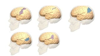
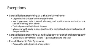
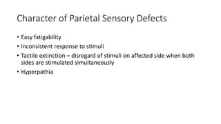
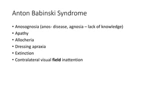
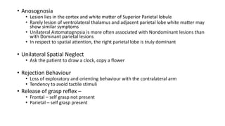
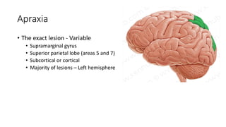
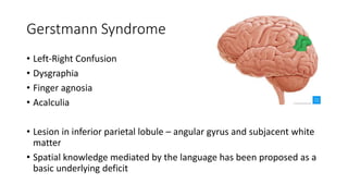
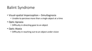
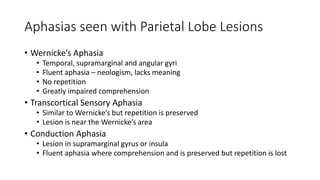
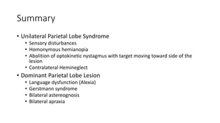
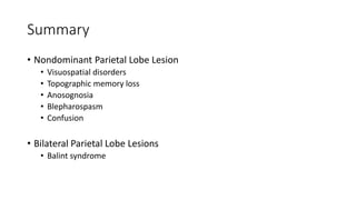
Parietal Lobe Signs
Dr. Mandeep Singh Kataria
Anatomy
• Limits
• Anteriorly – Central Sulcus
• Posteriorly
• Parieto-occipital sulcus superiorly
• Preoccipital notch inferiorly
• Inferiorly – Sylvian fissure
• Parts
• Two sulci
• Post-central sulcus
• Interparietal sulcus
• Inferior Parietal Lobule
• Supramarginal Gyrus (Area 40)
• Angular Gyrus (Area 39)
• Superior Parietal Lobule
• Bound by the post central gyrus,
interparietal gyrus and extends
onto the medial surface (areas 5
and 7)
• Post central gyrus
• Brodmans areas 1, 2 and 3
Brodman Areas In the Parietal Lobe
• Area 1, 2 and 3 – Postcentral Gyrus
• Primary sensory cortex
• Area 5 – Somatosensory association
cortex
• Area 7 – Somatosensory association
cortex
• Together form the superior parietal
lobule
• Area 39 – Wernicke’s area – Angular
Gyrus
• Area 40 – Supramarginal Gyrus
Unilateral Parietal Lobe Disease
• On either side
• Cortical Sensory syndrome
• Sensory extinction
• Homonymous hemianopia with visual inattention
• Contralateral neglect
• Abolition of optokinetic nystagmus to one side
Dominant lobe lesion
• Aphasias
• Broca’s
• Wernicke’s
• Global
• Conduction
• Alexia without agraphia (pure word blindness)
• Bilateral astereognosis
• Gerstmann’s syndrome
• Agraphia without alexia
• Right-left confusion
• Acalculia
• Finger agnosia
• Bilateral Ideomotor apraxia
Cortical Sensory Syndrome
• Loss of position sense and passive movement sense
• Inability to localize touch, temperature or pain
• Astereognosis
• Agraphesthesia
• Loss of two-point discrimination
• Easy fatigability of sensory perceptions
• Difficulty in distinguishing simultaneous stimulations
• Hyperpathia
• Touch hallucinations
• Preservation sensations – Pain, Pressure, Touch, Temperature, Vibration
(when the postcentral gyrus is not involved in the lesions)
Exceptions
• Cortical lesion presenting as a thalamic syndrome
• Dejerine and Mouzon’s sensory syndrome
• touch, pressure, pain, thermal, vibratory, and position sense are lost on one
side of the body or in a limb
• Typically seen with thalamic lesions
• May occur with acute lesions involving the central and subcortical region of
the parietal lobe
• Cortical lesion presenting as radiculopathy or peripheral neuropathy
• May be cause by smaller lesions – glancing blows to the skull
• Pseudothalamic Pain Syndrome
• Pain on the side deprived of sensations
Character of Parietal Sensory Defects
• Easy fatigability
• Inconsistent response to stimuli
• Tactile extinction – disregard of stimuli on affected side when both
sides are stimulated simultaneously
• Hyperpathia
Anton Babinski Syndrome
• Anosognosia (anos- disease, agnosia – lack of knowledge)
• Apathy
• Allocheria
• Dressing apraxia
• Extinction
• Contralateral visual field inattention
• Anosognosia
• Lesion lies in the cortex and white matter of Superior Parietal lobule
• Rarely lesion of ventrolateral thalamus and adjacent parietal lobe white matter may
show similar symptoms
• Unilateral Astomatognosia is more often associated with Nondominant lesions than
with Dominant parietal lesions
• In respect to spatial attention, the right parietal lobe is truly dominant
• Unilateral Spatial Neglect
• Ask the patient to draw a clock, copy a flower
• Rejection Behaviour
• Loss of exploratory and orienting behaviour with the contralateral arm
• Tendency to avoid tactile stimuli
• Release of grasp reflex –
• Frontal – self grasp not present
• Parietal – self grasp present
Motor disturbances with parietal lobe lesions
• Mild hemiparesis – significant contribution to the corticospinal tract
• May simulate a hemiplegia because of inability to move the limb
• Reduced movements of opposite side – Parietal lesions have been
reported to cause contralateral muscular atrophy and trophic skin
changes
• Optic ataxia – inability to grasp an object under visual guidance
• Pseudocerebellar syndrome – intention tremor and incoordination
• Pseudoathetosis – random finger movements in an outstretched
hand
• Apraxias
Apraxia
• Loss of the ability to execute previously learned activities in the
absence of weakness, ataxia, sensory loss, or extrapyramidal
derangement that would be adequate to explain the deficit.
• Planned or commanded action is normally first conceptualized in the
parietal lobe of the language-dominant hemisphere, where visual,
auditory, and somesthetic information is integrated – The space-time
plan
Apraxia
• Ideational apraxia – What to do?
• The failure to conceive or formulate an action to command
• Areas 5 and 7 in dominant parietal lobe + supplementary and premotor cortex of bilateral
frontal lobe
• Ideomotor apraxia – How to do?
• Impaired performance of skilled motor acts despite intact sensory, motor, and language
function.
• Patient may know how to do a task but is unable to execute it.
• Oral-buccal-lingual apraxia
• Inability to carry out facial movements on command
• May be able to imitate them
• Left supramarginal gyrus or the left motor association cortex
• Limb-kinetic apraxia
• Clumsiness due to inability to fluidly connect the various movements of the hand or limbs.
Apraxia
• The exact lesion - Variable
• Supramarginal gyrus
• Superior parietal lobe (areas 5 and 7)
• Subcortical or cortical
• Majority of lesions – Left hemisphere
Testing for Apraxia
1. Observe the actions of a patient while he engages in simulated
tasks
2. Ask he patient to carry out symbolic acts
• Wave goodbye
• Salute
• Blow a kiss
3. Ask the patient to imitate such acts if he cannot perform them on
command
4. Ask him to do more complex tasks – simulate taking out comb from
pocket and comb hair, brush teeth, hammer a nail…
Gerstmann Syndrome
• Left-Right Confusion
• Dysgraphia
• Finger agnosia
• Acalculia
• Lesion in inferior parietal lobule – angular gyrus and subjacent white
matter
• Spatial knowledge mediated by the language has been proposed as a
basic underlying deficit
• The right angular gyrus clearly participates in visuospatial processing,
and damage to it results in severe hemi-spatial neglect.
• The left angular gyrus participate in calculation abilities, reading/
writing, naming, and some type of body-knowledge (somatognosis).
Visual Disorders in Parietal Lobe Lesion
• Visual Field Defects
• Homonymous hemianopia
• Homonymous inferior quadrantanopia
• Visual neglect
• Posterior parietal lesions
• More prominent on the right side
• Loss of optokinetic nystagmus
• Abolished when following objects towards the lesion side
• Posterior Parietal Lesions
• Deficits in localization of visual stimuli
• Inability to compare the sizes of objects
• Failure to avoid objects when walking
• Inability to count objects
• Disturbances in smooth-pursuit eye movements
• Loss of stereoscopic vision
• Blepharospasm
• Disinclination to open eyelids when addressed
• Intense blepharospasm precluding ocular examination
• Topographagnosia
• Patients are unable to orient themselves to abstract space
• Loss of topographical memory
Auditory Neglect
• Less apparent than is visual neglect
• Displacement of the direction of the perceived origin of sounds
toward the opposite side
• Lesion in right superior lobule.
Balint Syndrome
• Visual-spatial Imperception – Simultagnosia
• Unable to percieve more than a single object at a time
• Optic Apraxia
• Difficulty in directing gaze to an object
• Optic Ataxia
• Difficulty in reaching out to an object under vision
Aphasias seen with Parietal Lobe Lesions
• Wernicke’s Aphasia
• Temporal, supramarginal and angular gyri
• Fluent aphasia – neologism, lacks meaning
• No repetition
• Greatly impaired comprehension
• Transcortical Sensory Aphasia
• Similar to Wernicke’s but repetition is preserved
• Lesion is near the Wernicke’s area
• Conduction Aphasia
• Lesion in supramarginal gyrus or insula
• Fluent aphasia where comprehension and is preserved but repetition is lost
Summary
• Unilateral Parietal Lobe Syndrome
• Sensory disturbances
• Homonymous hemianopia
• Abolition of optokinetic nystagmus with target moving toward side of the
lesion
• Contralateral Hemineglect
• Dominant Parietal Lobe Lesion
• Language dysfunction (Alexia)
• Gerstmann syndrome
• Bilateral astereognosis
• Bilateral apraxia
Summary
• Nondominant Parietal Lobe Lesion
• Visuospatial disorders
• Topographic memory loss
• Anosognosia
• Blepharospasm
• Confusion
• Bilateral Parietal Lobe Lesions
• Balint syndrome

Parietal Lobe SIgns 24_7_18

  • 1.
    Parietal Lobe Signs Dr.Mandeep Singh Kataria
  • 2.
    Anatomy • Limits • Anteriorly– Central Sulcus • Posteriorly • Parieto-occipital sulcus superiorly • Preoccipital notch inferiorly • Inferiorly – Sylvian fissure • Parts • Two sulci • Post-central sulcus • Interparietal sulcus • Inferior Parietal Lobule • Supramarginal Gyrus (Area 40) • Angular Gyrus (Area 39) • Superior Parietal Lobule • Bound by the post central gyrus, interparietal gyrus and extends onto the medial surface (areas 5 and 7) • Post central gyrus • Brodmans areas 1, 2 and 3
  • 4.
    Brodman Areas Inthe Parietal Lobe • Area 1, 2 and 3 – Postcentral Gyrus • Primary sensory cortex • Area 5 – Somatosensory association cortex • Area 7 – Somatosensory association cortex • Together form the superior parietal lobule • Area 39 – Wernicke’s area – Angular Gyrus • Area 40 – Supramarginal Gyrus
  • 6.
    Unilateral Parietal LobeDisease • On either side • Cortical Sensory syndrome • Sensory extinction • Homonymous hemianopia with visual inattention • Contralateral neglect • Abolition of optokinetic nystagmus to one side
  • 7.
    Dominant lobe lesion •Aphasias • Broca’s • Wernicke’s • Global • Conduction • Alexia without agraphia (pure word blindness) • Bilateral astereognosis • Gerstmann’s syndrome • Agraphia without alexia • Right-left confusion • Acalculia • Finger agnosia • Bilateral Ideomotor apraxia
  • 8.
    Cortical Sensory Syndrome •Loss of position sense and passive movement sense • Inability to localize touch, temperature or pain • Astereognosis • Agraphesthesia • Loss of two-point discrimination • Easy fatigability of sensory perceptions • Difficulty in distinguishing simultaneous stimulations • Hyperpathia • Touch hallucinations • Preservation sensations – Pain, Pressure, Touch, Temperature, Vibration (when the postcentral gyrus is not involved in the lesions)
  • 9.
    Exceptions • Cortical lesionpresenting as a thalamic syndrome • Dejerine and Mouzon’s sensory syndrome • touch, pressure, pain, thermal, vibratory, and position sense are lost on one side of the body or in a limb • Typically seen with thalamic lesions • May occur with acute lesions involving the central and subcortical region of the parietal lobe • Cortical lesion presenting as radiculopathy or peripheral neuropathy • May be cause by smaller lesions – glancing blows to the skull • Pseudothalamic Pain Syndrome • Pain on the side deprived of sensations
  • 10.
    Character of ParietalSensory Defects • Easy fatigability • Inconsistent response to stimuli • Tactile extinction – disregard of stimuli on affected side when both sides are stimulated simultaneously • Hyperpathia
  • 11.
    Anton Babinski Syndrome •Anosognosia (anos- disease, agnosia – lack of knowledge) • Apathy • Allocheria • Dressing apraxia • Extinction • Contralateral visual field inattention
  • 12.
    • Anosognosia • Lesionlies in the cortex and white matter of Superior Parietal lobule • Rarely lesion of ventrolateral thalamus and adjacent parietal lobe white matter may show similar symptoms • Unilateral Astomatognosia is more often associated with Nondominant lesions than with Dominant parietal lesions • In respect to spatial attention, the right parietal lobe is truly dominant • Unilateral Spatial Neglect • Ask the patient to draw a clock, copy a flower • Rejection Behaviour • Loss of exploratory and orienting behaviour with the contralateral arm • Tendency to avoid tactile stimuli • Release of grasp reflex – • Frontal – self grasp not present • Parietal – self grasp present
  • 14.
    Motor disturbances withparietal lobe lesions • Mild hemiparesis – significant contribution to the corticospinal tract • May simulate a hemiplegia because of inability to move the limb • Reduced movements of opposite side – Parietal lesions have been reported to cause contralateral muscular atrophy and trophic skin changes • Optic ataxia – inability to grasp an object under visual guidance • Pseudocerebellar syndrome – intention tremor and incoordination • Pseudoathetosis – random finger movements in an outstretched hand • Apraxias
  • 15.
    Apraxia • Loss ofthe ability to execute previously learned activities in the absence of weakness, ataxia, sensory loss, or extrapyramidal derangement that would be adequate to explain the deficit. • Planned or commanded action is normally first conceptualized in the parietal lobe of the language-dominant hemisphere, where visual, auditory, and somesthetic information is integrated – The space-time plan
  • 16.
    Apraxia • Ideational apraxia– What to do? • The failure to conceive or formulate an action to command • Areas 5 and 7 in dominant parietal lobe + supplementary and premotor cortex of bilateral frontal lobe • Ideomotor apraxia – How to do? • Impaired performance of skilled motor acts despite intact sensory, motor, and language function. • Patient may know how to do a task but is unable to execute it. • Oral-buccal-lingual apraxia • Inability to carry out facial movements on command • May be able to imitate them • Left supramarginal gyrus or the left motor association cortex • Limb-kinetic apraxia • Clumsiness due to inability to fluidly connect the various movements of the hand or limbs.
  • 17.
    Apraxia • The exactlesion - Variable • Supramarginal gyrus • Superior parietal lobe (areas 5 and 7) • Subcortical or cortical • Majority of lesions – Left hemisphere
  • 18.
    Testing for Apraxia 1.Observe the actions of a patient while he engages in simulated tasks 2. Ask he patient to carry out symbolic acts • Wave goodbye • Salute • Blow a kiss 3. Ask the patient to imitate such acts if he cannot perform them on command 4. Ask him to do more complex tasks – simulate taking out comb from pocket and comb hair, brush teeth, hammer a nail…
  • 19.
    Gerstmann Syndrome • Left-RightConfusion • Dysgraphia • Finger agnosia • Acalculia • Lesion in inferior parietal lobule – angular gyrus and subjacent white matter • Spatial knowledge mediated by the language has been proposed as a basic underlying deficit
  • 20.
    • The rightangular gyrus clearly participates in visuospatial processing, and damage to it results in severe hemi-spatial neglect. • The left angular gyrus participate in calculation abilities, reading/ writing, naming, and some type of body-knowledge (somatognosis).
  • 21.
    Visual Disorders inParietal Lobe Lesion • Visual Field Defects • Homonymous hemianopia • Homonymous inferior quadrantanopia • Visual neglect • Posterior parietal lesions • More prominent on the right side • Loss of optokinetic nystagmus • Abolished when following objects towards the lesion side
  • 22.
    • Posterior ParietalLesions • Deficits in localization of visual stimuli • Inability to compare the sizes of objects • Failure to avoid objects when walking • Inability to count objects • Disturbances in smooth-pursuit eye movements • Loss of stereoscopic vision • Blepharospasm • Disinclination to open eyelids when addressed • Intense blepharospasm precluding ocular examination • Topographagnosia • Patients are unable to orient themselves to abstract space • Loss of topographical memory
  • 23.
    Auditory Neglect • Lessapparent than is visual neglect • Displacement of the direction of the perceived origin of sounds toward the opposite side • Lesion in right superior lobule.
  • 24.
    Balint Syndrome • Visual-spatialImperception – Simultagnosia • Unable to percieve more than a single object at a time • Optic Apraxia • Difficulty in directing gaze to an object • Optic Ataxia • Difficulty in reaching out to an object under vision
  • 25.
    Aphasias seen withParietal Lobe Lesions • Wernicke’s Aphasia • Temporal, supramarginal and angular gyri • Fluent aphasia – neologism, lacks meaning • No repetition • Greatly impaired comprehension • Transcortical Sensory Aphasia • Similar to Wernicke’s but repetition is preserved • Lesion is near the Wernicke’s area • Conduction Aphasia • Lesion in supramarginal gyrus or insula • Fluent aphasia where comprehension and is preserved but repetition is lost
  • 26.
    Summary • Unilateral ParietalLobe Syndrome • Sensory disturbances • Homonymous hemianopia • Abolition of optokinetic nystagmus with target moving toward side of the lesion • Contralateral Hemineglect • Dominant Parietal Lobe Lesion • Language dysfunction (Alexia) • Gerstmann syndrome • Bilateral astereognosis • Bilateral apraxia
  • 27.
    Summary • Nondominant ParietalLobe Lesion • Visuospatial disorders • Topographic memory loss • Anosognosia • Blepharospasm • Confusion • Bilateral Parietal Lobe Lesions • Balint syndrome