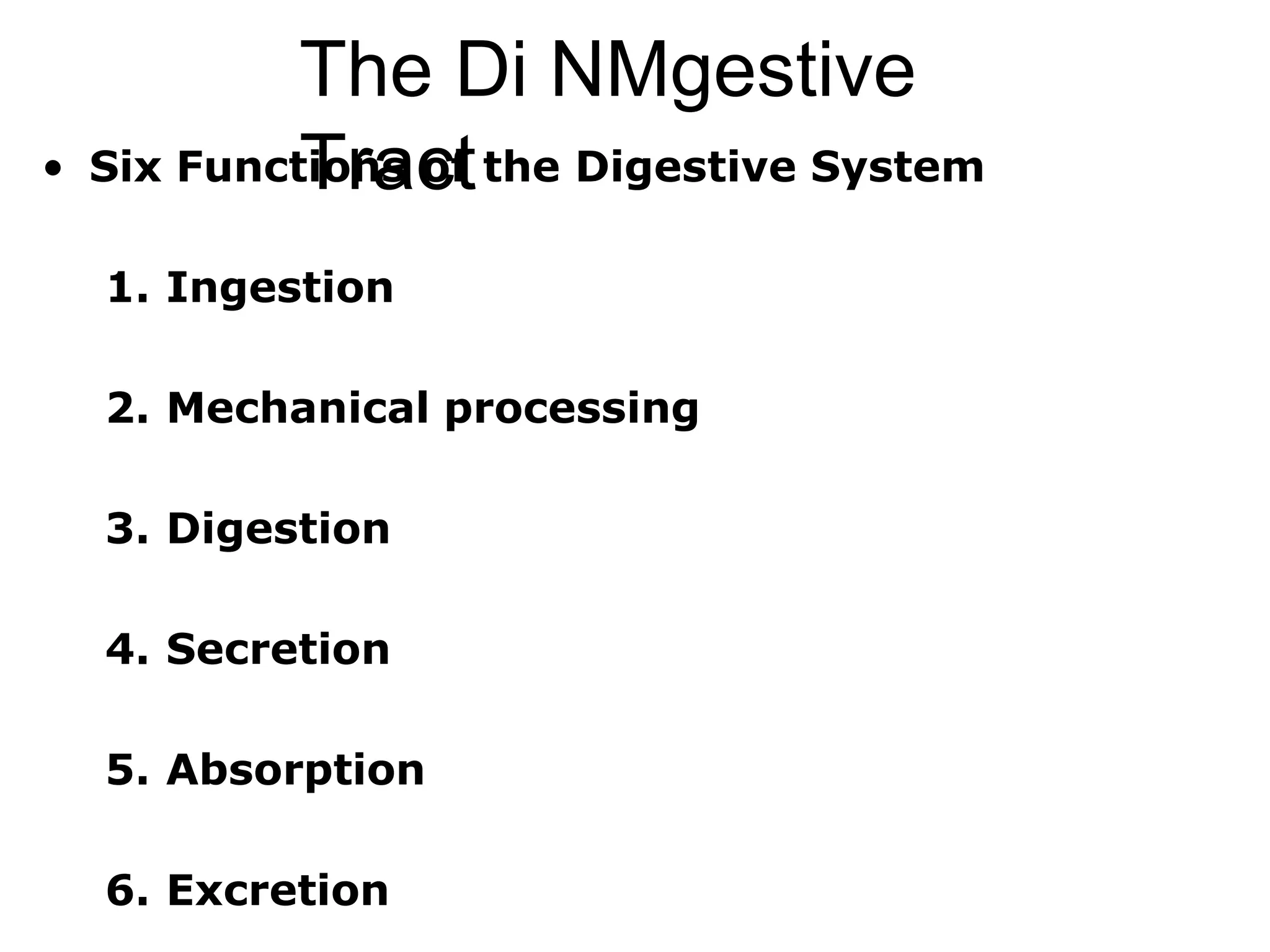
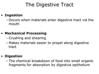
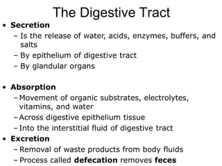
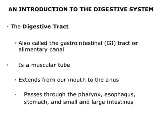
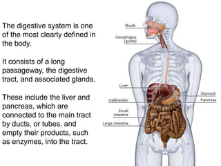
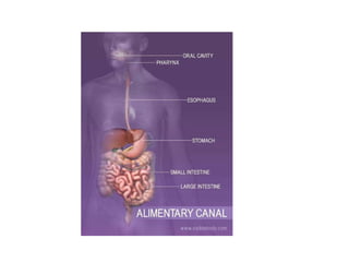
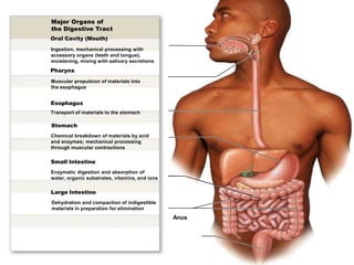
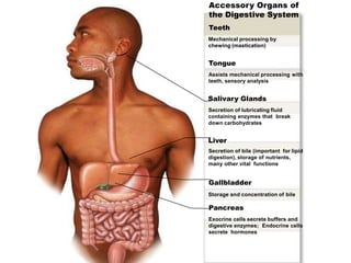
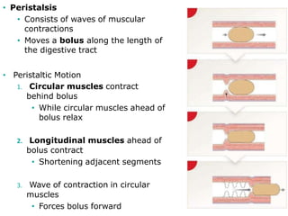
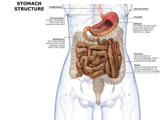
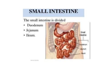
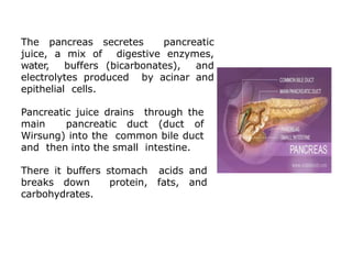
The Di NMgestive
Tract
• Six Functions of the Digestive System
1. Ingestion
2. Mechanical processing
3. Digestion
4. Secretion
5. Absorption
6. Excretion
The Digestive Tract
• Ingestion
– Occurs when materials enter digestive tract via the
mouth
• Mechanical Processing
– Crushing and shearing
– Makes materials easier to propel along digestive
tract
• Digestion
– The chemical breakdown of food into small organic
fragments for absorption by digestive epithelium
The Digestive Tract
• Secretion
– Is the release of water, acids, enzymes, buffers, and
salts
– By epithelium of digestive tract
– By glandular organs
• Absorption
– Movement of organic substrates, electrolytes,
vitamins, and water
– Across digestive epithelium tissue
– Into the interstitial fluid of digestive tract
• Excretion
– Removal of waste products from body fluids
– Process called defecation removes feces
AN INTRODUCTION TO THE DIGESTIVE SYSTEM
• The Digestive Tract
• Also called the gastrointestinal (GI) tract or
alimentary canal
• Is a muscular tube
• Extends from our mouth to the anus
• Passes through the pharynx, esophagus,
stomach, and small and large intestines
The digestive system is one
of the most clearly defined in
the body.
It consists of a long
passageway, the digestive
tract, and associated glands.
These include the liver and
pancreas, which are
connected to the main tract
by ducts, or tubes, and
empty their products, such
as enzymes, into the tract.
Major Organs of
the Digestive Tract
Oral Cavity (Mouth)
Ingestion, mechanical processing with
accessory organs (teeth and tongue),
moistening, mixing with salivary secretions
Pharynx
Muscular propulsion of materials into
the esophagus
Esophagus
Transport of materials to the stomach
Stomach
Chemical breakdown of materials by acid
and enzymes; mechanical processing
through muscular contractions
Small Intestine
Enzymatic digestion and absorption of
water, organic substrates, vitamins, and ions
Large Intestine
Dehydration and compaction of indigestible
materials in preparation for elimination
Anus
Accessory Organs of
the Digestive System
Teeth
Mechanical processing by
chewing (mastication)
Tongue
Assists mechanical processing with
teeth, sensory analysis
Salivary Glands
Secretion of lubricating fluid
containing enzymes that break
down carbohydrates
Liver
Secretion of bile (important for lipid
digestion), storage of nutrients,
many other vital functions
Gallbladder
Storage and concentration of bile
Pancreas
Exocrine cells secrete buffers and
digestive enzymes; Endocrine cells
secrete hormones
The Oral Cavity
• Functions of the Oral Cavity
1. Sensory analysis
• Of material before swallowing
2. Mechanical processing
• Through actions of teeth, tongue, and palatal
surfaces
3. Lubrication
• Mixing with mucus and salivary gland
secretions
4. Limited digestion
• Of carbohydrates and lipids
Figure 24-7a The Salivary Glands.
Parotid duct
Openings of
sublingual
ducts
Lingual
frenulum
Opening of left
submandibular
duct
Salivary Glands
Parotid salivary
gland
Sublingual
salivary gland
Submandibular
salivary gland
Submandibular
duct
The oral cavity is bounded by the teeth, tongue,
hard palate, and soft palate.
These structures make up the mouth and play a
key role in the first step of digestion, called
ingestion.
This is where the teeth and tongue work with
salivary glands to break down food into small
masses that can be swallowed, preparing them for
the journey through the alimentary canal.
Mouth and Salivary Glands
Mouth is otherwise known as oral cavity or buccal
cavity.
It is formed by cheeks, lips and palate. It encloses
the teeth, tongue and salivary glands.
Mouth opens anteriorly to the exterior through
lips and posteriorly through fauces into the
pharynx.
Digestive juice present in the mouth is saliva,
which is secreted by the salivary glands.
FUNCTIONS OF MOUTH
Primary function of mouth is eating and it
has few other important functions also.
Functions of mouth include:
1. Ingestion of food materials
2. Chewing the food and mixing it with
saliva
3. Appreciation of taste of the food
4. Transfer of food (bolus) to the
esophagus by swallowing
5. 5. Role in speech 6. Social functions
such as smiling and other expressions.
SALIVARY
GLANDS
PROPERTIES OF SALIVA
1. Volume: 1000 mL to 1500 mL of saliva is
secreted per day and it is approximately
about 1 mL/minute.
Reaction: Mixed saliva from all the glands is
slightly acidic with pH of 6.35 to 6.85 3.
Specific gravity: It ranges between 1.002 and
1.012 4.
Tonicity: Saliva is hypotonic to plasma
SALIVARY GLANDS
In humans, the saliva is secreted by three pairs of
major (larger) salivary glands and some minor (small)
salivary glands. Contribution by each major salivary
gland is: i. Parotid glands : 25% ii. Submaxillary glands :
70% iii. Sublingual glands : 5%
Major glands are: 1. Parotid glands 2. Submaxillary or
submandibular glands 3. Sublingual glands
Parotid glands are the largest of all salivary glands,
situated at the side of the face just below and in front
of the ear. Secretions from these glands are emptied
into the oral cavity by Stensen duct.
Submaxillary glands or submandibular glands are located in
medial to mandible. Saliva from these glands is emptied into the
oral cavity by Wharton duct,
Sublingual glands are the smallest salivary glands situated in the
mucosa at the floor of the mouth. Saliva from these glands is
poured into 5 to 15 small ducts called ducts of Rivinus. These
ducts open on small papillae beneath the tongue. One of the
ducts is larger and it is called Bartholin duct
Saliva Moistens Food & Begins the Process of Chemical
Digestion
Saliva secreted by salivary glands aids the mechanical and
chemical process of digestion.
Saliva is about 99% water and not only moistens food but
cleanses the mouth, dissolves food chemicals so they can be
tasted, and contains enzymes that start the chemical
breakdown of starchy foods.
Peristaltic Waves Move
Nutrients and Waste
Through the Intestines
MOVEMENTS IN GIT
• Peristalsis
• Consists of waves of muscular
contractions
• Moves a bolus along the length of
the digestive tract
• Peristaltic Motion
1. Circular muscles contract
behind bolus
• While circular muscles ahead of
bolus relax
2. Longitudinal muscles ahead of
bolus contract
• Shortening adjacent segments
3. Wave of contraction in circular
muscles
• Forces bolus forward
STOMACH
STRUCTURE
In general, wall of the GI tract is formed by four layers
which are from inside out:
1. Mucus layer 2. Submucus layer 3. Muscular layer 4.
Serous or fibrous layer.
This process begins moments after food enters the
stomach from the oesophagus, through the gastro-
oesophageal junction.
Gastric juices include digestive enzymes and hydrochloric
acid, which not only breaks down food but also kills
potentially harmful microbes
.
The stomach is the widest part of the digestive tube. It is a
muscular-walled, J- shaped sac in which food is stored,
churned, and mixed with gastric juices secreted by its
lining.
Stomach is a hollow organ situated just below the
diaphragm on the left side in the abdominal cavity. Volume
of empty stomach is 50 mL.
PARTS OF STOMACH
„FUNCTIONS OF STOMACH
1.MECHANICAL FUNCTION
i. Storage Function
. ii. Formation of Chyme
2. DIGESTIVE FUNCTION
3. PROTECTIVE FUNCTION
4. HEMOPOIETIC FUNCTION. „
5. EXCRETORY FUNCTION Many substances like toxins,
alkaloids and metals are excreted through gastric juice
GASTRIC JUICE
1. DIGESTIVE FUNCTION
Gastric juice acts mainly on proteins.
Proteolytic enzymes of the gastric juice are pepsin and
rennin .
Gastric juice also contains some other enzymes like gastric
lipase, gelatinase, urase and gastric amylase.
Pepsin is secreted as inactive pepsinogen.
Pepsinogen is converted into pepsin by hydrochloric acid.
Optimum pH for activation of pepsinogen I
Action of pepsin :
Pepsin converts proteins into proteoses,
peptones and polypeptides.
Pepsin also causes curdling and digestion of
milk (casein). Gastric Lipase Gastric lipase is a
weak lipolytic enzyme when compared to
pancreatic lipase. It is active only when the pH
is between 4 and 5
2. HEMOPOIETIC FUNCTION
3.PROTECTIVE FUNCTION – FUNCTION OF MUCUS
4. FUNCTIONS OF HYDROCHLORIC ACID
Hydrochloric acid is present in the gastric juice: i. Activates
pepsinogen into pepsin ii. Kills some of the bacteria entering the
stomach along with food substances. This action is called
bacteriolytic action iii. Provides acid medium, which is necessary for
the action of hormones.
FORMATION OF GASTRIC HCl
Pepsinogen is synthesized from amino acids in the
ribosomes attached to endoplasmic reticulum in
chief cells.
 Pepsinogen molecules are packed into zymogen
granules by Golgi apparatus.
When zymogen granule is secreted into stomach
from chief cells, the granule is dissolved and
pepsinogen is released into gastric juice.
Pepsinogen is activated into pepsin by
hydrochloric acid.
SECRETION OF HYDROCHLORIC
ACID
Carbon dioxide is derived from
metabolic activities of parietal cell.
Some amount of carbon dioxide is
obtained from blood also. It
combines with water to form
carbonic acid in the presence of
carbonic anhydrase. This enzyme is
present in high concentration in
parietal cells.
Carbonic acid is the most unstable compound and immediately
splits into hydrogen ion and bicarbonate ion. The hydrogen ion
is actively pumped into the canaliculus of parietal cell.
Simultaneously, the chloride ion is also pumped into canaliculus
actively.
The chloride is derived from sodium chloride in the blood. Now, the
hydrogen ion combines with chloride ion to form hydrochloric acid.
To compensate the loss of chloride ion, the bicarbonate ion from
parietal cell enters the blood and combines with sodium to form
sodium bicarbonate.
Factors Stimulating the Secretion of Hydrochloric Acid
1. Gastrin 2. Histamine 3. Vagal stimulation.
Factors Inhibiting the Secretion of Hydrochloric Acid
1. Secretin 2. Gastric inhibitory polypeptide 3. Peptide YY
Phases of gastric juice secretion
SMALL INTESTINE
The small intestine is part of your digestive
system. It makes up part of the long
pathway that food takes through your
body, called the gastrointestinal (GI) tract.
When food leaves your stomach, it enters
the small intestine, also called the small
bowel.
The small intestine has a beginning section, a middle section
and an end section. Although there is no real separation
between the parts, they do have slightly different
characteristics and roles to play.
Duodenum
The duodenum is the first part of the small intestine that the
stomach feeds into. It’s a short, descending chute (about 10
inches long) that curves around the pancreas in a “C” shape
before connecting to the rest of the coiled intestines.
functions
To help break food down, the small intestine receives
digestive juices from other organs in your digestive
system, including your liver, gallbladder and pancreas.
Hormone glands in the lining of the duodenum signal
these organs to release their chemicals when food is
present.
Jejunum
The remaining small intestine lays in many coils inside the
lower abdominal cavity. Its middle section, called the
jejunum, makes up a little less than half of this remaining
length. The jejunum is characterized by many blood vessels,
which give it a deep red color.
functions
After chemical digestion in the duodenum, food moves into
the jejunum, where the muscle work of digestion picks up.
Nerves in the intestinal walls trigger its muscles to churn food
back and forth (segmentation), mixing it with digestive juices.
Other muscle movements (peristalsis) keep the food moving
gradually forward.
Ileum
The ileum is the last and longest section of the small
intestine.
Here the walls of the small intestine begin to thin and
narrow, and blood supply is reduced.
Food spends the most time in the ileum, where the
most water and nutrients are absorbed.
•In the ileum, segmentation slows down and
peristalsis takes over, moving food waste
gradually toward the large intestine.
•The ileocecal valve separates the ileum
from the large intestine.
•Nerves and hormones signal the valve to
open to let food pass through and close to
keep bacteria out.
• Special immune cells line the ileum to
protect against bacteria.
functions
The small intestine is where most of the long process
of digestion takes place. It:
•Systematically breaks food down.
•Absorbs nutrients.
•Extracts water.
•Moves food along the gastrointestinal tract.
DIFFERENCES
The small intestine absorbs nutrients and water from your
food. If these functions are impaired, nutritional deficiencies
and watery stools (diarrhea) occurs.
The muscle movements of the small intestine help break food
down and process it through your body. If these movements
are impaired, you may
experience indigestion and constipation.
A variety of diseases and infections may cause inflammation
of the small intestine, which can cause abdominal
pain, nausea and vomiting.
Food that is chewed in the oral cavity
then swallowed ends up in the
stomach where it is further digested
so its nutrients can be absorbed in
the small intestine.
The salivary glands, liver and gall
bladder, and the pancreas aid the
processes of ingestion, digestion, and
absorption.
These accessory organs of digestion play
key roles in the digestive process. Each of
these organs either secretes or stores
substances that pass through ducts into
the alimentary canal.
DIGESTIVE-ACCESSORY-ORGANS
The liver is one of the largest organs
in the body and it is continuously
producing bile.
This yellowish-brown fluid aids
chemical digestion by emulsifying fats
in the duodenum.
Bile flows out of the liver into the right and
left hepatic ducts, into the common
hepatic ducts, and toward the small
intestine to help with digestion and the
absorption of fats.
The Liver Secretes Bile to Emulsify Fats
in the Small Intestine
The Gall Bladder Stores Bile
If bile is not immediately
needed for digestion, it flows
up the cystic duct to the
gall bladder.
The gall bladder is a green,
pear-shaped sac about 10
cm or 4 in. long that stores
and concentrates excess
bile secreted by the liver.
Bile is released by the gall
bladder as needed into the
small intestine.
The pancreas secretes pancreatic
juice, a mix of digestive enzymes,
water, buffers (bicarbonates), and
electrolytes produced by acinar and
epithelial cells.
Pancreatic juice drains through the
main pancreatic duct (duct of
Wirsung) into the common bile duct
and then into the small intestine.
There it buffers stomach acids and
breaks down protein, fats, and
carbohydrates.
Nutrients In, Waste Out:
How the Human Body Absorbs
Nutrients and Eliminates Waste
Villi that line the walls of the small
intestine absorb nutrients into
capillaries of the circulatory system
and lacteals of the lymphatic
system.
Villi contain capillary beds, as well
as lymphatic vessels called
lacteals.
Fatty acids absorbed from broken-
down chyme pass into the lacteals.
Other absorbed nutrients enter the
bloodstream through the capillary
beds and are taken directly to the
liver, via the hepatic vein, for
processing.
Nutrients In, Waste Out: How the
Human Body Absorbs Nutrients and
Eliminates Waste
Figure 24-3 The Structure of the Digestive Tract (Part 2 of 2).
Mucosa
Submucosa
Muscularis
externa
Serosa
(visceral
peritoneum)
Circular
fold
Mucosal
epithelium
Lamina
propria
Villi
Mucosal glands
Submucosal gland
Mucosa
Muscularis
mucosae
Lymphatic vessel
Artery and vein
Submucosal
plexus
Circular muscle
layer
Myenteric plexus
Longitudinal
muscle layer
Chyme passes from the small intestine
through the ileocecal valve and into the
cecum of the large intestine.
Any remaining nutrients and some water
are absorbed as peristaltic waves move
the chyme into the ascending and
transverse colons.
This dehydration, combined with
peristaltic waves, helps compact the
chyme.
The solid waste formed is called feces.
It continues to move through the
descending and sigmoid colons.
The large intestine temporarily stores the
feces prior to elimination.
The body expels waste products
from digestion through the rectum
and anus.
This process, called defecation,
involves contraction of rectal
muscles, relaxation of the internal
anal sphincter, and an initial
contraction of the skeletal muscle of
the external anal sphincter.
The defecation reflex is mostly
involuntary, under the command of
the autonomic nervous system.
But the somatic nervous system
also plays a role to control the timing
of elimination.

The Digestive System.pptx

  • 1.
    The Di NMgestive Tract •Six Functions of the Digestive System 1. Ingestion 2. Mechanical processing 3. Digestion 4. Secretion 5. Absorption 6. Excretion
  • 2.
    The Digestive Tract •Ingestion – Occurs when materials enter digestive tract via the mouth • Mechanical Processing – Crushing and shearing – Makes materials easier to propel along digestive tract • Digestion – The chemical breakdown of food into small organic fragments for absorption by digestive epithelium
  • 3.
    The Digestive Tract •Secretion – Is the release of water, acids, enzymes, buffers, and salts – By epithelium of digestive tract – By glandular organs • Absorption – Movement of organic substrates, electrolytes, vitamins, and water – Across digestive epithelium tissue – Into the interstitial fluid of digestive tract • Excretion – Removal of waste products from body fluids – Process called defecation removes feces
  • 4.
    AN INTRODUCTION TOTHE DIGESTIVE SYSTEM • The Digestive Tract • Also called the gastrointestinal (GI) tract or alimentary canal • Is a muscular tube • Extends from our mouth to the anus • Passes through the pharynx, esophagus, stomach, and small and large intestines
  • 5.
    The digestive systemis one of the most clearly defined in the body. It consists of a long passageway, the digestive tract, and associated glands. These include the liver and pancreas, which are connected to the main tract by ducts, or tubes, and empty their products, such as enzymes, into the tract.
  • 7.
    Major Organs of theDigestive Tract Oral Cavity (Mouth) Ingestion, mechanical processing with accessory organs (teeth and tongue), moistening, mixing with salivary secretions Pharynx Muscular propulsion of materials into the esophagus Esophagus Transport of materials to the stomach Stomach Chemical breakdown of materials by acid and enzymes; mechanical processing through muscular contractions Small Intestine Enzymatic digestion and absorption of water, organic substrates, vitamins, and ions Large Intestine Dehydration and compaction of indigestible materials in preparation for elimination Anus
  • 8.
    Accessory Organs of theDigestive System Teeth Mechanical processing by chewing (mastication) Tongue Assists mechanical processing with teeth, sensory analysis Salivary Glands Secretion of lubricating fluid containing enzymes that break down carbohydrates Liver Secretion of bile (important for lipid digestion), storage of nutrients, many other vital functions Gallbladder Storage and concentration of bile Pancreas Exocrine cells secrete buffers and digestive enzymes; Endocrine cells secrete hormones
  • 9.
    The Oral Cavity •Functions of the Oral Cavity 1. Sensory analysis • Of material before swallowing 2. Mechanical processing • Through actions of teeth, tongue, and palatal surfaces 3. Lubrication • Mixing with mucus and salivary gland secretions 4. Limited digestion • Of carbohydrates and lipids
  • 10.
    Figure 24-7a TheSalivary Glands. Parotid duct Openings of sublingual ducts Lingual frenulum Opening of left submandibular duct Salivary Glands Parotid salivary gland Sublingual salivary gland Submandibular salivary gland Submandibular duct
  • 11.
    The oral cavityis bounded by the teeth, tongue, hard palate, and soft palate. These structures make up the mouth and play a key role in the first step of digestion, called ingestion. This is where the teeth and tongue work with salivary glands to break down food into small masses that can be swallowed, preparing them for the journey through the alimentary canal.
  • 12.
    Mouth and SalivaryGlands Mouth is otherwise known as oral cavity or buccal cavity. It is formed by cheeks, lips and palate. It encloses the teeth, tongue and salivary glands. Mouth opens anteriorly to the exterior through lips and posteriorly through fauces into the pharynx. Digestive juice present in the mouth is saliva, which is secreted by the salivary glands.
  • 13.
    FUNCTIONS OF MOUTH Primaryfunction of mouth is eating and it has few other important functions also. Functions of mouth include: 1. Ingestion of food materials 2. Chewing the food and mixing it with saliva 3. Appreciation of taste of the food 4. Transfer of food (bolus) to the esophagus by swallowing 5. 5. Role in speech 6. Social functions such as smiling and other expressions.
  • 14.
  • 16.
    PROPERTIES OF SALIVA 1.Volume: 1000 mL to 1500 mL of saliva is secreted per day and it is approximately about 1 mL/minute. Reaction: Mixed saliva from all the glands is slightly acidic with pH of 6.35 to 6.85 3. Specific gravity: It ranges between 1.002 and 1.012 4. Tonicity: Saliva is hypotonic to plasma
  • 17.
    SALIVARY GLANDS In humans,the saliva is secreted by three pairs of major (larger) salivary glands and some minor (small) salivary glands. Contribution by each major salivary gland is: i. Parotid glands : 25% ii. Submaxillary glands : 70% iii. Sublingual glands : 5% Major glands are: 1. Parotid glands 2. Submaxillary or submandibular glands 3. Sublingual glands Parotid glands are the largest of all salivary glands, situated at the side of the face just below and in front of the ear. Secretions from these glands are emptied into the oral cavity by Stensen duct.
  • 18.
    Submaxillary glands orsubmandibular glands are located in medial to mandible. Saliva from these glands is emptied into the oral cavity by Wharton duct, Sublingual glands are the smallest salivary glands situated in the mucosa at the floor of the mouth. Saliva from these glands is poured into 5 to 15 small ducts called ducts of Rivinus. These ducts open on small papillae beneath the tongue. One of the ducts is larger and it is called Bartholin duct
  • 19.
    Saliva Moistens Food& Begins the Process of Chemical Digestion Saliva secreted by salivary glands aids the mechanical and chemical process of digestion. Saliva is about 99% water and not only moistens food but cleanses the mouth, dissolves food chemicals so they can be tasted, and contains enzymes that start the chemical breakdown of starchy foods.
  • 20.
    Peristaltic Waves Move Nutrientsand Waste Through the Intestines MOVEMENTS IN GIT
  • 21.
    • Peristalsis • Consistsof waves of muscular contractions • Moves a bolus along the length of the digestive tract • Peristaltic Motion 1. Circular muscles contract behind bolus • While circular muscles ahead of bolus relax 2. Longitudinal muscles ahead of bolus contract • Shortening adjacent segments 3. Wave of contraction in circular muscles • Forces bolus forward
  • 22.
  • 23.
    In general, wallof the GI tract is formed by four layers which are from inside out: 1. Mucus layer 2. Submucus layer 3. Muscular layer 4. Serous or fibrous layer. This process begins moments after food enters the stomach from the oesophagus, through the gastro- oesophageal junction. Gastric juices include digestive enzymes and hydrochloric acid, which not only breaks down food but also kills potentially harmful microbes .
  • 24.
    The stomach isthe widest part of the digestive tube. It is a muscular-walled, J- shaped sac in which food is stored, churned, and mixed with gastric juices secreted by its lining. Stomach is a hollow organ situated just below the diaphragm on the left side in the abdominal cavity. Volume of empty stomach is 50 mL. PARTS OF STOMACH
  • 25.
    „FUNCTIONS OF STOMACH 1.MECHANICALFUNCTION i. Storage Function . ii. Formation of Chyme 2. DIGESTIVE FUNCTION 3. PROTECTIVE FUNCTION 4. HEMOPOIETIC FUNCTION. „ 5. EXCRETORY FUNCTION Many substances like toxins, alkaloids and metals are excreted through gastric juice
  • 26.
  • 27.
    1. DIGESTIVE FUNCTION Gastricjuice acts mainly on proteins. Proteolytic enzymes of the gastric juice are pepsin and rennin . Gastric juice also contains some other enzymes like gastric lipase, gelatinase, urase and gastric amylase. Pepsin is secreted as inactive pepsinogen. Pepsinogen is converted into pepsin by hydrochloric acid. Optimum pH for activation of pepsinogen I
  • 28.
    Action of pepsin: Pepsin converts proteins into proteoses, peptones and polypeptides. Pepsin also causes curdling and digestion of milk (casein). Gastric Lipase Gastric lipase is a weak lipolytic enzyme when compared to pancreatic lipase. It is active only when the pH is between 4 and 5
  • 29.
    2. HEMOPOIETIC FUNCTION 3.PROTECTIVEFUNCTION – FUNCTION OF MUCUS 4. FUNCTIONS OF HYDROCHLORIC ACID Hydrochloric acid is present in the gastric juice: i. Activates pepsinogen into pepsin ii. Kills some of the bacteria entering the stomach along with food substances. This action is called bacteriolytic action iii. Provides acid medium, which is necessary for the action of hormones.
  • 30.
    FORMATION OF GASTRICHCl Pepsinogen is synthesized from amino acids in the ribosomes attached to endoplasmic reticulum in chief cells.  Pepsinogen molecules are packed into zymogen granules by Golgi apparatus. When zymogen granule is secreted into stomach from chief cells, the granule is dissolved and pepsinogen is released into gastric juice. Pepsinogen is activated into pepsin by hydrochloric acid.
  • 31.
    SECRETION OF HYDROCHLORIC ACID Carbondioxide is derived from metabolic activities of parietal cell. Some amount of carbon dioxide is obtained from blood also. It combines with water to form carbonic acid in the presence of carbonic anhydrase. This enzyme is present in high concentration in parietal cells. Carbonic acid is the most unstable compound and immediately splits into hydrogen ion and bicarbonate ion. The hydrogen ion is actively pumped into the canaliculus of parietal cell.
  • 32.
    Simultaneously, the chlorideion is also pumped into canaliculus actively. The chloride is derived from sodium chloride in the blood. Now, the hydrogen ion combines with chloride ion to form hydrochloric acid. To compensate the loss of chloride ion, the bicarbonate ion from parietal cell enters the blood and combines with sodium to form sodium bicarbonate. Factors Stimulating the Secretion of Hydrochloric Acid 1. Gastrin 2. Histamine 3. Vagal stimulation. Factors Inhibiting the Secretion of Hydrochloric Acid 1. Secretin 2. Gastric inhibitory polypeptide 3. Peptide YY
  • 34.
    Phases of gastricjuice secretion
  • 37.
    SMALL INTESTINE The smallintestine is part of your digestive system. It makes up part of the long pathway that food takes through your body, called the gastrointestinal (GI) tract. When food leaves your stomach, it enters the small intestine, also called the small bowel.
  • 39.
    The small intestinehas a beginning section, a middle section and an end section. Although there is no real separation between the parts, they do have slightly different characteristics and roles to play. Duodenum The duodenum is the first part of the small intestine that the stomach feeds into. It’s a short, descending chute (about 10 inches long) that curves around the pancreas in a “C” shape before connecting to the rest of the coiled intestines.
  • 40.
    functions To help breakfood down, the small intestine receives digestive juices from other organs in your digestive system, including your liver, gallbladder and pancreas. Hormone glands in the lining of the duodenum signal these organs to release their chemicals when food is present.
  • 41.
    Jejunum The remaining smallintestine lays in many coils inside the lower abdominal cavity. Its middle section, called the jejunum, makes up a little less than half of this remaining length. The jejunum is characterized by many blood vessels, which give it a deep red color. functions After chemical digestion in the duodenum, food moves into the jejunum, where the muscle work of digestion picks up. Nerves in the intestinal walls trigger its muscles to churn food back and forth (segmentation), mixing it with digestive juices. Other muscle movements (peristalsis) keep the food moving gradually forward.
  • 42.
    Ileum The ileum isthe last and longest section of the small intestine. Here the walls of the small intestine begin to thin and narrow, and blood supply is reduced. Food spends the most time in the ileum, where the most water and nutrients are absorbed.
  • 43.
    •In the ileum,segmentation slows down and peristalsis takes over, moving food waste gradually toward the large intestine. •The ileocecal valve separates the ileum from the large intestine. •Nerves and hormones signal the valve to open to let food pass through and close to keep bacteria out. • Special immune cells line the ileum to protect against bacteria.
  • 44.
    functions The small intestineis where most of the long process of digestion takes place. It: •Systematically breaks food down. •Absorbs nutrients. •Extracts water. •Moves food along the gastrointestinal tract.
  • 45.
  • 47.
    The small intestineabsorbs nutrients and water from your food. If these functions are impaired, nutritional deficiencies and watery stools (diarrhea) occurs. The muscle movements of the small intestine help break food down and process it through your body. If these movements are impaired, you may experience indigestion and constipation. A variety of diseases and infections may cause inflammation of the small intestine, which can cause abdominal pain, nausea and vomiting.
  • 61.
    Food that ischewed in the oral cavity then swallowed ends up in the stomach where it is further digested so its nutrients can be absorbed in the small intestine. The salivary glands, liver and gall bladder, and the pancreas aid the processes of ingestion, digestion, and absorption. These accessory organs of digestion play key roles in the digestive process. Each of these organs either secretes or stores substances that pass through ducts into the alimentary canal. DIGESTIVE-ACCESSORY-ORGANS
  • 62.
    The liver isone of the largest organs in the body and it is continuously producing bile. This yellowish-brown fluid aids chemical digestion by emulsifying fats in the duodenum. Bile flows out of the liver into the right and left hepatic ducts, into the common hepatic ducts, and toward the small intestine to help with digestion and the absorption of fats. The Liver Secretes Bile to Emulsify Fats in the Small Intestine
  • 63.
    The Gall BladderStores Bile If bile is not immediately needed for digestion, it flows up the cystic duct to the gall bladder. The gall bladder is a green, pear-shaped sac about 10 cm or 4 in. long that stores and concentrates excess bile secreted by the liver. Bile is released by the gall bladder as needed into the small intestine.
  • 64.
    The pancreas secretespancreatic juice, a mix of digestive enzymes, water, buffers (bicarbonates), and electrolytes produced by acinar and epithelial cells. Pancreatic juice drains through the main pancreatic duct (duct of Wirsung) into the common bile duct and then into the small intestine. There it buffers stomach acids and breaks down protein, fats, and carbohydrates.
  • 66.
    Nutrients In, WasteOut: How the Human Body Absorbs Nutrients and Eliminates Waste Villi that line the walls of the small intestine absorb nutrients into capillaries of the circulatory system and lacteals of the lymphatic system. Villi contain capillary beds, as well as lymphatic vessels called lacteals. Fatty acids absorbed from broken- down chyme pass into the lacteals. Other absorbed nutrients enter the bloodstream through the capillary beds and are taken directly to the liver, via the hepatic vein, for processing. Nutrients In, Waste Out: How the Human Body Absorbs Nutrients and Eliminates Waste
  • 67.
    Figure 24-3 TheStructure of the Digestive Tract (Part 2 of 2). Mucosa Submucosa Muscularis externa Serosa (visceral peritoneum) Circular fold Mucosal epithelium Lamina propria Villi Mucosal glands Submucosal gland Mucosa Muscularis mucosae Lymphatic vessel Artery and vein Submucosal plexus Circular muscle layer Myenteric plexus Longitudinal muscle layer
  • 68.
    Chyme passes fromthe small intestine through the ileocecal valve and into the cecum of the large intestine. Any remaining nutrients and some water are absorbed as peristaltic waves move the chyme into the ascending and transverse colons. This dehydration, combined with peristaltic waves, helps compact the chyme. The solid waste formed is called feces. It continues to move through the descending and sigmoid colons. The large intestine temporarily stores the feces prior to elimination.
  • 69.
    The body expelswaste products from digestion through the rectum and anus. This process, called defecation, involves contraction of rectal muscles, relaxation of the internal anal sphincter, and an initial contraction of the skeletal muscle of the external anal sphincter. The defecation reflex is mostly involuntary, under the command of the autonomic nervous system. But the somatic nervous system also plays a role to control the timing of elimination.