KLINTAPS COLLEGE OFHEALTH
AND ALLIED SCIENCES
The Digestive system
KUC 118: HUMAN ANATOMY II
Mr. Eshun
2.
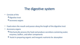
The digestive system
•Consists of the
digestive tract
accessory organs
• Food enters the mouth and passes along the length of the digestive tract
• Accessory organs
Mechanically process the food and produce secretions containing water,
enzymes, buffers, and other components
Assist in preparing organic and inorganic nutrients for absorption
3.
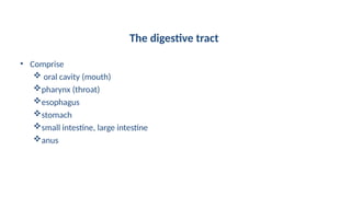
The digestive tract
•Comprise
oral cavity (mouth)
pharynx (throat)
esophagus
stomach
small intestine, large intestine
anus
4.
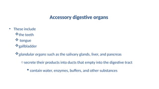
Accessory digestive organs
•These include
the teeth
tongue
gallbladder
glandular organs such as the salivary glands, liver, and pancreas
osecrete their products into ducts that empty into the digestive tract
contain water, enzymes, buffers, and other substances
5.
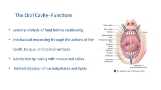
The Oral Cavity-Functions
• sensory analysis of food before swallowing
• mechanical processing through the actions of the
teeth, tongue, and palatal surfaces
• lubrication by mixing with mucus and saliva
• limited digestion of carbohydrates and lipids
6.
The Oral Cavity
•Lined by the oral mucosa
• Keratinized stratified squamous epithelium
superior surface of the tongue and hard palate
oexposed to severe abrasion
• Non-keratinized stratified squamous epithelium
cheeks, lips, and inferior surface of the tongue
7.
The Oral Cavity
•Nutrients are not absorbed in the oral cavity
• Mucosa inferior to the tongue is thin enough and vascular enough to permit the
rapid absorption of lipid-soluble drugs
Nitroglycerin may be administered by this route to treat acute angina pectoris
7
8.
Some terminologies
• Thevestibule is the space between the cheeks (or lips) and the teeth
• The gingivae (gums) are ridges of oral mucosa that surround the base of each
tooth on the alveolar processes of the maxillary bones and mandible
• The frenulum of lower lip is a fold of mucosa that extends from the gingiva to
the lower lip, attaching the lip to the gum
• The hard and soft palates form the roof of the oral cavity
• Uvula a dangling process that helps prevent food from entering the pharynx
too soon
9.
The tongue
• manipulatesfood inside the mouth and occasionally brings in foods (such as ice
cream on a cone)
• The primary functions of the tongue are
mechanical processing by compression, abrasion, and distortion
manipulation to assist in chewing and to prepare food for swallowing
sensory analysis by touch, temperature, and taste receptors
10.
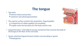
The tongue
• Twoparts
anterior body (oral portion)
posterior root (pharyngeal portion)
• The superior surface contains fine projections lingual papillae
• A V-shaped line of vallate papillae (circumvallate)
roughly marks the boundary b/n the body and the root
• Lingual frenulum- a thin fold of mucous membrane that connects the body of
the tongue to the floor of the oral cavity
• Overly restrictive lingual frenulum hinders normal eating or speech
Ankyloglossia
11.
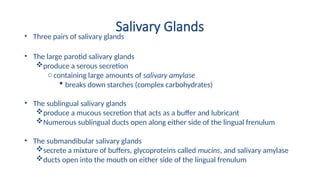
Salivary Glands
• Threepairs of salivary glands
• The large parotid salivary glands
produce a serous secretion
o containing large amounts of salivary amylase
breaks down starches (complex carbohydrates)
• The sublingual salivary glands
produce a mucous secretion that acts as a buffer and lubricant
Numerous sublingual ducts open along either side of the lingual frenulum
• The submandibular salivary glands
secrete a mixture of buffers, glycoproteins called mucins, and salivary amylase
ducts open into the mouth on either side of the lingual frenulum
12.
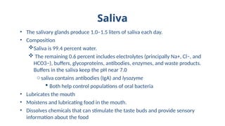
Saliva
• The salivaryglands produce 1.0–1.5 liters of saliva each day.
• Composition
Saliva is 99.4 percent water.
The remaining 0.6 percent includes electrolytes (principally Na+, Cl−, and
HCO3−), buffers, glycoproteins, antibodies, enzymes, and waste products.
Buffers in the saliva keep the pH near 7.0
osaliva contains antibodies (IgA) and lysozyme
Both help control populations of oral bacteria
• Lubricates the mouth
• Moistens and lubricating food in the mouth.
• Dissolves chemicals that can stimulate the taste buds and provide sensory
information about the food
13.
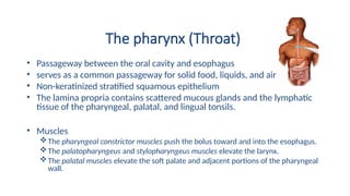
The pharynx (Throat)
•Passageway between the oral cavity and esophagus
• serves as a common passageway for solid food, liquids, and air
• Non-keratinized stratified squamous epithelium
• The lamina propria contains scattered mucous glands and the lymphatic
tissue of the pharyngeal, palatal, and lingual tonsils.
• Muscles
The pharyngeal constrictor muscles push the bolus toward and into the esophagus.
The palatopharyngeus and stylopharyngeus muscles elevate the larynx.
The palatal muscles elevate the soft palate and adjacent portions of the pharyngeal
wall.
14.
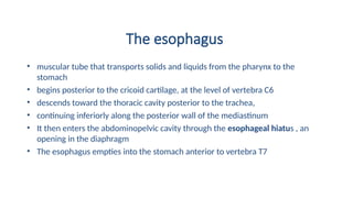
The esophagus
• musculartube that transports solids and liquids from the pharynx to the
stomach
• begins posterior to the cricoid cartilage, at the level of vertebra C6
• descends toward the thoracic cavity posterior to the trachea,
• continuing inferiorly along the posterior wall of the mediastinum
• It then enters the abdominopelvic cavity through the esophageal hiatus , an
opening in the diaphragm
• The esophagus empties into the stomach anterior to vertebra T7
15.
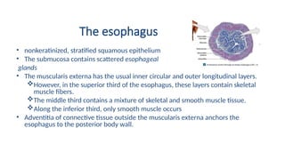
The esophagus
• nonkeratinized,stratified squamous epithelium
• The submucosa contains scattered esophageal
glands
• The muscularis externa has the usual inner circular and outer longitudinal layers.
However, in the superior third of the esophagus, these layers contain skeletal
muscle fibers.
The middle third contains a mixture of skeletal and smooth muscle tissue.
Along the inferior third, only smooth muscle occurs
• Adventitia of connective tissue outside the muscularis externa anchors the
esophagus to the posterior body wall.
16.
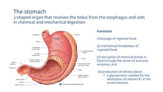
Functions
(1)storage of ingestedfood;
(2) mechanical breakdown of
ingested food;
(3) disruption of chemical bonds in
food through the action of acid and
enzymes; and
(4) production of intrinsic factor,
a glycoprotein needed for the
absorption of vitamin B12 in the
small intestine.
The stomach
J-shaped organ that receives the bolus from the esophagus and aids
in chemical and mechanical digestion
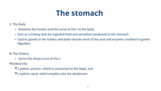
17.
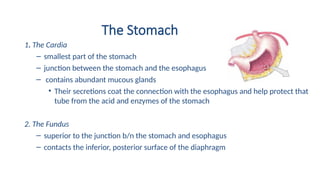
The Stomach
1. TheCardia
– smallest part of the stomach
– junction between the stomach and the esophagus
– contains abundant mucous glands
• Their secretions coat the connection with the esophagus and help protect that
tube from the acid and enzymes of the stomach
2. The Fundus
– superior to the junction b/n the stomach and esophagus
– contacts the inferior, posterior surface of the diaphragm
18.
The stomach
3. TheBody
– between the fundus and the curve of the J is the body,
– Acts as a mixing tank for ingested food and secretions produced in the stomach
– Gastric glands in the fundus and body secrete most of the acid and enzymes involved in gastric
digestion
4. The Pylorus
– forms the sharp curve of the J
•Divided into
a pyloric antrum, which is connected to the body, and
a pyloric canal, which empties into the duodenum
18
19.
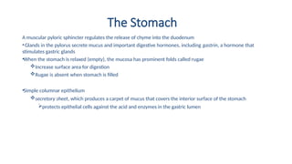
The Stomach
A muscularpyloric sphincter regulates the release of chyme into the duodenum
•Glands in the pylorus secrete mucus and important digestive hormones, including gastrin, a hormone that
stimulates gastric glands
•When the stomach is relaxed (empty), the mucosa has prominent folds called rugae
Increase surface area for digestion
Rugae is absent when stomach is filled
•Simple columnar epithelium
secretory sheet, which produces a carpet of mucus that covers the interior surface of the stomach
protects epithelial cells against the acid and enzymes in the gastric lumen
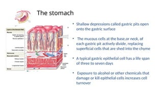
20.
• Shallow depressionscalled gastric pits open
onto the gastric surface
• The mucous cells at the base,or neck, of
each gastric pit actively divide, replacing
superficial cells that are shed into the chyme
• A typical gastric epithelial cell has a life span
of three to seven days
• Exposure to alcohol or other chemicals that
damage or kill epithelial cells increases cell
turnover
The stomach
21.
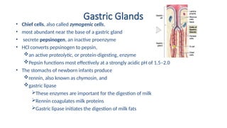
Gastric Glands
• Twotypes of secretory cells:
parietal cells and chief cells
• Parietal cells are common along the proximal portions of
each gastric gland
secrete intrinsic factor
This glycoprotein helps absorb vitamin B12 across
the intestinal lining
secrete hydrochloric acid (HCl)
The secretions of the parietal cells can keep the
stomach contents at pH 1.5–2.0
22.
Gastric Glands
• Chiefcells, also called zymogenic cells,
• most abundant near the base of a gastric gland
• secrete pepsinogen, an inactive proenzyme
• HCl converts pepsinogen to pepsin,
an active proteolytic, or protein-digesting, enzyme
Pepsin functions most effectively at a strongly acidic pH of 1.5–2.0
• The stomachs of newborn infants produce
rennin, also known as chymosin, and
gastric lipase
These enzymes are important for the digestion of milk
Rennin coagulates milk proteins
Gastric lipase initiates the digestion of milk fats
23.
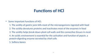
Functions of HCl
•Some important functions of HCl;
1. The acidity of gastric juice kills most of the microorganisms ingested with food
2. The acidity denatures proteins and inactivates most of the enzymes in food
3. The acidity helps break down plant cell walls and the connective tissues in meat
4. An acidic environment is essential for the activation and function of pepsin, a
protein-digesting enzyme secreted by chief cells
5. Softens bones
23
24.
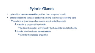
Pyloric Glands
• primarilya mucous secretion, rather than enzymes or acid
• enteroendocrine cells are scattered among the mucus-secreting cells
produce at least seven hormones, most notably gastrin
Gastrin is produced by G cells,
Gastrin stimulates secretion by both parietal and chief cells
D cells, which release somatostatin,
inhibits the release of gastrin
25.
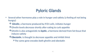
Pyloric Glands
• Severalother hormones play a role in hunger and satiety (a feeling of not being
hungry)
Ghrelin, a hormone produced by P/D1 cells, initiates hunger
Ghrelin levels decrease shortly after eating to curb appetite
Ghrelin is also antagonistic to leptin, a hormone derived from fat tissue that
induces satiety
Obestatin, is thought to decrease appetite and inhibit thirst
The same gene encodes both ghrelin and obestatin
25
26.
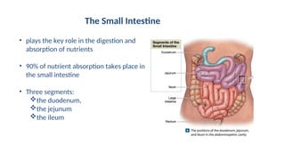
• plays thekey role in the digestion and
absorption of nutrients
• 90% of nutrient absorption takes place in
the small intestine
• Three segments:
the duodenum,
the jejunum
the ileum
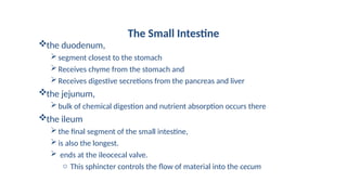
The Small Intestine
27.
the duodenum,
segment closestto the stomach
Receives chyme from the stomach and
Receives digestive secretions from the pancreas and liver
the jejunum,
bulk of chemical digestion and nutrient absorption occurs there
the ileum
the final segment of the small intestine,
is also the longest.
ends at the ileocecal valve.
o This sphincter controls the flow of material into the cecum
The Small Intestine
28.
The Small Intestine
•Intestinal lining has a series of transverse folds called circular folds or plicae
circulares
• The circular folds are permanent features
• The mucosa of the small intestine has a series of fingerlike projections called
intestinal villi
The villi are covered by simple columnar epithelium that is carpeted with
microvilli or brush border
29.
The Small Intestine
•Capillaries carry absorbed nutrients to the
hepatic portal circulation for delivery to the
liver
• A lymphatic capillary called a lacteal transport
absorbed fatty acids are too large to diffuse
into the bloodstream
30.
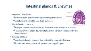
Intestinal glands &Enzymes
• crypts of Lieberkuhn
Mucous cells between the columnar epithelial cells
eject mucins onto the intestinal surfaces
• Brush border enzymes
integral membrane proteins on the surfaces of intestinal microvilli
These enzymes break down materials that come in contact with the
brush border
• Enteropeptidase
one brush border enzyme that enters the lumen in this way
it activates a key pancreatic proenzyme, trypsinogen
31.
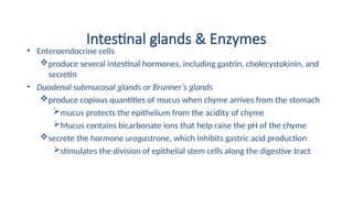
Intestinal glands &Enzymes
• Enteroendocrine cells
produce several intestinal hormones, including gastrin, cholecystokinin, and
secretin
• Duodenal submucosal glands or Brunner’s glands
produce copious quantities of mucus when chyme arrives from the stomach
mucus protects the epithelium from the acidity of chyme
Mucus contains bicarbonate ions that help raise the pH of the chyme
secrete the hormone urogastrone, which inhibits gastric acid production
stimulates the division of epithelial stem cells along the digestive tract
32.
The Pancreas
• Primarilyan exocrine organ
• It produces digestive enzymes and buffers
• The large pancreatic duct (duct of Wirsung) delivers
these secretions to the duodenum
• The pancreatic duct extends to the duodenum, where
it meets the common bile duct from the liver and
gallbladder.
• The two ducts then empty into the duodenal ampulla
(ampulla of Vater)
33.
Pancreatic secretions
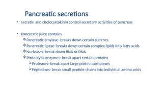
• secretinand cholecystokinin control secretory activities of pancreas
• Pancreatic juice contains
Pancreatic amylase- breaks down certain starches
Pancreatic lipase- breaks down certain complex lipids into fatty acids
Nucleases- break down RNA or DNA
Proteolytic enzymes- break apart certain proteins
Proteases- break apart large protein complexes
Peptidases- break small peptide chains into individual amino acids
34.
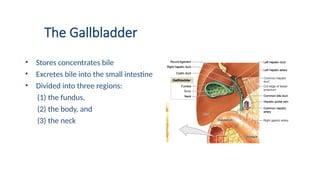
The Gallbladder
• Storesconcentrates bile
• Excretes bile into the small intestine
• Divided into three regions:
(1) the fundus,
(2) the body, and
(3) the neck
35.
The Gallbladder
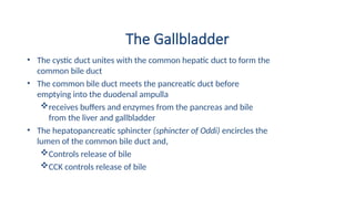
• Thecystic duct unites with the common hepatic duct to form the
common bile duct
• The common bile duct meets the pancreatic duct before
emptying into the duodenal ampulla
receives buffers and enzymes from the pancreas and bile
from the liver and gallbladder
• The hepatopancreatic sphincter (sphincter of Oddi) encircles the
lumen of the common bile duct and,
Controls release of bile
CCK controls release of bile
36.
The Gallbladder
• Thegallbladder also functions in bile modification
• The composition of bile gradually changes as it remains in the gallbladder:
Much of the water is absorbed
the bile salts and other components of bile become increasingly concentrated
crystals of insoluble minerals and salts begin to form
These deposits are called gallstones
Large gallstones can damage the wall of the gallbladder or block the cystic duct or
common bile duct
ocholecystitis
37.
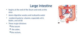
Large Intestine
• beginsat the end of the ileum and ends at the
anus
• stores digestive wastes and reabsorbs water
• resident bacteria- vitamin, especially vit K,
biotin, and vit B5
• Three major divisions
the cecum
the colon,
the rectum,
38.
Large Intestine
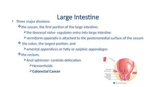
• Threemajor divisions
the cecum, the first portion of the large intestine;
the ileocecal valve- regulates entry into large intestine
vermiform appendix is attached to the posteromedial surface of the cecum
the colon, the largest portion; and
omental appendices or fatty or epiploic appendages
the rectum,
Anal sphincter- controls defecation
Hemorrhoids
Colorectal Cancer


![CTEV [ clubfoot] DR ARUN LAL ,DR MOHAMED ASHRAF travancore medical college k...](https://cdn.slidesharecdn.com/ss_thumbnails/ctevclubfootdrarunlaldrmohamedashraftravancoremedicalcollegekollamkeralaindia-260208063247-18fc466c-thumbnail.jpg?width=640&height=640&fit=bounds)


