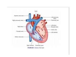
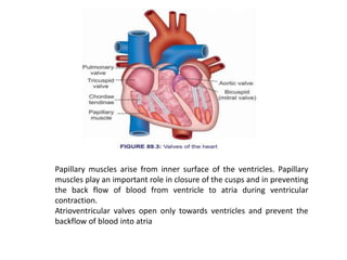
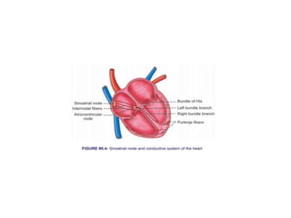
The heart is a muscular organ that pumps blood through the circulatory system. It has four chambers - two upper atria and two lower ventricles. The right side receives deoxygenated blood and pumps it to the lungs, while the left side receives oxygenated blood from the lungs and pumps it out to the body. The heart's rhythm is controlled by the sinoatrial node, while electrical signals are conducted through the atrioventricular node and Purkinje fibers to coordinate contractions. Valves ensure blood flows in only one direction through the heart and vessels.
Cardiovascular physiology for university studentsItsOnyii
A detailed pdf document on cardiovascular physiology for university students including structure and functions of heart, Electrocardiogram, echocardiography, chest and limb leads, Diseases and disorders of the heart.
This presentation is a combination of different slides which I re-purposed. I included a reference of all the slides I used at the end of my presentation.
A powerpoint designed for the South African Life Sciences syllabus for grade 11. Includes information about blood and it's transportation, the human heart, the lymph system etc. Hope it helps :)
CARDIO VASCULAR SYSTEM.pdf for bsc nursing studentsshanmukhadevi
The cardiovascular system refers to the heart, blood vessels and the blood.
Blood contains oxygen and other nutrients, which your body needs to survive. The body takes these essential nutrients from the blood.
At the same time, the body dumps waste products like carbon dioxide, back into the blood, so they can be removed.
The main function of the cardiovascular system is therefore to maintain blood flow to all parts of the body, to allow it to survive.
Veins deliver used blood from the body back to the heart. Blood in the veins is low in oxygen (as it has been taken out by the body) and high in carbon dioxide (as the body has unloaded it back into the blood).
All the veins drain into the superior and inferior vena cava, which then drain into the right atrium.
The right atrium pumps blood into the right ventricle. Then the right ventricle pumps blood to the pulmonary trunk, through the pulmonary arteries and into the lungs.
In the lungs the blood picks up oxygen that we breathe in and gets rid of carbon dioxide, which we breathe out. The blood is becomes rich in oxygen, which the body can use.
From the lungs, blood drains into the left atrium and is then pumped into the left ventricle. The left ventricle then pumps this oxygen-rich blood out into the aorta, which then distributes it to the rest of the body through other arteries.
This blood will again return back to the heart through the veins and the cycle continues.
The cardiovascular system can be thought of as the transport system of the body.
This system has three main components: the heart, the blood vessel and the blood itself.
The heart is the system's pump and the blood vessels are like the delivery routes.
Blood can be thought of as a fluid, which contains the oxygen and nutrients the body needs and carries the wastes, which need to be removed.
Structure & Function of Heart and its parts. Heart walls, pericardium, heart valves, septa, nodal tissues, coronary circulation, blood vessels of heart, AV bundle, bundle of his, purkinje fibers, myogenic nature of heart, action potential generation.
This presentation provides a clear understanding of the physiology of the circulatory system. It focus lies on the division and component of the circulatory system, the three major function of the circulatory system, blood composition, structure of the heart, blood circulation; pulmonary and systemic circuit, valves of the heart, the pathway of blood flow through the heart, the cardiac cycle, pressure changes during the cardiac cycle; systole and diastole, cardiac output, heart sounds among others.
This presentation was designed by Fasama H. Kollie and presented by Benetta N. Kekulah, Cordelia Capehart and Abraham Peters.
Factory Supply Best Quality Pmk Oil CAS 28578–16–7 PMK Powder in Stockrebeccabio
Factory Supply Best Quality Pmk Oil CAS 28578–16–7 PMK Powder in Stock
Telegram: bmksupplier
signal: +85264872720
threema: TUD4A6YC
You can contact me on Telegram or Threema
Communicate promptly and reply
Free of customs clearance, Double Clearance 100% pass delivery to USA, Canada, Spain, Germany, Netherland, Poland, Italy, Sweden, UK, Czech Republic, Australia, Mexico, Russia, Ukraine, Kazakhstan.Door to door service
Hot Selling Organic intermediates
Explore natural remedies for syphilis treatment in Singapore. Discover alternative therapies, herbal remedies, and lifestyle changes that may complement conventional treatments. Learn about holistic approaches to managing syphilis symptoms and supporting overall health.
Report Back from SGO 2024: What’s the Latest in Cervical Cancer?bkling
Are you curious about what’s new in cervical cancer research or unsure what the findings mean? Join Dr. Emily Ko, a gynecologic oncologist at Penn Medicine, to learn about the latest updates from the Society of Gynecologic Oncology (SGO) 2024 Annual Meeting on Women’s Cancer. Dr. Ko will discuss what the research presented at the conference means for you and answer your questions about the new developments.
These simplified slides by Dr. Sidra Arshad present an overview of the non-respiratory functions of the respiratory tract.
Learning objectives:
1. Enlist the non-respiratory functions of the respiratory tract
2. Briefly explain how these functions are carried out
3. Discuss the significance of dead space
4. Differentiate between minute ventilation and alveolar ventilation
5. Describe the cough and sneeze reflexes
Study Resources:
1. Chapter 39, Guyton and Hall Textbook of Medical Physiology, 14th edition
2. Chapter 34, Ganong’s Review of Medical Physiology, 26th edition
3. Chapter 17, Human Physiology by Lauralee Sherwood, 9th edition
4. Non-respiratory functions of the lungs https://academic.oup.com/bjaed/article/13/3/98/278874
MANAGEMENT OF ATRIOVENTRICULAR CONDUCTION BLOCK.pdfJim Jacob Roy
Cardiac conduction defects can occur due to various causes.
Atrioventricular conduction blocks ( AV blocks ) are classified into 3 types.
This document describes the acute management of AV block.
Ethanol (CH3CH2OH), or beverage alcohol, is a two-carbon alcohol
that is rapidly distributed in the body and brain. Ethanol alters many
neurochemical systems and has rewarding and addictive properties. It
is the oldest recreational drug and likely contributes to more morbidity,
mortality, and public health costs than all illicit drugs combined. The
5th edition of the Diagnostic and Statistical Manual of Mental Disorders
(DSM-5) integrates alcohol abuse and alcohol dependence into a single
disorder called alcohol use disorder (AUD), with mild, moderate,
and severe subclassifications (American Psychiatric Association, 2013).
In the DSM-5, all types of substance abuse and dependence have been
combined into a single substance use disorder (SUD) on a continuum
from mild to severe. A diagnosis of AUD requires that at least two of
the 11 DSM-5 behaviors be present within a 12-month period (mild
AUD: 2–3 criteria; moderate AUD: 4–5 criteria; severe AUD: 6–11 criteria).
The four main behavioral effects of AUD are impaired control over
drinking, negative social consequences, risky use, and altered physiological
effects (tolerance, withdrawal). This chapter presents an overview
of the prevalence and harmful consequences of AUD in the U.S.,
the systemic nature of the disease, neurocircuitry and stages of AUD,
comorbidities, fetal alcohol spectrum disorders, genetic risk factors, and
pharmacotherapies for AUD.
TEST BANK for Operations Management, 14th Edition by William J. Stevenson, Ve...kevinkariuki227
TEST BANK for Operations Management, 14th Edition by William J. Stevenson, Verified Chapters 1 - 19, Complete Newest Version.pdf
Anti ulcer drugs and their Advance pharmacology ||
Anti-ulcer drugs are medications used to prevent and treat ulcers in the stomach and upper part of the small intestine (duodenal ulcers). These ulcers are often caused by an imbalance between stomach acid and the mucosal lining, which protects the stomach lining.
||Scope: Overview of various classes of anti-ulcer drugs, their mechanisms of action, indications, side effects, and clinical considerations.
- Video recording of this lecture in English language: https://youtu.be/lK81BzxMqdo
- Video recording of this lecture in Arabic language: https://youtu.be/Ve4P0COk9OI
- Link to download the book free: https://nephrotube.blogspot.com/p/nephrotube-nephrology-books.html
- Link to NephroTube website: www.NephroTube.com
- Link to NephroTube social media accounts: https://nephrotube.blogspot.com/p/join-nephrotube-on-social-media.html
Title: Sense of Taste
Presenter: Dr. Faiza, Assistant Professor of Physiology
Qualifications:
MBBS (Best Graduate, AIMC Lahore)
FCPS Physiology
ICMT, CHPE, DHPE (STMU)
MPH (GC University, Faisalabad)
MBA (Virtual University of Pakistan)
Learning Objectives:
Describe the structure and function of taste buds.
Describe the relationship between the taste threshold and taste index of common substances.
Explain the chemical basis and signal transduction of taste perception for each type of primary taste sensation.
Recognize different abnormalities of taste perception and their causes.
Key Topics:
Significance of Taste Sensation:
Differentiation between pleasant and harmful food
Influence on behavior
Selection of food based on metabolic needs
Receptors of Taste:
Taste buds on the tongue
Influence of sense of smell, texture of food, and pain stimulation (e.g., by pepper)
Primary and Secondary Taste Sensations:
Primary taste sensations: Sweet, Sour, Salty, Bitter, Umami
Chemical basis and signal transduction mechanisms for each taste
Taste Threshold and Index:
Taste threshold values for Sweet (sucrose), Salty (NaCl), Sour (HCl), and Bitter (Quinine)
Taste index relationship: Inversely proportional to taste threshold
Taste Blindness:
Inability to taste certain substances, particularly thiourea compounds
Example: Phenylthiocarbamide
Structure and Function of Taste Buds:
Composition: Epithelial cells, Sustentacular/Supporting cells, Taste cells, Basal cells
Features: Taste pores, Taste hairs/microvilli, and Taste nerve fibers
Location of Taste Buds:
Found in papillae of the tongue (Fungiform, Circumvallate, Foliate)
Also present on the palate, tonsillar pillars, epiglottis, and proximal esophagus
Mechanism of Taste Stimulation:
Interaction of taste substances with receptors on microvilli
Signal transduction pathways for Umami, Sweet, Bitter, Sour, and Salty tastes
Taste Sensitivity and Adaptation:
Decrease in sensitivity with age
Rapid adaptation of taste sensation
Role of Saliva in Taste:
Dissolution of tastants to reach receptors
Washing away the stimulus
Taste Preferences and Aversions:
Mechanisms behind taste preference and aversion
Influence of receptors and neural pathways
Impact of Sensory Nerve Damage:
Degeneration of taste buds if the sensory nerve fiber is cut
Abnormalities of Taste Detection:
Conditions: Ageusia, Hypogeusia, Dysgeusia (parageusia)
Causes: Nerve damage, neurological disorders, infections, poor oral hygiene, adverse drug effects, deficiencies, aging, tobacco use, altered neurotransmitter levels
Neurotransmitters and Taste Threshold:
Effects of serotonin (5-HT) and norepinephrine (NE) on taste sensitivity
Supertasters:
25% of the population with heightened sensitivity to taste, especially bitterness
Increased number of fungiform papillae
2. HEART
• Heart is a muscular organ that pumps blood
throughout the circulatory system.
• It is situated in between two lungs in the
mediastinum.
• It is made up of four chambers, two atria and
two ventricles.
• The musculature of ventricles is thicker than
that of atria. Force of contraction of heart
depends upon the muscles.
3.
4. RIGHT SIDE OF THE HEART
• Right side of the heart has two chambers, right atrium
and right ventricle.
• Right atrium is a thin walled and low pressure chamber.
It has got the pacemaker known as sinoatrial node that
produces cardiac impulses and atrioventricular node
that conducts the impulses to the ventricles.
• Right atrium receives venous (deoxygenated) blood via
two large veins:
• 1. Superior vena cava that returns venous blood from
the head, neck and upper limbs
• 2. Inferior vena cava that returns venous blood from
lower parts of the body.
5. Right atrium communicates with right ventricle
through tricuspid valve.
Wall of right ventricle is thick. Venous blood
from the right atrium enters the right ventricle
through this valve.
From the right ventricle, pulmonary artery
arises. It carries the venous blood from right
ventricle to lungs. In the lungs, the
deoxygenated blood is oxygenated.
6. LEFT SIDE OF THE HEART
• Left side of the heart has two chambers, left
atrium and left ventricle.
• Left atrium is a thin walled and low pressure
chamber. It receives oxygenated blood from the
lungs through pulmonary veins.
• This is the only exception in the body, where an
artery carries venous blood and vein carries the
arterial blood. Blood from left atrium enters the
left ventricle through mitral valve (bicuspid
valve).
• Wall of the left ventricle is very thick. Left
ventricle pumps the arterial blood to different
parts of the body through systemic aorta.
7. SEPTA OF THE HEART
• Right and left atria are separated from one
another by a fibrous septum called interatrial
septum.
• Right and left ventricles are separated from
one another by interventricular septum. The
upper part of this septum is a membranous
structure, whereas the lower part of it is
muscular in nature
8. VALVES OF THE HEART
There are four valves in human heart.
• Two valves are in between atria and the
ventricles called atrioventricular valves.
• Other two are the semilunar valves, placed at
the opening of blood vessels arising from
ventricles, namely systemic aorta and
pulmonary artery.
• Valves of the heart permit the flow of blood
through heart in only one direction
9. Atrioventricular Valves
• Left atrioventricular valve is otherwise known
as mitral valve or bicuspid valve. It is formed
by two valvular cusps or flaps.
• Right atrioventricular valve is known as
tricuspid valve and it is formed by three cusps.
• Brim of the atrioventricular valves is attached
to atrioventricular ring, which is the fibrous
connection between the atria and ventricles.
• Cusps of the valves are attached to papillary
muscles by means of chordae tendineae.
10. Papillary muscles arise from inner surface of the ventricles. Papillary
muscles play an important role in closure of the cusps and in preventing
the back flow of blood from ventricle to atria during ventricular
contraction.
Atrioventricular valves open only towards ventricles and prevent the
backflow of blood into atria
11. • Semilunar Valves
Semilunar valves are present at the openings of
systemic aorta and pulmonary artery and are
known as aortic valve and pulmonary valve
respectively.
Because of the half moon shape, these two valves
are called semilunar valves. Semilunar valves are
made up of three flaps.
Semilular valves open only towards the aorta and
pulmonary artery and prevent the backflow of
blood into the ventricles
12.
13.
14.
15.
16.
17.
18. Properties of Cardiac Muscle
• EXCITABILITY „
• Excitability is defined as the ability of a living tissue to give
response to a stimulus. In all the tissues, initial response to
a stimulus is electrical activity in the form of action
potential. It is followed by mechanical activity in the form
of contraction, secretion, etc.
• Action Potential:
• Action potential in cardiac muscle is different from that of
other tissues such as skeletal muscle, smooth muscle and
nervous tissue. Duration of the action potential in cardiac
muscle is 250 to 350 msec (0.25 to 0.35 sec).
• Phases of action potential Action potential in a single
cardiac muscle fiber occurs in four phases:
• 1. Initial depolarization 2. Initial repolarization 3. A plateau
or final depolarization 4. Final repolarization
19. • 1. Initial Depolarization Initial depolarization is
very rapid and it lasts for about 2 msec (0.002
sec). Amplitude of depolarization is about + 20
mV
IONIC BASIS
• 1. Initial Depolarization Initial depolarization (first
phase) is because of rapid opening of fast sodium
channels and the rapid influx of sodium ions, as in
the case of skeletal muscle fiber suddenly and
slow sodium channels open, resulting in slow
influx of low quantity of sodium ions
20. • IONIC BASIS
• 1. Initial Depolarization Initial depolarization
(first phase) is because of rapid opening of fast
sodium channels and the rapid influx of sodium
ions, as in the case of skeletal muscle fiber
• 2. Initial Repolarization Immediately after
depolarization, there is an initial rapid
repolarization for a short period of about 2
msec. The end of rapid repolarization is
represented by a notch.
21. 3. Plateau or Final Depolarization
Afterwards, the muscle fiber remains in
depolarized state for sometime before further
repolarization. It forms the plateau (stable
period) in action potential curve.
The plateau lasts for about 200 msec in atrial
muscle fibers and for about 300 msec in
ventricular muscle fibers.
Due to long plateau in action potential, the
contraction time is also longer in cardiac muscle
by 5 to 15 times than in skeletal muscle.
22. • Plateau is due to the slow opening of calcium
channels. These channels are kept open for a
longer period and cause influx of large number of
calcium ions. Already the slow sodium channels
are opened, through which slow influx of sodium
ions continues. Because of the entry of calcium
and sodium ions into the muscle fiber, positivity is
maintained inside the muscle fiber, producing
prolonged depolarization, i.e. plateau. Calcium
ions entering the muscle fiber play an important
role in the contractile process
23. • 4. Final Repolarization
• Final repolarization occurs after the plateau. It is
a slow process and it lasts for about 50 to 80
msec before the re-establishment of resting
membrane potential
• Final repolarization is due to efflux of potassium
ions. Number of potassium ions moving out of
the muscle fiber exceeds the number of calcium
ions moving in. It makes negativity inside,
resulting in final repolarization. Potassium efflux
continues until the end of repolarization.
24. • RHYTHMICITY „
• Rhythmicity is the ability of a tissue to
produce its own impulses regularly. It is also
called autorhythmicity or self-excitation.
Property of rhythmicity is present in all the
tissues of heart. However, heart has a
specialized excitatory structure, from which
the discharge of impulses is rapid. This
specialized structure is called pacemaker.
From here, the impulses spread to other parts
through the specialized conductive system.
25. CONDUCTIVITY
• Human heart has a specialized conductive
system, through which impulses from SA node
are transmitted to all other parts of the heart
26. CONDUCTIVE SYSTEM IN HUMAN
HEART
• Conductive system of the heart is formed by the
modified cardiac muscle fibers. These fibers are
the specialized cells, which conduct the impulses
rapidly from SA node to the ventricles.
Conductive tissues of the heart are also called the
junctional tissues.
• Components of Conductive System in Human
Heart
• 1. AV node 2. Bundle of His 3. Right and left
bundle branches 4. Purkinje fibers
27.
28. • SA node is situated in right atrium, just below
the opening of superior vena cava.
• AV node is situated in right posterior portion
of intra-atrial septum. Impulses from SA node
are conducted throughout right and left atria.
Impulses also reach the AV node via some
specialized fibers called internodal fibers.
29. • There are three types of internodal fibers: 1.
Anterior internodal fibers of Bachman 2. Middle
internodal fibers of Wenckebach 3. Posterior
internodal fibers of Thorel.
• All these fibers from SA node converge on AV
node and interdigitate with fibers of AV node.
From AV node, the bundle of His arises.
• It divides into right and left branches, which run
on either side of the interventricular septum.
From each branch of bundle of His, many Purkinje
fibers arise and spread all over the ventricular
myocardium
33. Cardiac Cycle
• Cardiac cycle is defined as the succession of
(sequence of) coordinated events taking place in
the heart during each beat.
• Each heartbeat consists of two major periods
called systole and diastole.
• During systole, heart contracts and pumps the
blood through arteries.
• During diastole, heart relaxes and blood is filled
in the heart. All these changes are repeated
during every heartbeat, in a cyclic manner.
34.
35. EVENTS OF CARDIAC CYCLE
Events of cardiac cycle are classified into two:
1. Atrial events 2. Ventricular events. „
• DIVISIONS AND DURATION OF CARDIAC CYCLE
When the heart beats at a normal rate of 72duration of each cardiac cycle is about 0.8
second. „
/minute
ATRIAL EVENTS
Atrial events are divided into two divisions: 1. Atrial systole = 0.11 (0.1) sec
2. Atrial diastole = 0.69 (0.7) sec. „
VENTRICULAR EVENTS
Ventricular events are divided into two divisions:
1. Ventricular systole = 0.27 (0.3) sec
2. Ventricular diastole = 0.53 (0.5) sec.
In clinical practice, the term ‘systole’ refers to ventricular systole and ‘diastole’ refers
to ventricular diastole. Ventricular systole is divided into two subdivisions and
ventricular diastole is divided into five subdivisions. Ventricular Systole Time
(second)
1. Isometric contraction = 0.05 2. Ejection period = 0.22 0.27 Ventricular
Diastole 1. Protodiastole = 0.04
2. Isometric relaxation = 0.08
3. Rapid filling = 0.11
4. Slow filling = 0.19
5. Last rapid filling = 0.1
36. DESCRIPTION OF ATRIAL EVENTS
1. ATRIAL SYSTOLE
• Atrial systole is also known as last rapid filling phase or
presystole.
• It is usually considered as the last phase of ventricular
diastole. Its duration is 0.11 second.
• During this period, only a small amount, i.e. 10% of
blood is forced from atria into ventricles.
• Atrial systole is not essential for the maintenance of
circulation. Many persons with atrial fibrillation survive
for years, without suffering from circulatory
insufficiency. However, such persons feel difficult to
cope up with physical stress like exercise.
37. 2. ATRIAL DIASTOLE
• After atrial systole, the atrial diastole starts.
Simultaneously, ventricular systole also starts.
Atrial diastole lasts for about 0.7 sec (accurate
duration is 0.69 sec).
• This long atrial diastole is necessary because, this
is the period during which atrial filling takes
place.
• Right atrium receives deoxygenated blood from
all over the body through superior and inferior
venae cavae. Left atrium receives oxygenated
blood from lungs through pulmonary veins.
38. DESCRIPTION OF VENTRICULAR
EVENTS
„
3. ISOMETRIC CONTRACTION PERIOD
• Isometric contraction period in cardiac cycle
is the first phase of ventricular systole.
• It lasts for 0.05 second. Isometric contraction
is the type of muscular contraction
characterized by increase in tension, without
any change in the length of muscle fibers.
Isometric contraction of ventricular muscle is
also called isovolumetric contraction.
39. „4. EJECTION PERIOD
• Due to the opening of semilunar valves and isotonic contraction of
ventricles, blood is ejected out of both the ventricles. Hence, this
period is called ejection period. Duration of this period is 0.22
second.
Ejection period is of two stages:
A. First Stage or Rapid Ejection Period : First stage starts immediately
after the opening of semilunar valves. During this stage, a large
amount of blood is rapidly ejected from both the ventricles. It lasts
for 0.13 second.
B. Second Stage or Slow Ejection Period: During this stage, the blood is
ejected slowly with much less force. Duration of this period is 0.09
second.
End-systolic Volume: Ventricles are not emptied at the end of ejection
period and some amount of blood remains in each ventricle.
Amount of blood remaining in ventricles at the end of called end
systolic volume. It is 60 to 80 mL per ventricle.
Ejection Fraction
Ejection fraction refers to the fraction (or portion) of end diastolic
volume that is ejected out by each ventricle per beat.
40.
41. 5. PROTODIASTOLE
• Protodiastole is the first stage of ventricular
diastole, hence the name protodiastole.
Duration of this period is 0.04 second.
• Due to the ejection of blood, the pressure in
aorta and pulmonary artery increases and
pressure in ventricles drops.
42. 6. ISOMETRIC RELAXATION PERIOD
• Isometric relaxation is the type of muscular relaxation,
characterized by decrease in tension without any change in
the length of muscle fibers.
• Isometric relaxation of ventricular muscle is also called
isovolumetric relaxation.
• During isometric relaxation period, once again all the valves
of the heart are closed.
• Now, both the ventricles relax as closed cavities without
any change in volume or length of the muscle fiber.
• Intraventricular pressure decreases during this period.
Duration of isometric relaxation period is 0.08 second
43. 7. RAPID FILLING PHASE
When atrionventricular valves are opened,
there is a sudden rush of blood (which is
accumulated in atria during atrial diastole)
from atria into ventricles. So, this period is
called the first rapid filling period. Ventricles
also relax isotonically. About 70% of filling
takes place during this phase, which lasts for
0.11 second.
44. 8. SLOW FILLING PHASE
• After the sudden rush of blood, the ventricular filling
becomes slow. Now, it is called the slow filling. It is also
called diastasis. About 20% of filling occurs in this
phase. Duration of slow filling phase is 0.19 second.
9. „LAST RAPID FILLING PHASE
• Last rapid filling phase occurs because of atrial systole.
After slow filling period, the atria contract and push a
small amount of blood into ventricles. About 10% of
ventricular filling takes place during this period. Flow of
additional amount of blood into ventricle due to atrial
systole is called atrial kick. End-diastolic Volume End-
diastolic volume is the amount of blood remaining in
each ventricle at the end of diastole. It is about 130 to
150 mL per ventricle.
47. Questions
• 1. Explain in detail about structure and
functions of heart with the help of neat
diagram.
• 2.What are components in conducting system
of the heart. How the conduction of impulses
takes place in heart.
• ADOBE SCANER
• Mailid:dskphcol@gmail.com