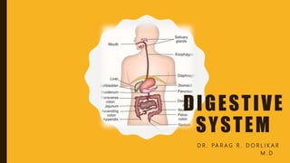
Gastrointestinal Tract By Dr. Parag Dorlikar M.D
•Download as PPTX, PDF•
2 likes•155 views

This document provides information on the digestive system. It begins with an overview of digestion and the functions of the digestive system. It then describes the functional anatomy of the digestive system, including the primary and accessory digestive organs. The document focuses on the mouth and salivary glands, providing details on their anatomy, functions of saliva, and properties of saliva. It also discusses the stomach, including the anatomy and functions of the stomach and gastric juice. The document concludes with information on mechanisms of gastric secretion and an overview of gastritis.

Recommended
More Related Content
What's hot
What's hot (20)
Similar to Gastrointestinal Tract By Dr. Parag Dorlikar M.D
Similar to Gastrointestinal Tract By Dr. Parag Dorlikar M.D (20)
Recently uploaded
Recently uploaded (20)
Gastrointestinal Tract By Dr. Parag Dorlikar M.D
- 1. DIGESTIVE SYSTEM D R . PA R A G R . D O R L I K A R M . D
- 2. DIGESTION • Digestion is defined as the process by which food is broken down into simple chemical substances that can be absorbed and used as Important Nutrients by the body. • Digestive process is done by Mechanical and Enzymatic breakdown of food into simple chemical compounds. Thus, the functions of digestive system include: • 1. Ingestion or consumption of food substances. • 2. Break down of food into small particles. • 3. Transport of small particles to different areas of the digestive tract • 4. Secretion of necessary enzymes and other substances for digestion • 5. Digestion of the food particles • 6. Absorption of the digestive products (nutrients) • 7. Removal of unwanted substances from the body.
- 3. FUNCTIONAL ANATOMY OF DIGESTIVE SYSTEM • Digestive system is made up of Gastrointestinal tract (GI tract) or Alimentary canal and accessory organs, which help in the process of digestion and absorption. • GI tract is formed by two types of organs: • 1. Primary digestive organs. Primary digestive organs are the organs where actual digestion takes place. Primary digestive organs are: i. Mouth ii. Pharynx iii. Esophagus iv. Stomach v. Small intestine
- 4. • 2. Accessory digestive organs: • Those organs which helps primary digestive organs in the process of digestion. • Accessory digestive organs are: i. Teeth ii. Tongue iii. Salivary glands iv. Exocrine part of pancreas v. Liver vi. Gallbladder
- 5. MOUTH AND SALIVARY GLANDS • FUNCTIONAL ANATOMY OF MOUTH : • Mouth is also known as oral cavity or buccal cavity. It is formed by cheeks, lips and palate. It encloses the teeth, tongue and salivary glands. • FUNCTIONS OF MOUTH • Functions of mouth include: 1. Ingestion of food materials 2. Chewing the food and mixing it with saliva 3. Stimulation for taste of the food 4. Transfer of food (bolus) to the esophagus by swallowing 5. Role in speech 6. Communicative functions such as expressions.
- 6. SALIVARY GLANDS • In humans, the saliva is secreted by three pairs of major (larger) salivary glands and some minor (small) salivary glands. • Major glands are: 1. Parotid glands 2. Submaxillary or submandibular glands 3. Sublingual glands. • Minor glands are: 1. Lingual Mucus Glands 2. Lingual Serous Glands 3. Buccal Glands 4. Labial Glands 5. Palatal Glands
- 7. PROPERTIES AND COMPOSITION OF SALIVA • PROPERTIES OF SALIVA: • Volume: 1000 mL to 1500 mL of saliva is secreted per day and it is approximately about 1 mL/minute. • Reaction: Mixed saliva from all the glands is slightly acidic with pH of 6.35 to 6.85 • Specific gravity: It ranges between 1.002 and 1.012 • Tonicity: Saliva is hypotonic to plasma.
- 9. FUNCTIONS OF SALIVA • PREPARATION OF FOOD FOR SWALLOWING : • When food is taken into the mouth, it is moistened and dissolved by saliva. • The mucus membrane of mouth is also moistened by saliva. It facilitates chewing. • By the movement of tongue, the moistened and masticated food is rolled into a bolus.
- 10. PERCEPTION OF TASTE • Taste is a chemical sensation. • By its solvent action, saliva dissolves the solid food substances, so that the dissolved substances can stimulate the taste buds. • The stimulated taste buds recognize the taste of food.
- 11. DIGESTIVE FUNCTION • Saliva has three digestive enzymes, namely • Salivary Amylase • Maltase And • Lingual Lipase • Salivary Amylase: • Salivary amylase is a carbohydrate-digesting (amylolytic) enzyme. It acts on cooked or boiled starch and converts it into dextrin and maltose. • Maltase • Maltase is present only in traces in human saliva and it converts maltose into glucose. • Lingual lipase • Lingual lipase is a lipid-digesting (lipolytic) enzyme. It is secreted from serous glands situated on the posterior aspect of tongue.
- 13. CLARENCE AND PROTECTIVE FUNCTIONS • Due to the constant secretion of saliva, the mouth and teeth are rinsed and kept free off food debris, abnormal epithelial cells and foreign particles. • Enzyme lysozyme of saliva kills some bacteria such as staphylococcus, streptococcus and brucella by its phagocytic action. • Proline-rich proteins present in saliva posses antimicrobial property and neutralize the toxic substances. • Immunoglobulin IgA in saliva also has antibacterial and antiviral actions.
- 14. ROLE IN SPEECH • By moistening and lubricating soft parts of mouth and lips, saliva helps in speech. • If the mouth becomes dry (Hyposalivation), articulation and pronunciation becomes difficult.
- 15. EXCRETORY FUNCTION • Many substances, both organic and inorganic, are excreted in saliva. It excretes substances like mercury, potassium iodide, lead etc. • Saliva also excretes some viruses such as those causing rabies and mumps. • Excessive urea is excreted in saliva during nephritis and excess calcium is excreted during hyperparathyroidism.
- 16. REGULATION OF WATER BALANCE • When the body water content decreases, salivary secretion also decreases. This causes dryness of the mouth and induces thirst.
- 17. REGULATION OF SALIVARY SECRETION • Salivary secretion is regulated only by nervous mechanism. • Autonomic nervous system is involved in the regulation of salivary secretion. • Salivary glands are supplied by both parasympathetic and sympathetic divisions of autonomic nervous system.
- 18. APPLIED PHYSIOLOGY • HYPOSALIVATION • Reduction in the secretion of saliva is called hyposalivation. It is of two types, namely temporary hyposalivation and permanent hyposalivation. • 1. Temporary hyposalivation occurs in: • i. Emotional conditions like fear. • ii. Fever. • iii. Dehydration. • 2. Permanent hyposalivation occurs in: • i. Obstruction of salivary duct • ii. Congenital absence of salivary glands. • iii. Bells palsy (paralysis of facial nerve)
- 19. HYPERSALIVATION • Excess secretion of saliva is known as hypersalivation. • Hypersalivation occurs in the following pathological conditions: • 1. Decay of tooth or neoplasm (abnormal new growth or tumor) in mouth or tongue due to continuous irritation of nerve endings in the mouth. • 2. Disease of esophagus, stomach and intestine. • 3. Neurological disorders such as cerebral palsy, mental retardation, cerebral stroke and parkinsonism. • 4. Some psychological and psychiatric conditions. • 5. Nausea and vomiting.
- 20. STOMACH FUNCTIONAL ANATOMY OF STOMACH • Stomach is a hollow muscular organ located just below the diaphragm on the left side in the abdominal cavity. • Volume of empty stomach is 50 mL. Under normal conditions, it can expand to accommodate 1 L to 1.5 L of solids and liquids. • PARTS OF STOMACH • Stomach has four parts: • 1. Cardiac region • 2. Fundus • 3. Body or corpus • 4. Pyloric region.
- 22. GLANDS OF STOMACH • Glands of the stomach or gastric glands are tubular structures made up of different types of cells. • CLASSIFICATION OF GLANDS OF THE STOMACH • Gastric glands are classified into three types, on the basis of their location in the stomach: • 1. Fundic glands or main gastric gland: Situated in body and fundus of stomach • 2. Pyloric glands: Present in the pyloric part of the stomach • 3. Cardiac glands: Located in the cardiac region of the stomach.
- 23. FUNCTIONS OF GASTRIC GLANDS • Function of the gastric gland is to secrete gastric juice. • Secretory activities of different cells of gastric glands and enteroendocrine cells are as follows:
- 24. FUNCTIONS OF STOMACH • 1. MECHANICAL FUNCTION: • i. Storage Function: Food is stored in the stomach for a long period, i.e. for 3 to 4 hours and emptied into the intestine slowly. The maximum capacity of stomach is up to 1.5 L. • ii. Formation of Chyme: Peristaltic movements of stomach mix the bolus with gastric juice and convert it into the semisolid material known as chyme. • 2. DIGESTIVE FUNCTION • Gastric juice acts mainly on proteins. Proteolytic enzymes of the gastric juice are pepsin and rennin. Gastric juice also contains other enzymes like gastric lipase, gelatinase, urase and gastric amylase.
- 25. • 3. PROTECTIVE FUNCTION • Mucus is a mucoprotein, secreted by mucus cells of the gastric glands and surface mucus cells in fundus, body and other parts of stomach. • Mucus protects the stomach wall from irritation or mechanical injury. • It also protects the gastric mucosa from hydrochloric acid of gastric juice because of its alkaline nature.
- 26. 4. HEMOPOIETIC FUNCTION • Intrinsic factor of Castle, secreted by parietal cells of gastric glands in stomach plays an important role in erythropoiesis. • Intrinsic factor also necessary for the absorption of vitamin B12 (Extrinsic factor) from GI tract into the blood. • Vitamin B12 is an important maturation factor during erythropoiesis. • Absence of intrinsic factor in gastric juice causes deficiency of vitamin B12, leading to pernicious anemia
- 27. 5. EXCRETORY FUNCTION • Many unwanted substances like toxins, alkaloids and metals are excreted through gastric juice.
- 28. GASTRIC JUICE • PROPERTIES AND COMPOSITION OF GASTRIC JUICE • Gastric juice is a mixture of secretions from different gastric glands. • PROPERTIES : • Volume : 1200 mL/day to 1500 mL/day. • Reaction : Gastric juice is highly acidic with a pH of 0.9 to 1.2. Acidity of gastric juice is due to the presence of hydrochloric acid. • Specific gravity : 1.002 to 1.004
- 29. • COMPOSITION OF GASTRIC JUICE • Gastric juice contains 99.5% of water and 0.5% solids. • Solids are organic and inorganic substances.
- 30. FUNCTIONS OF GASTRIC JUICE • 1. DIGESTIVE FUNCTION • Gastric juice acts mainly on proteins. Proteolytic enzymes of the gastric juice are pepsin and rennin. • Gastric juice also contains some other enzymes like gastric lipase, gelatinase, urase and gastric amylase. • Pepsin • Pepsin is secreted as inactive pepsinogen. Pepsinogen is converted pepsin by hydrochloric acid. Optimum pH for activation of pepsinogen is below 6. • Action of pepsin • Pepsin converts proteins into proteoses, peptones and
- 31. • Gastric Lipase • Gastric lipase is a weak lipolytic enzyme when compared to pancreatic lipase. It is active only when the pH is between 4 and 5 and becomes inactive at a pH below 2.5. • Gastric lipase is mostly used for digestion of Lipids as it converts fats into Fatty acids and Glycerol. • Actions of Other Enzymes of Gastric Juice : • i . Gelatinase: It Degrades gelatin and collagen into peptides. • ii. Urase: Acts on urea and produces ammonia • iii. Gastric amylase: It Degrades starch • iv. Rennin: It Precipitate milk
- 32. • 2. HEMOPOIETIC FUNCTION • Intrinsic factor of Castle, secreted by parietal cells of gastric glands plays an important role in erythropoiesis. • 3. FUNCTIONS OF HYDROCHLORIC ACID • Hydrochloric acid is present in the gastric juice: • i. Activates pepsinogen into pepsin • ii. Kills some of the bacteria entering the stomach along with food substances. This action is called bacteriolytic action • iii. Provides acid medium, which is necessary for the action of
- 33. MECHANISM OF GASTRIC SECRETION • Secretion of gastric juice is a continuous process. But the quantity of gastric juice varies, depending upon Condition and stimulus. • Accordingly, gastric secretion occurs in three different phases: • I. Cephalic phase • II. Gastric phase • III. Intestinal phase • IV. Interdigestive phase (Phase of Secretion of Gastric Juice in between meals or During Fast)
- 34. CEPHALIC PHASE • Secretion of gastric juice by the stimuli arising from head region (cephalus) is called cephalic phase. • This phase of gastric secretion is regulated by nervous mechanism. The gastric juice secreted during this phase is called appetite juice. • During this phase, gastric secretion occurs even without the presence of food in stomach. • The quantity of the juice is less but it is rich in enzymes and hydrochloric acid. • Nervous mechanism regulates cephalic phase through reflex action. • Two types of reflexes occur: • 1. Unconditioned reflex • 2. Conditioned reflex.
- 35. food in the mouth stimulates the taste buds Sensory (afferent) impulses to nerve fibers of glossopharyngeal and facial nerves Stimulates Appetite Centre Stimulation of Vagus secrete acetylcholine, which stimulates gastric secretion. UNCONDITIONED REFLEX CONDITIONED REFLEX Impulses from the special sensory organs (eye, ear and nose) Thinking of food stimulates the cerebral cortex directly From cerebral cortex, the impulses pass through dorsal nucleus of vagus and vagal efferent and reach the stomach wall Vagal nerve endings secrete acetylcholine, which stimulates the gastric secretion.
- 37. GASTRIC PHASE • Secretion of gastric juice when food enters the stomach is called gastric phase. • This phase is regulated by both Nervous and Hormonal control. • Gastric juice secreted during this phase is rich in Pepsinogen & Hydrochloric acid. • Mechanisms involved in gastric phase are: • 1. Nervous mechanism through local myenteric reflex and vago-vagal reflex • 2. Hormonal mechanism through gastrin Stimuli, which initiate these two mechanisms are: • 1. Distention of stomach • 2. Mechanical stimulation of gastric mucosa by bulk of food. • 3. Chemical stimulation of gastric mucosa by the food contents.
- 38. INTESTINAL PHASE • Intestinal phase is the secretion of gastric juice when chyme enters the intestine. • When chyme enters the intestine, initially, the gastric secretion increases but later it stops. • Intestinal phase of gastric secretion is regulated by nervous and hormonal control. • Initial Stage of Intestinal Phase • Chyme that enters the intestine stimulates the duodenal mucosa to release gastrin, which is transported to stomach by blood. There it increases gastric secretion. • Later Stage of Intestinal Phase • After the initial increase, there is a decrease or complete stoppage of gastric secretion. • Gastric secretion is inhibited by two factors: • 1. Enterogastric reflex
- 39. INTERDIGESTIVE PHASE • Secretion of small amount of gastric juice in between meals (or during period of fasting) is called interdigestive phase. • Gastric secretion during this phase is mainly due to the hormones like gastrin.
- 40. APPLIED PHYSIOLOGY GASTRITIS • Inflammation of gastric mucosa is called gastritis. • It may be acute or chronic. • Acute gastritis is characterized by inflammation of superficial layers of mucus membrane and infiltration with leukocytes, mostly neutrophils. • Chronic gastritis involves inflammation of even the deeper and infiltration with more lymphocytes. • It results in the atrophy of the gastric mucosa, with loss of chief cells and parietal cells of glands. Therefore, the secretion of gastric juice decreases.
- 41. • Causes of Acute Gastritis • i. Infection with bacterium Helicobacter pylori • ii. Excess consumption of alcohol • iii. Excess administration of Aspirin and other nonsteroidal antiinflammatory drugs (NSAIDs) • iv. Trauma by nasogastric tubes • v. Repeated exposure to radiation (rare). • Causes of Chronic Gastritis • i. Chronic infection with Helicobacter pylori • ii. Long term intake of excess alcohol • iii. Long term use of NSAIDs • iv. Autoimmune disease.
- 42. • Clinical Features • Features of gastritis are nonspecific. • Common feature is abdominal upset or pain felt as a diffused burning sensation. • It is often referred to epigastric pain. • Other features are: • i. Nausea • ii. Vomiting • iii. Anorexia (loss of appetite) • iv. Indigestion • v. Discomfort or feeling of fullness in the epigastric region • vi. Belching (process to relieve swallowed air that is accumulated in stomach).
- 43. PANCREAS • FUNCTIONAL ANATOMY OF PANCREAS • Pancreas is a dual organ having two functions, namely • Endocrine function and Exocrine function. • Endocrine function is concerned with the production of hormones. • The Exocrine function is concerned with the secretion of digestive juice called pancreatic juice
- 44. PROPERTIES AND COMPOSITION OF PANCREATIC JUICE PROPERTIES OF PANCREATIC JUICE Volume : 500 to 800 mL/day Reaction : Highly alkaline with a pH of 8 to 8.3 Specific gravity : 1.010 to 1.018
- 45. COMPOSITION OF PANCREATIC JUICE
- 46. FUNCTIONS OF PANCREATIC JUICE • DIGESTIVE FUNCTIONS OF PANCREATIC JUICE • Pancreatic juice plays an important role in the digestion of proteins and lipids. It also has mild digestive action on carbohydrates. • 1) DIGESTION OF PROTEINS • Major proteolytic enzymes of pancreatic juice are trypsin and chymotrypsin. • Other proteolytic enzymes are - • Carboxypeptidases, • Nuclease, • Elastase and • Collagenase.
- 47. • 2) DIGESTION OF LIPIDS • Lipolytic enzymes present in pancreatic juice are • Pancreatic lipase, • Cholesterol ester hydrolase, • Phospholipase A, • Phospholipase B, • Colipase and • Bile-salt-activated lipase. • DIGESTION OF CARBOHYDRATES • Pancreatic amylase is the amylolytic enzyme present in pancreatic juice. • Like salivary amylase, the pancreatic amylase also converts starch into
- 48. • NEUTRALIZING ACTION OF PANCREATIC JUICE • When acid chyme enters intestine from stomach, pancreatic juice with large quantity of bicarbonate is released into intestine. • Presence of large quantity of bicarbonate ions makes the pancreatic juice highly alkaline. This alkaline pancreatic juice neutralizes acidity of chyme in the intestine. • Neutralizing action is an important function of pancreatic juice because it protects the intestine from the destructive action of acid in the chyme.
- 49. MECHANISM OF PANCREATIC SECRETION • SECRETION OF PANCREATIC ENZYMES • The exocrine pancreas secretes a variety of digestive enzymes specialized for digestion of all the different basic biological macromolecules. • Carbohydrates are digested by Pancreatic amylase. • Proteins are digested by trypsin, chymotrypsin, elastase, and carboxypeptidase. • Fats are digested by lipase, phospholipase, Cholesterol Esterase. • Nucleic Acids are digested by a variety of nucleases.
- 50. • Pancreatic enzymes are synthesized in ribosomes, which are attached to the endoplasmic reticulum of acinar cells in pancreas. • The raw materials for the synthesis of pancreatic enzymes are the amino acids, which are derived from the blood. • After synthesis, the enzymes are packed into different zymogen granules by Golgi apparatus and stored in cytoplasm. • When there is stimulation, the acinar cells release zymogen granules into the pancreatic duct. • From the granules the pancreatic enzymes are liberated into intestine.
- 51. • Pancreatic secretions are primarily regulated by three factors: • Acetylcholine: Released by vagal efferent, which primarily stimulates synthesis of digestive enzymes by pancreatic acinar cells. • Cholecystokinin: It is released by Cells in the duodenum and jejunum upon entry of food and primarily stimulates synthesis of digestive enzymes by pancreatic acinar cells. • Secretin: It is released by S Cells of duodenum in response to entry of low pH stomach acid and primarily stimulates production of aqueous sodium bicarbonate solution by pancreatic ductal cells.
- 52. REGULATION OF PANCREATIC SECRETION • Secretion of pancreatic juice is regulated by both nervous and hormonal factors. • STAGES OF PANCREATIC SECRETION : • Pancreatic juice is secreted in three stages like the gastric juice: • 1. Cephalic phase • 2. Gastric phase • 3. Intestinal phase.
- 53. CEPHALIC PHASE • As in case of gastric secretion, cephalic phase is regulated by nervous mechanism through reflex action. • Two types of reflexes occur: • 1. Unconditioned reflex • 2. Conditioned reflex. • 1.UNCONDITIONED REFLEX. • Unconditioned reflex is the inborn reflex. • When food is placed in the mouth, salivary secretion and gastric secretion are induced. Simultaneously, pancreatic secretion also occurs.
- 54. • CONDITIONED REFLEX • Conditioned reflex is the reflex response acquired by experience. • Presence of food in the mouth is not necessary to elicit this reflex. • The sight, smell, hearing or thought of food, which induce salivary secretion and gastric secretion induce pancreatic secretion also.
- 55. GASTRIC PHASE • Secretion of pancreatic juice when food enters the stomach is known as gastric phase. • This phase of pancreatic secretion is under hormonal control. • The hormone gastrin is involved in secretion of pancreatic juice in stomach. • When food enters the stomach, gastrin is secreted from stomach. • When gastrin is transported to pancreas through blood, it stimulates the pancreatic secretion. • The pancreatic juice secreted during gastric phase is rich in enzymes.
- 56. INTESTINAL PHASE • Intestinal phase is the secretion of pancreatic juice when the chyme enters the intestine. • This phase is also under hormonal control. • When chyme enters the intestine, many hormones are released. • Some hormones stimulate the pancreatic secretion and some hormones inhibit the pancreatic secretion.
- 58. • HORMONES STIMULATING PANCREATIC SECRETION • I . Secretin • II. Cholecystokinin (CCK) • HORMONES INHIBITING PANCREATIC SECRETION • I. Pancreatic polypeptide (PP) • II. Somatostatin • III. Peptide YY
- 59. APPLIED PHYSIOLOGY PANCREATITIS • Pancreatitis is the inflammation of pancreatic acini. • It is a rare but dangerous disease. • It is of two types: • 1. Acute pancreatitis • 2. Chronic pancreatitis.
- 60. ACUTE PANCREATITIS • Acute pancreatitis is more severe and it occurs because of heavy alcohol intake or gallstones. • Features of acute pancreatitis: • i. Abdominal pain is the major symptom of acute pancreatitis. • Pain may vary from a mild and tolerable discomfort to severe, constant. Characteristically, the pain, which is steady and boring in character, it is located in the epigastrium and periumbilical region and often radiates to the back as well as to the chest, flanks, and lower abdomen. • ii. Nausea and vomiting • iii. Loss of appetite and Loss of weight • iv. Fever • v. Shock
- 61. CHRONIC PANCREATITIS • Chronic pancreatitis develops due to repeated acute inflammation or chronic damage to pancreas. • Causes of chronic pancreatitis • i. Chronic alcoholism • ii. Chronic obstruction of ampulla of Vater by gallstone • iii. Hereditary cause (passed on genetically from one generation to another) • iv. Congenital abnormalities of pancreatic duct • v. Cystic fibrosis, a generalized disorder affecting the functions of many organs such as lungs (due to excessive fibrin), exocrine glands like pancreas, biliary system and immune system • vi. Malnutrition • vii. Idiopathic pancreatitis (due to unknown cause).
- 62. • Clinical Features of chronic pancreatitis: • Complete destruction of pancreas • Absence of pancreatic enzymes • Severe pain in upper abdominal region, which radiates to back • Fever • Nausea and vomiting • Tender and swollen abdomen • Weight loss.
- 63. STEATORRHEA • Steatorrhea is the formation of bulky, foul smelling, frothy and clay colored stools with large quantity of undigested fat because of impaired digestion and absorption of fat. • Causes of Steatorrhea • Any condition that causes indigestion or malabsorption of fat leads to steatorrhea. • Various causes of steatorrhea are: • 1. Lack of pancreatic lipase: Since most of the fat is digested only by pancreatic lipase, its deficiency leads to steatorrhea • 2. Liver disease affecting secretion of bile: Bile salts are essential for the digestion of fat by lipase and absorption of fat from intestine. Absence of bile salts results in excretion of fatty stool • 3. Celiac disease: Atrophy of intestinal villi leads to malabsorption, resulting in steatorrhea • 4. Cystic fibrosis (see above).
- 64. BILE JUICE • PROPERTIES AND COMPOSITION OF BILE • PROPERTIES OF BILE • Volume : 800 to 1,200 mL/day • Reaction : Alkaline • pH : 8 to 8.6 • Specific gravity : 1.010 to 1.011 • Color : Golden yellow or green
- 65. COMPOSITION OF BILE • Bile contains 97.6% of water and 2.4% of solids. Solids include organic and inorganic substances.
- 66. SECRETION OF BILE • Bile is secreted by hepatocytes. • The initial bile secreted by hepatocytes contains large quantity of bile acids, bile pigments, cholesterol, lecithin and fatty acids. • From hepatocytes, bile is released into canaliculi. • From here, it passes through small ducts and hepatic ducts and reaches the common hepatic duct. • From common hepatic duct, bile is diverted either directly into the intestine or into the gallbladder. • Sodium, bicarbonate and water are added to bile when it passes through the ducts. • These substances are secreted by the epithelial cells of the ducts. • Addition of sodium, bicarbonate and water increases the total quantity of bile.
- 67. STORAGE OF BILE • Most of the bile from liver enters the gallbladder, where it is stored. • It is released from gallbladder into the intestine whenever it is required. When bile is stored in gallbladder,
- 68. BILE SALTS • Bile salts are the sodium and potassium salts of bile acids, which are conjugated with glycine or taurine. • FORMATION OF BILE SALTS: • Bile salts are formed from bile acids. • There are two primary bile acids in human, namely cholic acid and chenodeoxycholic acid, which are formed in liver and enter the intestine through bile. • Due to the bacterial action in the intestine, the primary bile acids are converted into secondary bile acids: • Cholic acid → deoxycholic acid • Chenodeoxycholic acid → lithocholic acid
- 69. • Secondary bile acids from intestine are transported back to liver through enterohepatic circulation. • In liver, the secondary bile acids are conjugated with glycine (amino acid) or taurin (derivative of an amino acid) and form conjugated bile acids, namely glycocholic acid and taurocholic acids. • These bile acids combine with sodium or potassium ions to form the salts, sodium or potassium glycocholate and sodium or potassium taurocholate.
- 71. FUNCTIONS OF BILE SALTS • Bile salts are required for digestion and absorption of fats in the intestine. The functions of bile salts are: • 1.Emulsification of Fats • Emulsification is the process by which the fat globules are broken down into minute droplets and made in the form of a milky fluid called emulsion in small intestine, by the action of bile salts. • Lipolytic enzymes of GI tract cannot digest the fats directly because the fats are insoluble in water due to the surface tension. • Bile salts emulsify the fats by reducing the surface tension due to their detergent action. Thus the fats can be easily digested by lipolytic enzymes. • Emulsification of fats by bile salts needs the presence of lecithin from
- 72. 2. ABSORPTION OF FATS • Bile salts help in the absorption of digested fats from intestine into blood. • Bile salts combine with fats and make complexes of fats called micelles. • The fats in the form of micelles can be absorbed easily.
- 73. 3. CHOLERETIC ACTION • Bile salts stimulate the secretion of bile from liver. • This action is called choleretic action.
- 74. 4. CHOLAGOGUE ACTION • Cholagogue is an agent which causes contraction of gallbladder and release of bile into the intestine. • Bile salts act as cholagogues indirectly by stimulating the secretion of hormone cholecystokinin. • This hormone causes contraction of gallbladder, resulting in release of bile.
- 75. 5. LAXATIVE ACTION • Laxative is an agent which induces defecation. • Bile salts act as laxatives by stimulating peristaltic movements of the intestine.
- 76. 6. PREVENTION OF GALLSTONE • Formation Bile salts prevent the formation of gallstone by keeping the cholesterol and lecithin in solution. • In the absence of bile salts, cholesterol precipitates along with lecithin and forms gallstone.
- 77. BILE PIGMENTS • Bile pigments are the excretory products in bile. Bilirubin and biliverdin are the two bile pigments and bilirubin is the major bile pigment in human beings. • Bile pigments are formed during the breakdown of hemoglobin, which is released from the destroyed RBCs in the reticuloendothelial system
- 78. FORMATION AND EXCRETION OF BILE PIGMENTS • Stages of formation and circulation of bile pigments: • 1. Senile erythrocytes are destroyed in reticuloendothelial system and hemoglobin is released from them. • 2. Hemoglobin is broken into globin and heme. • 3. Heme is split into iron and the pigment biliverdin • 4. Iron goes to iron pool and is reused • 5. First formed pigment biliverdin is reduced to bilirubin. • 6. Bilirubin is released into blood from the reticuloendothelial cells. • 7. In blood, the bilirubin is transported by the plasma protein, albumin. Bilirubin circulating in the blood is called free bilirubin or unconjugated bilirubin. • 8. Within few hours after entering the circulation, the free bilirubin is taken up by the liver cells. • 9. In the liver, it is conjugated with glucuronic acid to form conjugated bilirubin.
- 80. GALLBLADDER • Bile secreted from liver is stored in gallbladder. • The capacity of gallbladder is approximately 50 mL.
- 81. FUNCTIONS OF GALLBLADDER • Major functions of gallbladder are the storage and concentration of bile. • 1.STORAGE OF BILE • Bile is continuously secreted from liver. • But it is released into intestine only intermittently and most of the bile is stored in gallbladder till it is required. • 2.CONCENTRATION OF BILE • Bile is concentrated while it is stored in gallbladder. • The mucosa of gallbladder rapidly reabsorbs water and electrolytes, except calcium and potassium. But the bile salts, bile pigments, cholesterol and lecithin are not reabsorbed. So, the concentration of these substances in bile increases 5 to 10 times.
- 82. • 3.ALTERATION OF PH OF BILE • The pH of bile decreases from 8 – 8.6 to 7 – 7.6 and it becomes less alkaline when it is stored in gallbladder. • 4.SECRETION OF MUCIN • Gallbladder secretes mucin and adds it to bile. • When bile is released into the intestine, mucin acts as a lubricant for movement of chyme in the intestine. • 5.MAINTENANCE OF PRESSURE IN BILIARY SYSTEM • Due to the concentrating capacity, gallbladder maintains a pressure of about 7 cm H2O in biliary system. This pressure the biliary system is essential for the release of bile into the intestine.
- 83. REGULATION OF BILE SECRETION • Bile secretion is a continuous process though the amount is less during fasting. It starts increasing after meals and continues for three hours. Secretion of bile from liver and release of bile from the gallbladder are influenced by some chemical factors, which are categorized into three groups: • 1. Choleretics • 2. Cholagogue • 3. Hydrocholeretic agents. • 1. Choleretics Substances • which increase the secretion of bile from liver are known as choleretics. • Effective choleretic agents are: • i. Acetylcholine • ii. Secretin • iii. Cholecystokinin • iv. Acid chyme in intestine • v. Bile salts.
- 84. • 2. Cholagogues: • Cholagogue is an agent which increases the release of bile into the intestine by contracting gallbladder. • Common cholagogues are: • i. Bile salts • ii. Calcium • iii. Fatty acids • iv. Amino acids • v. Inorganic acids • All these substances stimulate the secretion of cholecystokinin, which in turn causes contraction of gallbladder and flow of bile into intestine.
- 85. • 3. Hydrocholeretic Agents: • Hydrocholeretic agent is a substance which causes the secretion of bile from liver, with large amount of water and less amount of solids. • Hydrochloric acid is a hydrocholeretic agent.
- 86. APPLIED PHYSIOLOGY • JAUNDICE OR ICTERUS • Jaundice or icterus is the condition characterized by yellow coloration of the skin, mucous membrane and deeper due to increased bilirubin level in blood. • The normal serum bilirubin level is 0.5 to 1.5 mg/dL. Jaundice occurs when bilirubin level exceeds 2 mg/dL. • Types of Jaundice • Jaundice is classified into three types: • 1. Prehepatic or hemolytic jaundice • 2. Hepatic or hepatocellular jaundice • 3. Post hepatic or obstructive jaundice.
- 87. 1.PREHEPATIC OR HEMOLYTIC JAUNDICE • Hemolytic jaundice is the type of jaundice that occurs because of excessive destruction of RBCs resulting in increased blood level of free (unconjugated) bilirubin. • In this condition, the excretory function of liver is normal. But the quantity of bilirubin increases enormously. • The liver cells cannot excrete that much excess bilirubin rapidly. • Unconjugated bilirubin is insoluble in water and is not excreted in urine. So, it accumulates in the blood resulting in jaundice. • Formation of urobilinogen also increases resulting in the
- 88. •Causes • Any condition that causes hemolytic anemia can lead to hemolytic jaundice. • Common causes of hemolytic jaundice are: • i. Renal disorder • ii. Hypersplenism • iii. Burns • iv. Infections such as malaria • v. Hemoglobin abnormalities such as sickle cell anemia or thalassemia • vi. Drugs or chemical substances causing red cell damage • vii. Autoimmune diseases.
- 89. 2. HEPATIC OR HEPATOCELLULAR OR CHOLESTATIC JAUNDICE • Hepatic jaundice is the type of jaundice that occurs due to the damage of hepatic cells. • Because of the damage, the conjugated bilirubin from liver cannot be excreted and it returns to blood. • Causes • i. Infection (infective jaundice) by virus, resulting in hepatitis (viral hepatitis) • ii. Alcoholic hepatitis • iii. Cirrhosis of liver • iv. Exposure to toxic materials.
- 90. 3. POST HEPATIC OR OBSTRUCTIVE OR EXTRAHEPATIC JAUNDICE • Post hepatic type of jaundice occurs because of the obstruction of bile flow at any level of the biliary system. • The bile cannot be excreted into small intestine. So, bile salts and bile pigments enter the circulation. • The blood contains more amount of conjugated bilirubin • Causes • i. Gallstones • ii. Cancer of biliary system or pancreas
- 91. MOVEMENTS OF GASTROINTESTINAL TRACT • MASTICATION • DEGLUTITION • MOVEMENTS OF STOMACH • FILLING AND EMPTYING OF STOMACH • VOMITING • MOVEMENTS OF SMALL INTESTINE • MOVEMENTS OF LARGE INTESTINE • DEFECATION • EVACUATION OF GASES FROM GASTROINTESTINAL TRACT
- 92. MASTICATION • Mastication or chewing is the first mechanical process in the gastrointestinal (GI) tract, by which the food substances are torn or cut into small particles and crushed or ground into a soft bolus. • Significances of mastication • 1. Breakdown of foodstuffs into smaller particles • 2. Mixing of saliva with food substances thoroughly • 3. Lubrication and moistening of dry food by saliva, so that the bolus can be easily swallowed • 4. Appreciation of taste of the food.
- 93. • MUSCLES AND THE MOVEMENTS OF MASTICATION • Muscles of Mastication • 1. Masseter muscle • 2. Temporal muscle • 3. Pterygoid muscles • 4. Buccinator muscle • Movements of Mastication • 1. Opening and closure of mouth • 2. Rotational movements of jaw • 3. Protraction and retraction of jaw.
- 94. • CONTROL OF MASTICATION • Action of mastication is mostly a reflex process. It is carried out voluntarily also. • The center for mastication is situated in medulla and cortex. • Muscles of mastication are supplied by mandibular division 5th cranial (trigeminal) nerve.
- 95. DEGLUTITION • Definition : • Deglutition or swallowing is the process by which food or bolus moves from mouth into stomach. • Stages of Deglutition : • Deglutition occurs in three stages: • I. Oral stage, when food moves from mouth to pharynx • II. Pharyngeal stage, when food moves from pharynx to esophagus • III. Esophageal stage, when food moves from esophagus to
- 96. ORAL STAGE OR FIRST STAGE • Oral stage of deglutition is a voluntary stage. In this stage, the bolus from mouth passes into pharynx by means of series of actions. • Sequence of Events during Oral Stage • 1. Bolus is placed over postero-dorsal surface of the tongue. It is called the preparatory position • 2. Anterior part of tongue is retracted and depressed. • 3. Posterior part of tongue is elevated and retracted against the hard palate. This pushes the bolus backwards into the pharynx • 4. Forceful contraction of tongue against the palate produces a positive pressure in the posterior part of oral cavity. This also pushes the food into pharynx.
- 98. PHARYNGEAL STAGE OR SECOND STAGE • Pharyngeal stage is an involuntary stage. • In this stage, the bolus is pushed from pharynx into the esophagus. • Pharynx is a common passage for food and air. It divides into larynx and esophagus. • Larynx lies anteriorly and continues as respiratory passage. • Esophagus lies behind the larynx and continues as GI tract. • Since pharynx communicates with mouth, nose, larynx and esophagus, during this stage of deglutition, bolus from the pharynx can enter into four paths: • 1. Back into mouth • 2. Upward into nasopharynx • 3. Forward into larynx • 4. Downward into esophagus. However, due to various coordinated movements. bolus is made to enter only the esophagus. Entrance of bolus through other paths is prevented as follows:
- 99. • 1. Back into Mouth • Return of bolus back into the mouth is prevented by: • i. Position of tongue against the soft palate (roof of the mouth) • ii. High intraoral pressure, developed by the movement of tongue. • 2. Upward into Nasopharynx • Movement of bolus into the nasopharynx from pharynx is prevented by elevation of soft palate along with its extension called uvula. • 3. Forward into Larynx • Movement of bolus into the larynx is prevented by the following actions: • i. Approximation of the vocal cords • ii. Forward and upward movement of larynx • iii. Backward movement of epiglottis to seal the opening of the larynx (glottis)
- 100. • 4. Entrance of Bolus into Esophagus • As the other three paths are closed, the bolus has to pass only through the esophagus. • This occurs by the combined effects of various factors: • i. Upward movement of larynx stretches the opening of esophagus • ii. Simultaneously, upper 3 to 4 cm of esophagus relaxes. • This part of esophagus is formed by the cricopharyngeal and it is called upper esophageal sphincter or pharyngoesophageal sphincter • iii. At the same time, peristaltic contractions start in the due to the contraction of pharyngeal muscles • iv. Elevation of larynx also lifts the glottis away from the food
- 101. ESOPHAGEAL STAGE OR THIRD STAGE • Esophageal stage is also an involuntary stage. • In this stage, food from esophagus enters the stomach. Esophagus forms the passage for movement of bolus from pharynx to the stomach. • Movements of esophagus are specifically organized for this function and the movements are called peristaltic waves. • Peristalsis means a wave of contraction, followed by the wave of relaxation of muscle fibers of GI tract, which travel in aboral direction (away from mouth). • By this type of movement, the contents are propelled down along the GI tract. • When bolus reaches the esophagus, the peristaltic waves are initiated. Usually, two types of peristaltic contractions are produced in esophagus. • 1. Primary peristaltic contractions
- 102. • 1. Primary Peristaltic Contractions • When bolus reaches the upper part of esophagus, the peristalsis starts. This is known as primary peristalsis. • After origin, the peristaltic contractions pass down through the rest of the esophagus, propelling the bolus towards stomach. • Pressure developed during the primary peristaltic is important to propel the bolus.
- 103. • 2. Secondary Peristaltic Contractions • If the primary peristaltic contractions are unable to propel bolus into the stomach, the secondary peristaltic appear and push the bolus into stomach. • Secondary peristaltic contractions are induced by the distention of upper esophagus by the bolus.
- 104. MOVEMENTS OF STOMACH • Activities of smooth muscles of stomach increase during gastric digestion (when stomach is filled with food) and when the stomach is empty. • Types of movements in stomach: • 1. Hunger contractions • 2. Receptive relaxation • 3. Peristalsis.
- 105. HUNGER CONTRACTIONS • Hunger contractions are the movements of empty stomach. These contractions are related to the sensations of hunger. • Hunger contractions are the peristaltic waves superimposed over the contractions of gastric smooth muscle. • Peristaltic contractions of empty stomach involve the entire stomach. • Hunger contractions are of three types: • Type I Hunger Contractions (first contractions to appear in the empty stomach) • Type II Hunger Contractions(contractions appear when the tone of stomach is stronger) • Type III Hunger Contractions(contractions appear when the hunger
- 106. RECEPTIVE RELAXATION • Receptive relaxation is the relaxation of the upper portion of the stomach when bolus enters the stomach from esophagus. • It involves the fundus and upper part of the body of stomach. • Its significance is to accommodate the food easily, without much increase in pressure inside the stomach. This process is called accommodation of stomach.
- 107. PERISTALSIS • When food enters the stomach, the peristaltic contraction or peristaltic wave appears with a frequency of 3 per minute. • It starts from the lower part of the body of stomach, passes through the pylorus till the pyloric sphincter. • Initially, the contraction appears as a slight downward toon the greater and lesser curvatures and travels towards pylorus. • The contraction becomes deeper while traveling. Finally, it ends with the constriction of pyloric sphincter. Some of the waves disappear before reaching the sphincter. • Each peristaltic wave takes about one minute to travel from the point of origin to the point of ending. • This type of peristaltic contraction is called digestive peristalsis because it is responsible for the grinding of food particles and mixing them with gastric juice for digestive activities
- 108. FILLING AND EMPTYING OF STOMACH • FILLING OF STOMACH • While taking food, it arranges itself in the stomach in layers. • The first eaten food is placed against the greater curvature the fundus and body of the stomach. • The successive layers of food particles lie nearer, the lesser curvature, until the last portion of food eaten lies near the upper end of lesser curvature, adjacent to cardiac sphincter.
- 109. EMPTYING OF STOMACH • Gastric emptying is the process by which the chyme from stomach is emptied into intestine. • Food that is swallowed enters the stomach and remains there for about 3 hours. • During this period, digestion takes place. Partly digested food in stomach becomes the chyme.
- 110. MOVEMENTS OF SMALL INTESTINE • Movements of small intestine are essential for mixing the chyme with digestive juices, propulsion of food and absorption. • Types of Movements of Small Intestine Movements of small intestine are of four types: • 1. Mixing movements: • i. Segmentation movements • ii. Pendular movements. • 2. Propulsive movements: • i. Peristaltic movements • ii. Peristaltic rush. • 3. Peristalsis in fasting – migrating motor complex • 4. Movements of villi.