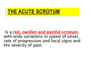
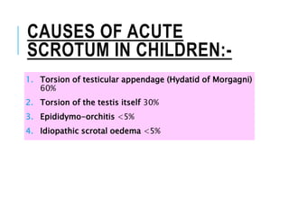
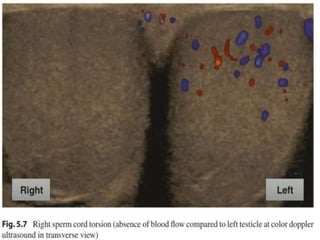
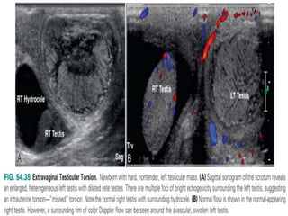
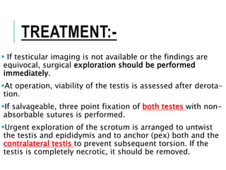
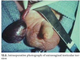
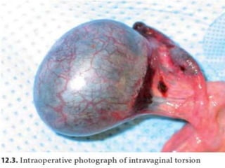
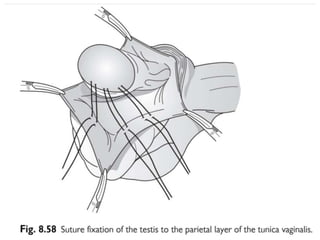
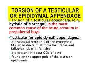
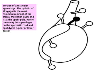
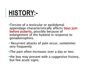
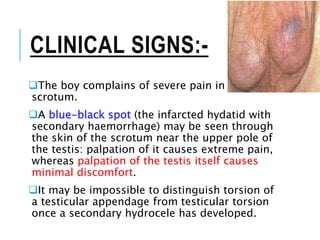
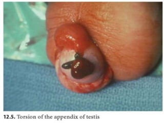
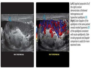
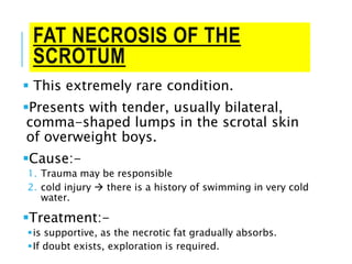
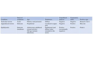
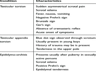
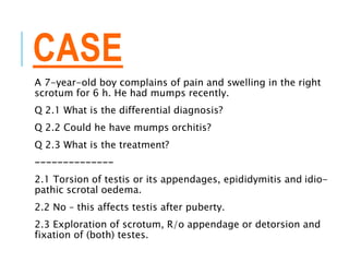
THE ACUTE SCROTUM Jwan Ali
Ahmed AlSofi
CONTENTS:-
1. The acute scrotum – definition and causes
with differential diagnosis
2. Management of the acute scrotum
3. Testicular torsion
4. Torsion of a testicular or epididymal
appendage
5. Epididymitis or epididymo-orchitis
6. Idiopathic scrotal oedema
7. Fat necrosis of the scrotum
THE ACUTE SCROTUM
Is a red, swollen and painful scrotum,
with wide variations in speed of onset,
rate of progression and local signs and
the severity of pain.
CAUSES OF ACUTE
SCROTUM IN CHILDREN:-
1. Torsion of testicular appendage (Hydatid of Morgagni)
60%
2. Torsion of the testis itself 30%
3. Epididymo-orchitis <5%
4. Idiopathic scrotal oedema <5%
DIFFERENTIAL
DIAGNOSIS:-
1. Testicular torsion
2. Torsion of a testicular or epididymal
appendage
3. epididymo-orchitis
4. idiopathic scrotal oedema (typically painless,
erythematous scrotal swelling in a young boy
extending off the scrotum into the groin and
towards the anus),
5. incarcerated inguinal hernia,
6. vasculitis - Henoch-Schonlein purpura
7. scrotal haematoma
INCIDENCE OF COMMON
CAUSES OF ACUTE SCROTAL
PROBLEMS IN INFANTS AND
CHILDREN.
MANAGEMENT OF THE ACUTE
SCROTUM
 As a general rule, an urgent exploration is
required in all cases of acute scrotum in which the
possibility of testicular torsion cannot be positively
excluded.
 The diagnosis of epididymitis or orchitis is
unlikely, unless there is:-
1. a history of urinary tract infection,
2. a known developmental anomaly of the renal tract
3. significant pyobacteriuria.
A midline scrotal incision has advantages:
1. when torsion of the testis is found, the testis may be untwisted
and fixed
2. exploration and fixation of the opposite testis are done through
the same incision.
TESTICULAR TORSION
 Testicular torsion is not the most common cause of an
acute scrotum, but it is the most important.
Testicular torsion is the rotation of the testis along its
longitudinal axis.
This results in torsion of the spermatic cord with an initial
blockage of venous drainage and a subsequent reduction in
arterial supply to the testis if complete rotation (>360°)
persists.
Venous blockage causes oedema and haemorrhage, followed
by ischaemia and necrosis when arterial inflow is
significantly reduced.
The rapid drop in blood flow is due to the fact that the testis
is supplied by three arterial vessels that divide in the
Those who are at risk of testicular
torsion:-
1. Just after the testis enlarges at puberty in
12–16-year-olds.
2. In unoperated undescended testes
Testicular torsion is most common in
adolescents,
-may occur at any age
Intratunical (or
intravaginal)
Extratunical (or
extravaginal)
More common Rare
Unoperated undescended testes are at
an increased risk, as their fixation
within the tunica is commonly tenuous.
The predisposing abnormality is
almost always present on the
contralateral side as well, and this
testis should be fixed at the time of
operation to prevent metachronous
torsion.
Typically occurs either just before birth
or in the early neonatal period
The testis is almost always necrotic by
the time the diagnosis is made
is made possible by an abnormally
narrow base of the mesenteric
attachment of the testis and
During testicular descent, a plane of
mobility between the tunica vaginalis
and surrounding areolar tissue per-
HISTORY:-
The onset is usually sudden, with pain in the testis
and/or ipsilateral iliac fossa, nausea and vomiting.
The pain is not always scrotal and may be felt in the groin
or lower abdomen.
Sometimes, the onset is more gradual, without severe
pain.
A previous history of previous transient episodes
:-
Similar but short-lived, even momentary, pain is
suggestive of episodes of prior incomplete and
spontaneously resolving torsion.
Torsion of the testis must be relieved within 6–8
CLINICAL EXAMINATION
 The swollen testis and epididymis are exquisitely tender
(unless already necrotic)
Reactive hydrocele  partially obscuring the tenderness.
The amount of swelling depends on the time that has elapsed and the rate of
progression.
The hydrocele and the exquisite tenderness may make
precise palpation of the testis difficult.
As the pathology is contained within the peritoneal
membrane of the tunica vaginalis, the inflammatory signs
are confined to the ipsilateral hemi-scrotum.
 the scrotum may appear swollen and reddened.
 Scrotal oedema and erythema can be absent.
CLINICAL EXAMINATION
1. Ger’s sign: + pitting of the skin at the base of the
scrotum
2. Brunzel’s sign / bell-clapper testis: + the testis lies
higher and horizontal in the scrotum with the patient
standing)
3. Prehn’s sign is negative (the elevation of the testis fails
to relieve pain, as it normally does in inflammatory
conditions).
4. The presence/absence of the cremasteric reflex can also
help in the differential diagnosis.
-This reflex is elicited by stroking or gently pinching the skin of the upper inner
thigh while observing the scrotum.
-A normal response is contraction of the cremasteric muscles on the ipsilateral
side with unilateral elevation of the testis.
INVESTIGATIONS:-
Urinalysis
• should be
performed in all
patients
presenting with
acute hemiscrotal
pain.
• With testicular
torsion, the
urinalysis is
usually clear.
• The presence of
pyuria and
bacteriuria is
Colour duplex
Doppler
ultrasonography.
• Is The imaging
modality of choice
for diagnosing
testicular torsion
• Colour Doppler
ultrasound will
show reduced
arterial blood flow
to the involved
testicle.
Radionuclide
scintigraphy
• Is an alternative
to Doppler.
• shows decreased
uptake of the
radioisotope in
the affected
testis, an
indication of
absent blood flow
to that testis.
• Useful though
these tests may
be, they are not
TREATMENT:-
 If testicular imaging is not available or the findings are
equivocal, surgical exploration should be performed
immediately.
At operation, viability of the testis is assessed after derota-
tion.
If salvageable, three point fixation of both testes with non-
absorbable sutures is performed.
Urgent exploration of the scrotum is arranged to untwist
the testis and epididymis and to anchor (pex) both and the
contralateral testis to prevent subsequent torsion. If the
testis is completely necrotic, it should be removed.
TORSION OF A TESTICULAR
OR EPIDIDYMAL APPENDAGE
 Torsion of a testicular appendage (e.g.
hydatid of Morgagni) is the most
common cause of the acute scrotum in
prepubertal boys.
Testicular (or epididymal) appendages:-
-are vestigial remnants of the embryonic
Mullerian ducts (that form the uterus and
fallopian tubes in females)
-are present in about 90% of boys
-found on the upper pole of the testis or
epididymis.
Torsion of a testicular
appendage. The hydatid of
Morgagni is the most
common (remnant of the
cranial Mu ̋llerian duct) and
is at the upper pole. Rarely,
there may be appendages
on the spermatic cord and
epididymis (upper or lower
poles).
HISTORY:-
Torsion of a testicular or epididymal
appendage characteristically affects boys just
before puberty, possibly because of
enlargement of the hydatid in response to
gonadotrophins.
 Recurrent attacks of pain occur, sometimes
very frequently
The pain often increases over a day or two.
the boy may present with a suggestive history,
but few acute signs.
CLINICAL SIGNS:-
The boy complains of severe pain in his
scrotum.
A blue-black spot (the infarcted hydatid with
secondary haemorrhage) may be seen through
the skin of the scrotum near the upper pole of
the testis: palpation of it causes extreme pain,
whereas palpation of the testis itself causes
minimal discomfort.
It may be impossible to distinguish torsion of
a testicular appendage from testicular torsion
once a secondary hydrocele has developed.
TREATMENT:-
 If testicular torsion cannot be excluded on
clinical examination:-
-urgent exploration is mandatory.
-At operation, the torted appendix testis is removed, which
provides relief of symptoms and prevents recurrence.
-Excision of the appendage leads to rapid resolution of
symptoms.
If a torted appendage can be diagnosed on
the basis of clinical findings:-
-non-operative treatment with effective analgesia is a valid
alternative.
EPIDIDYMITIS OR
EPIDIDYMO-ORCHITIS
 Epididymo-orchitis is rare in childhood and
virtually never occurs between 6 months of
age and puberty.
Although it is common practice to refer to
inflammatory conditions in the scrotum as
epididymo-orchitis, the inflammation is
usually confined to the epididymis.
Viral or bacterial epididymo-orchitis may
cause an acute scrotum in infants and
toddlers but this diagnosis is often only made
after scrotal exploration.
Epididymitis
• The most common causative
bacterium in children is
Escherichia coli,
• Infection is carried by
retrograde flow along a
patent vas deferens from the
urinary tract.
• Predisposing factors for
bacterial infection include
• abnormalities of the
urinary tract
• urethral instrumentation.
True acute orchitis
• is very uncommon
• may occur in
• mumps,
• Henoch–Schönlein purpura
(HSP)
• septicaemia.
• Mumps orchitis is extremely
rare prior to puberty, and
where the tell-tale parotid
swelling is not obvious may
be suspected due to a testis,
which is larger and harder
than expected in epididymo-
orchitis.
CLINICAL SIGNS:-
 The usual findings are those of an acute scrotum in a baby
or adolescent.
 A lax secondary hydrocele is common.
Bilateral signs are particularly suggestive of epididymitis.
Fever, Prehn sign +, associated LUTS, urethral discharge.
Examination of the urine may show pyobacteriuria.
Doppler Ultrasound of the testis  Hypervasularity
Young children with epididymitis due to urinary organisms
should have a renal ultrasound scan after the epididymitis
has subsided, and some may require also a micturating
cystourethrogram.
• These investigations aim to identify anomalies of the lower urinary
TREATMENT:-
 Treatment of epididymitis consists of
1. rest,
2. antibiotics (e.g. co-trimoxazole,
nitrofurantoin),
3. a high fluid intake
4. alkalinisation of the urine.
 Severe or repeated infections may
lead to:-
1. an abscess
2. progressive destruction of the testis
3. sterility is rare when only one side is
affected.
IDIOPATHIC SCROTAL
OEDEMA
 In this condition, there is rapidly
developing scrotal oedema, which may
then spread to the inguinal region, penis
and foreskin and/or the perineum.
 The pathology involves the skin (and
therefore spreads beyond the tunica
vaginalis).
Cause:-
1. a history of allergy
2. History of playing outside at the onset
3. a bite from an insect or a spider
4. may represent allergic inflammation
CLINICAL FEATURES:-
 The scrotum is symmetrically swollen, pale pink
or red.
 There is slight discomfort rather than acute pain.
Careful palpation reveals non-tender testes that
are normal in size and position.
The oedema subsides in 1–2 days, but may
occasionally recur some weeks later.
It may be distinguished from other causes of the
acute scrotum by:-
1. The spread of oedema beyond the confines of the hemi-scrotum
2. by the complete absence of tenderness in the epididymis or testis.
 Discomfort due to oedema of the scrotum per se may masquerade as testicular
FAT NECROSIS OF THE
SCROTUM
 This extremely rare condition.
Presents with tender, usually bilateral,
comma-shaped lumps in the scrotal skin
of overweight boys.
Cause:-
1. Trauma may be responsible
2. cold injury  there is a history of swimming in very cold
water.
Treatment:-
is supportive, as the necrotic fat gradually absorbs.
If doubt exists, exploration is required.
CASE
A 7-year-old boy complains of pain and swelling in the right
scrotum for 6 h. He had mumps recently.
Q 2.1 What is the differential diagnosis?
Q 2.2 Could he have mumps orchitis?
Q 2.3 What is the treatment?
--------------
2.1 Torsion of testis or its appendages, epididymitis and idio-
pathic scrotal oedema.
2.2 No – this affects testis after puberty.
2.3 Exploration of scrotum, R/o appendage or detorsion and
fixation of (both) testes.
THANKS

The Acute Scrotum.pptx

  • 1.
    THE ACUTE SCROTUMJwan Ali Ahmed AlSofi
  • 2.
    CONTENTS:- 1. The acutescrotum – definition and causes with differential diagnosis 2. Management of the acute scrotum 3. Testicular torsion 4. Torsion of a testicular or epididymal appendage 5. Epididymitis or epididymo-orchitis 6. Idiopathic scrotal oedema 7. Fat necrosis of the scrotum
  • 3.
    THE ACUTE SCROTUM Isa red, swollen and painful scrotum, with wide variations in speed of onset, rate of progression and local signs and the severity of pain.
  • 4.
    CAUSES OF ACUTE SCROTUMIN CHILDREN:- 1. Torsion of testicular appendage (Hydatid of Morgagni) 60% 2. Torsion of the testis itself 30% 3. Epididymo-orchitis <5% 4. Idiopathic scrotal oedema <5%
  • 5.
    DIFFERENTIAL DIAGNOSIS:- 1. Testicular torsion 2.Torsion of a testicular or epididymal appendage 3. epididymo-orchitis 4. idiopathic scrotal oedema (typically painless, erythematous scrotal swelling in a young boy extending off the scrotum into the groin and towards the anus), 5. incarcerated inguinal hernia, 6. vasculitis - Henoch-Schonlein purpura 7. scrotal haematoma
  • 6.
    INCIDENCE OF COMMON CAUSESOF ACUTE SCROTAL PROBLEMS IN INFANTS AND CHILDREN.
  • 7.
    MANAGEMENT OF THEACUTE SCROTUM  As a general rule, an urgent exploration is required in all cases of acute scrotum in which the possibility of testicular torsion cannot be positively excluded.  The diagnosis of epididymitis or orchitis is unlikely, unless there is:- 1. a history of urinary tract infection, 2. a known developmental anomaly of the renal tract 3. significant pyobacteriuria. A midline scrotal incision has advantages: 1. when torsion of the testis is found, the testis may be untwisted and fixed 2. exploration and fixation of the opposite testis are done through the same incision.
  • 8.
    TESTICULAR TORSION  Testiculartorsion is not the most common cause of an acute scrotum, but it is the most important. Testicular torsion is the rotation of the testis along its longitudinal axis. This results in torsion of the spermatic cord with an initial blockage of venous drainage and a subsequent reduction in arterial supply to the testis if complete rotation (>360°) persists. Venous blockage causes oedema and haemorrhage, followed by ischaemia and necrosis when arterial inflow is significantly reduced. The rapid drop in blood flow is due to the fact that the testis is supplied by three arterial vessels that divide in the
  • 9.
    Those who areat risk of testicular torsion:- 1. Just after the testis enlarges at puberty in 12–16-year-olds. 2. In unoperated undescended testes Testicular torsion is most common in adolescents, -may occur at any age
  • 10.
    Intratunical (or intravaginal) Extratunical (or extravaginal) Morecommon Rare Unoperated undescended testes are at an increased risk, as their fixation within the tunica is commonly tenuous. The predisposing abnormality is almost always present on the contralateral side as well, and this testis should be fixed at the time of operation to prevent metachronous torsion. Typically occurs either just before birth or in the early neonatal period The testis is almost always necrotic by the time the diagnosis is made is made possible by an abnormally narrow base of the mesenteric attachment of the testis and During testicular descent, a plane of mobility between the tunica vaginalis and surrounding areolar tissue per-
  • 12.
    HISTORY:- The onset isusually sudden, with pain in the testis and/or ipsilateral iliac fossa, nausea and vomiting. The pain is not always scrotal and may be felt in the groin or lower abdomen. Sometimes, the onset is more gradual, without severe pain. A previous history of previous transient episodes :- Similar but short-lived, even momentary, pain is suggestive of episodes of prior incomplete and spontaneously resolving torsion. Torsion of the testis must be relieved within 6–8
  • 13.
    CLINICAL EXAMINATION  Theswollen testis and epididymis are exquisitely tender (unless already necrotic) Reactive hydrocele  partially obscuring the tenderness. The amount of swelling depends on the time that has elapsed and the rate of progression. The hydrocele and the exquisite tenderness may make precise palpation of the testis difficult. As the pathology is contained within the peritoneal membrane of the tunica vaginalis, the inflammatory signs are confined to the ipsilateral hemi-scrotum.  the scrotum may appear swollen and reddened.  Scrotal oedema and erythema can be absent.
  • 14.
    CLINICAL EXAMINATION 1. Ger’ssign: + pitting of the skin at the base of the scrotum 2. Brunzel’s sign / bell-clapper testis: + the testis lies higher and horizontal in the scrotum with the patient standing) 3. Prehn’s sign is negative (the elevation of the testis fails to relieve pain, as it normally does in inflammatory conditions). 4. The presence/absence of the cremasteric reflex can also help in the differential diagnosis. -This reflex is elicited by stroking or gently pinching the skin of the upper inner thigh while observing the scrotum. -A normal response is contraction of the cremasteric muscles on the ipsilateral side with unilateral elevation of the testis.
  • 17.
    INVESTIGATIONS:- Urinalysis • should be performedin all patients presenting with acute hemiscrotal pain. • With testicular torsion, the urinalysis is usually clear. • The presence of pyuria and bacteriuria is Colour duplex Doppler ultrasonography. • Is The imaging modality of choice for diagnosing testicular torsion • Colour Doppler ultrasound will show reduced arterial blood flow to the involved testicle. Radionuclide scintigraphy • Is an alternative to Doppler. • shows decreased uptake of the radioisotope in the affected testis, an indication of absent blood flow to that testis. • Useful though these tests may be, they are not
  • 20.
    TREATMENT:-  If testicularimaging is not available or the findings are equivocal, surgical exploration should be performed immediately. At operation, viability of the testis is assessed after derota- tion. If salvageable, three point fixation of both testes with non- absorbable sutures is performed. Urgent exploration of the scrotum is arranged to untwist the testis and epididymis and to anchor (pex) both and the contralateral testis to prevent subsequent torsion. If the testis is completely necrotic, it should be removed.
  • 24.
    TORSION OF ATESTICULAR OR EPIDIDYMAL APPENDAGE  Torsion of a testicular appendage (e.g. hydatid of Morgagni) is the most common cause of the acute scrotum in prepubertal boys. Testicular (or epididymal) appendages:- -are vestigial remnants of the embryonic Mullerian ducts (that form the uterus and fallopian tubes in females) -are present in about 90% of boys -found on the upper pole of the testis or epididymis.
  • 25.
    Torsion of atesticular appendage. The hydatid of Morgagni is the most common (remnant of the cranial Mu ̋llerian duct) and is at the upper pole. Rarely, there may be appendages on the spermatic cord and epididymis (upper or lower poles).
  • 26.
    HISTORY:- Torsion of atesticular or epididymal appendage characteristically affects boys just before puberty, possibly because of enlargement of the hydatid in response to gonadotrophins.  Recurrent attacks of pain occur, sometimes very frequently The pain often increases over a day or two. the boy may present with a suggestive history, but few acute signs.
  • 27.
    CLINICAL SIGNS:- The boycomplains of severe pain in his scrotum. A blue-black spot (the infarcted hydatid with secondary haemorrhage) may be seen through the skin of the scrotum near the upper pole of the testis: palpation of it causes extreme pain, whereas palpation of the testis itself causes minimal discomfort. It may be impossible to distinguish torsion of a testicular appendage from testicular torsion once a secondary hydrocele has developed.
  • 29.
    TREATMENT:-  If testiculartorsion cannot be excluded on clinical examination:- -urgent exploration is mandatory. -At operation, the torted appendix testis is removed, which provides relief of symptoms and prevents recurrence. -Excision of the appendage leads to rapid resolution of symptoms. If a torted appendage can be diagnosed on the basis of clinical findings:- -non-operative treatment with effective analgesia is a valid alternative.
  • 30.
    EPIDIDYMITIS OR EPIDIDYMO-ORCHITIS  Epididymo-orchitisis rare in childhood and virtually never occurs between 6 months of age and puberty. Although it is common practice to refer to inflammatory conditions in the scrotum as epididymo-orchitis, the inflammation is usually confined to the epididymis. Viral or bacterial epididymo-orchitis may cause an acute scrotum in infants and toddlers but this diagnosis is often only made after scrotal exploration.
  • 31.
    Epididymitis • The mostcommon causative bacterium in children is Escherichia coli, • Infection is carried by retrograde flow along a patent vas deferens from the urinary tract. • Predisposing factors for bacterial infection include • abnormalities of the urinary tract • urethral instrumentation. True acute orchitis • is very uncommon • may occur in • mumps, • Henoch–Schönlein purpura (HSP) • septicaemia. • Mumps orchitis is extremely rare prior to puberty, and where the tell-tale parotid swelling is not obvious may be suspected due to a testis, which is larger and harder than expected in epididymo- orchitis.
  • 32.
    CLINICAL SIGNS:-  Theusual findings are those of an acute scrotum in a baby or adolescent.  A lax secondary hydrocele is common. Bilateral signs are particularly suggestive of epididymitis. Fever, Prehn sign +, associated LUTS, urethral discharge. Examination of the urine may show pyobacteriuria. Doppler Ultrasound of the testis  Hypervasularity Young children with epididymitis due to urinary organisms should have a renal ultrasound scan after the epididymitis has subsided, and some may require also a micturating cystourethrogram. • These investigations aim to identify anomalies of the lower urinary
  • 34.
    TREATMENT:-  Treatment ofepididymitis consists of 1. rest, 2. antibiotics (e.g. co-trimoxazole, nitrofurantoin), 3. a high fluid intake 4. alkalinisation of the urine.  Severe or repeated infections may lead to:- 1. an abscess 2. progressive destruction of the testis 3. sterility is rare when only one side is affected.
  • 35.
    IDIOPATHIC SCROTAL OEDEMA  Inthis condition, there is rapidly developing scrotal oedema, which may then spread to the inguinal region, penis and foreskin and/or the perineum.  The pathology involves the skin (and therefore spreads beyond the tunica vaginalis). Cause:- 1. a history of allergy 2. History of playing outside at the onset 3. a bite from an insect or a spider 4. may represent allergic inflammation
  • 36.
    CLINICAL FEATURES:-  Thescrotum is symmetrically swollen, pale pink or red.  There is slight discomfort rather than acute pain. Careful palpation reveals non-tender testes that are normal in size and position. The oedema subsides in 1–2 days, but may occasionally recur some weeks later. It may be distinguished from other causes of the acute scrotum by:- 1. The spread of oedema beyond the confines of the hemi-scrotum 2. by the complete absence of tenderness in the epididymis or testis.  Discomfort due to oedema of the scrotum per se may masquerade as testicular
  • 38.
    FAT NECROSIS OFTHE SCROTUM  This extremely rare condition. Presents with tender, usually bilateral, comma-shaped lumps in the scrotal skin of overweight boys. Cause:- 1. Trauma may be responsible 2. cold injury  there is a history of swimming in very cold water. Treatment:- is supportive, as the necrotic fat gradually absorbs. If doubt exists, exploration is required.
  • 41.
    CASE A 7-year-old boycomplains of pain and swelling in the right scrotum for 6 h. He had mumps recently. Q 2.1 What is the differential diagnosis? Q 2.2 Could he have mumps orchitis? Q 2.3 What is the treatment? -------------- 2.1 Torsion of testis or its appendages, epididymitis and idio- pathic scrotal oedema. 2.2 No – this affects testis after puberty. 2.3 Exploration of scrotum, R/o appendage or detorsion and fixation of (both) testes.
  • 42.