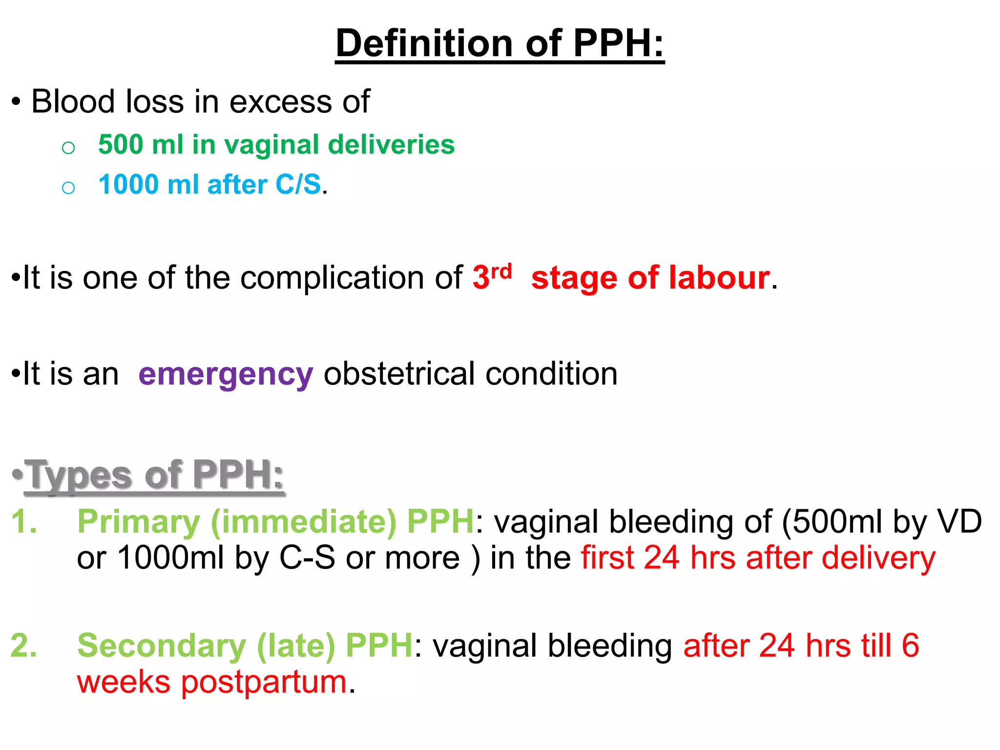
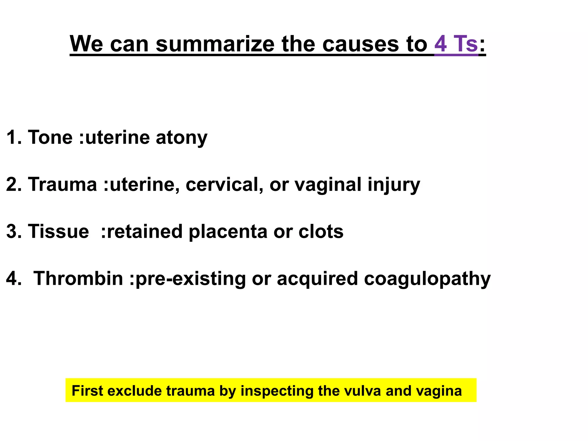
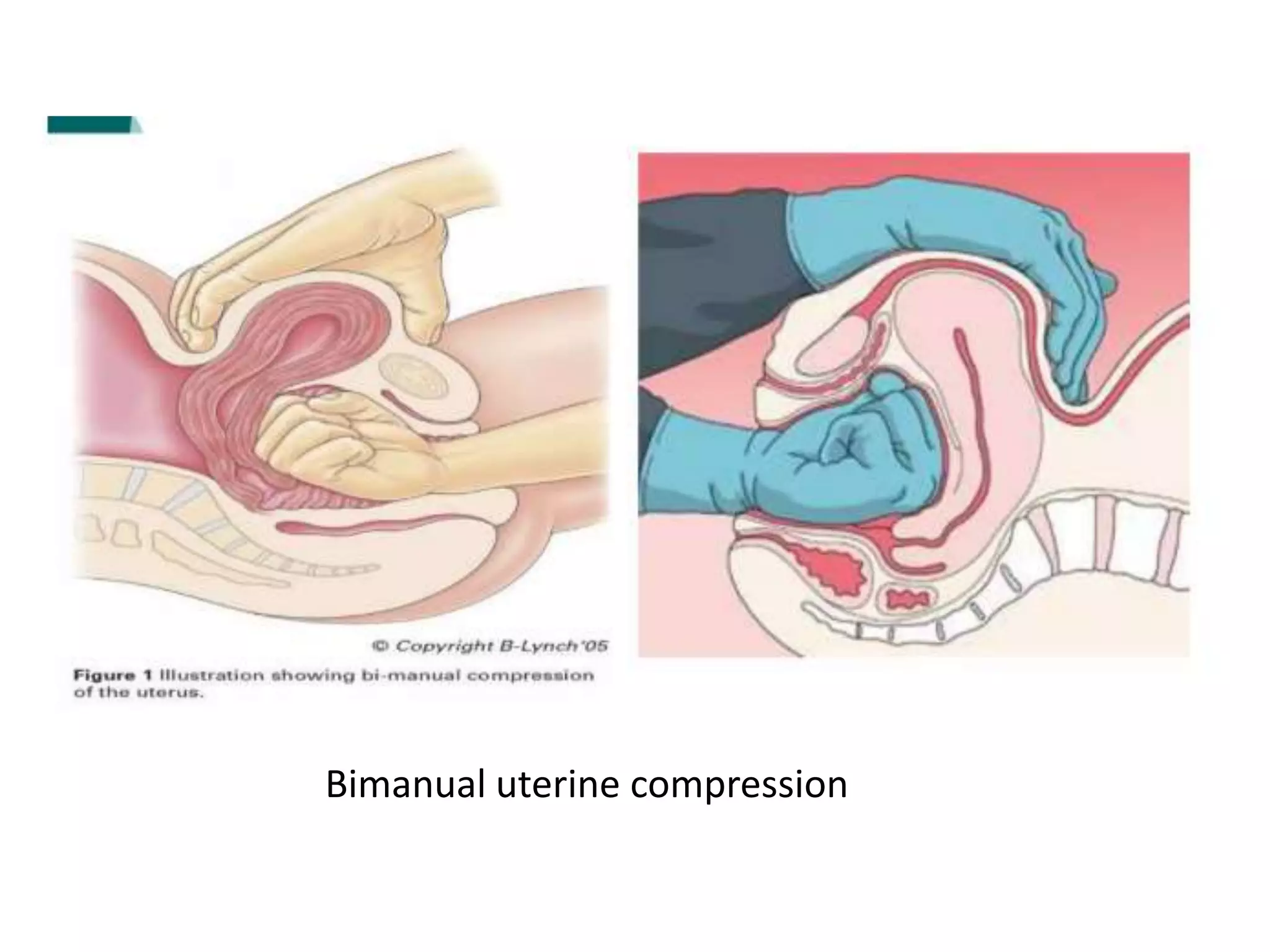
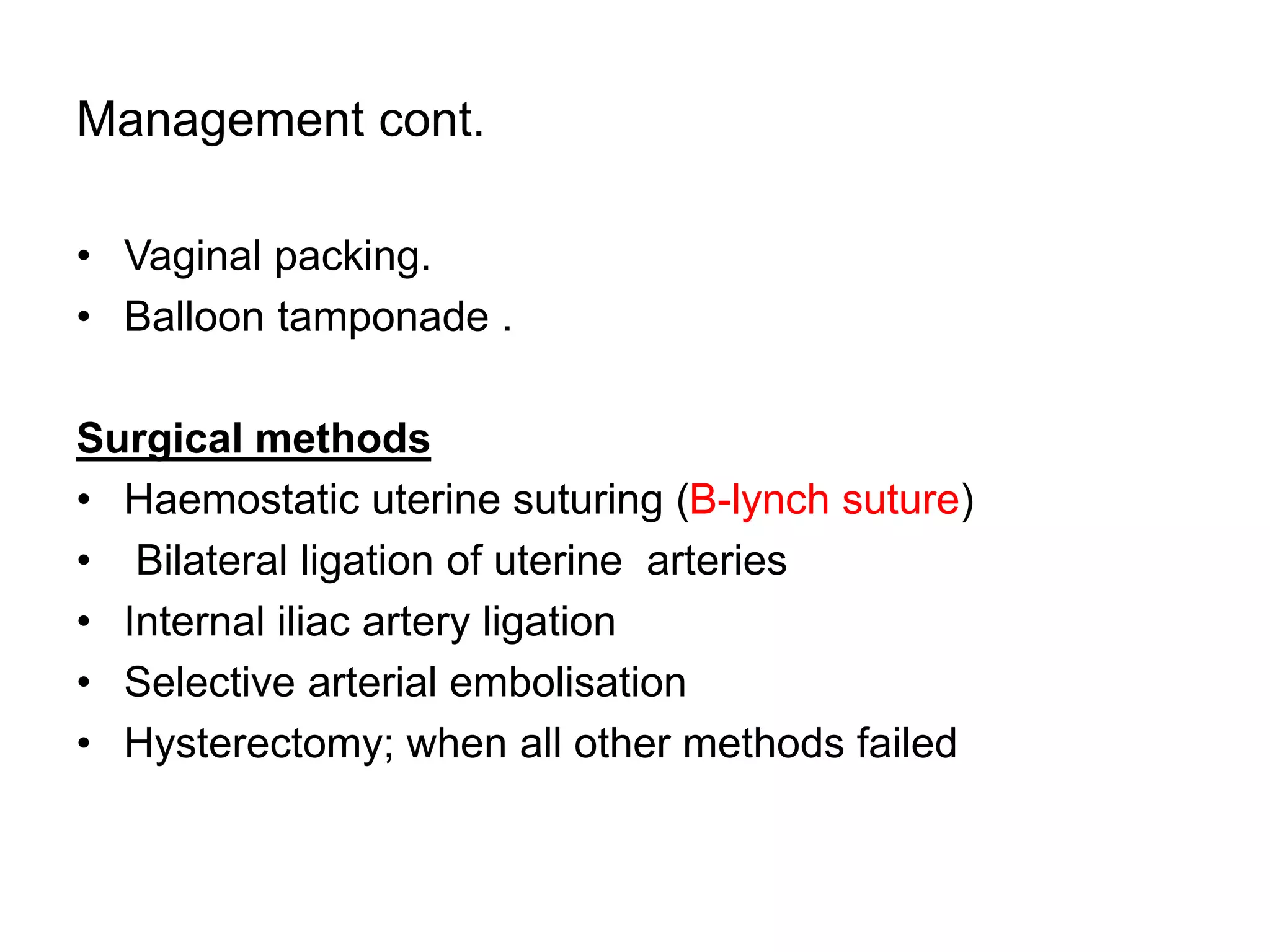
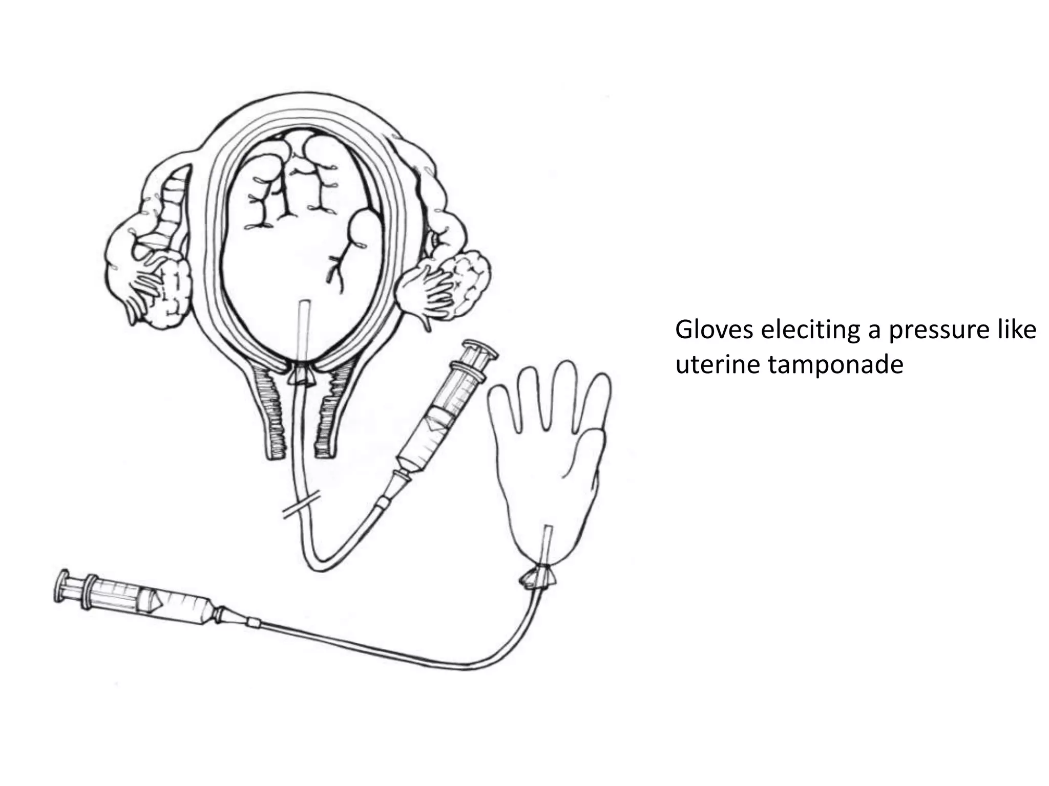
Post Partum Hemorrhage (PPH)
By:-
Jwan Ali Ahmed AlSofi
Contents
•Definition
•Types and classification
•Incidence and Etiology
• Predisposing factor
•Management of underlined etiology
•Conclusion and Question
Objectives
Identify the common causes and risk factors for PPH
Identify appropriate care management to the woman
experiencing PPH
PLACENTAL SEPARATION
• Mechanism of separation:
– Marked retraction reduces effectively the surface area at the placental site to about
its half.
– But as the placenta is inelastic, it cannot keep pace with such an extent of
diminution resulting in its buckling .
– A shearing force is instituted between the placenta and the placental site which
brings about its ultimate separation.
– The plane of separation runs through deep spongy layer of decidua basalis so that
a variable thickness of decidua covers the maternal surface of the separated
placenta.
• There are two ways of separation of placenta :
1. Central separation (Schultze):
– Detachment of placenta from its uterine attachment starts at the center resulting in
opening up of few uterine sinuses and accumulation of blood behind the placenta
(retroplacental hematoma).
– With increasing contraction, more and more detachment occurs facilitated by
weight of the placenta and retroplacental blood until whole of the placenta gets
detached.
2. Marginal separation (Mathews-Duncan):
– Separation starts at the margin as it is mostly unsupported.
– With progressive uterine contraction, more and more areas of the placenta get
Haemostasis post- placental separation
1. Retraction of the oblique uterine muscle fibres in the upper uterine
segment through which the tortuous blood vessels intertwine – the
resultant thickening of the muscles exerts pressure on the torn vessels,
acting as clamps, and preventing haemorrhage.
– It is the absence of oblique fibres in the lower uterine segment that explains the
greatly increased blood loss usually accompanying placental separation in
placenta praevia.
2. The presence of vigorous uterine contraction following separation –
this brings the walls into apposition so that further pressure is exerted on
the placental site.
3. The achievement of haemostasis – there is a transitory activation of the
coagulation and fibrinolytic systems during, and immediately following,
placental separation. It is believed that this protective response is
especially active at the placental site so that clot formation in the torn
vessels is intensified. Following separation, the placental site is rapidly
covered by a fibrin mesh.
4. Breast-feeding – the release of oxytocin from the posterior pituitary in
response to skin-to-skin contact between mother and baby, and the baby’s
nuzzling at the breast, causes uterine contractions.
Definition of PPH:
• Blood loss in excess of
o 500 ml in vaginal deliveries
o 1000 ml after C/S.
•It is one of the complication of 3rd stage of labour.
•It is an emergency obstetrical condition
•Types of PPH:
1. Primary (immediate) PPH: vaginal bleeding of (500ml by VD
or 1000ml by C-S or more ) in the first 24 hrs after delivery
2. Secondary (late) PPH: vaginal bleeding after 24 hrs till 6
weeks postpartum.
Classification
1. Minor PPH:
– blood loss 500–1000 ml,
– no clinical shock
– loss of <20% of blood volume
2. Major PPH
– blood loss>1000 ml
– continuing to bleed
– clinical shock
– Major PPH could be divided to:
• Moderate: 20-40% loss of blood volume (1000–2000 ml)
• Severe: >40% loss of blood volume (> 2000 ml).
Incidence
• The 4th cause of maternal mortality in the developed
countries.
• In developing countries remains the leading cause of
maternal mortality.
• The incidence is 4-8% of deliveries in developed
counties.
• Recurrence risk of about 20–25%.
Prediposing factor of PPH:-
• Hemodynamic compromise is more likely in:
 Anemia (Iron deficiency,sickle cell,and thalassemia).
Volume contracted states (e.g. dehydration, gestational
hypertension with proteinuria).
PPH : prevention/ Prophylaxis
• haemoglobin levels below the normal range for pregnancy
should be investigated
• iron supplementation considered if indicated to optimize
haemoglobin prior to delivery.
• Prophylactic use of oxytocin agents for high- risk patients.
• During labour, good management practices during the first and
second stages are important to prevent prolonged labour and
ketoacidosis.
• A mother should not enter the second or third stage with a full
bladder.
• AMTSL
• Two units of cross-matched blood should be kept available for
any woman known to have a placenta praevia or other major
predisposing risk factors for PPH.
Active management of the third stage of labour decreases
the risk of PPH so:
1. Administration of oxytocic drugs during or immediately
after delivery.
2. Massage of uterus & early cord clamping & cutting .
3. Controlled cord traction to deliver the placenta.
Active management of 3rd stage of labour
Etiology of Primary PPH:
• Atonic uterus.
• Retained placenta
• Genital tract trauma.
• Placenta accreta
• Uterine inversion
• Coagulation disorders; DIC, ITP, leukemia, Von-willebrand
disease
We can summarize the causes to 4 Ts:
1. Tone :uterine atony
2. Trauma :uterine, cervical, or vaginal injury
3. Tissue :retained placenta or clots
4. Thrombin :pre-existing or acquired coagulopathy
First exclude trauma by inspecting the vulva and vagina
Uterine atony
Definition
 This is a failure of the myometrium at the placental
site to contract and retract and to compress torn
blood vessels and control blood loss by a living ligature
action.
 Failure of uterus to contract effectively after delivery of
placenta.
 Most common cause of PPH and can cause major
PPH , about 70%.
• Distended bladder and over distended uterus.
• prolonged and Instrumental delivery
• Multiparity & scared uterus
• In a case of APH and previous history of PPH
• Obstetrical complications: PE,anemia,chorioamnionitis
• Leiomyoma
•Drugs: sedatives, anesthetics, tocolytics.
Risk factors for the uterine atony
Clinical features
• If hemorrhage continues sign and symptome of shock will
develop.
 Pallor
 rising pulse rate
 falling blood pressure
 altered level of consciousness; the mother may become
restless or drowsy
• Abdominal palpation the fundus of uterus:
 Abnormally high in the abdomen
 The non contracted uterus distended with blood
 Has a boggy consistency (i.e. soft and distended and lacking
tone).
Management of PPH
• Shout for help
• AbC – take vitals
• Put 2-wide bore IV cannula
• Take blood for – CBC, cross match and Prepare
at least 2-6 pints of blood, coagulation profile and
basic biochemistry
• Start bolus fluid crystalloids – RL or NS
• Ensure bladder is empty (catheter leave in situe)
Management of uterine atony
• Examine the vulva/vagina for any trauma
• Uterine massage: External bimanual uterine massage.
• Bimanual uterine compression – wear sterile gloves
Bimanual uterine compression
Management (cont.)
• Oxytocic drugs: the drug of choice is:
 Ergometrin (except in hypertension or cardiac disease or
asthma).
 No more than two doses of ergometrine should be given (including any
dose of combined ergometrine/oxytocin) as it may cause pulmonary
hypertension.
 Oxytocin 5 units by slow IV injection, oxytocin infusion (40
units in 500mls Hartmann’s at 125 mls / hr)
 Carboprost (Hemabate).
 Misoprostol (PGE1 analogue) 800mcg rectally & 400 mcg
sublingually.
Management cont.
• Vaginal packing.
• Balloon tamponade .
Surgical methods
• Haemostatic uterine suturing (B-lynch suture)
• Bilateral ligation of uterine arteries
• Internal iliac artery ligation
• Selective arterial embolisation
• Hysterectomy; when all other methods failed
pack for vaginal packing
Folly's catheter eliciting a pressure like uterine
tamponade
Gloves eleciting a pressure like
uterine tamponade
Balloon tamponade
B-lynch suture
Retained placenta & membrane:
 Failure of expulsion of the placenta and membrane.
 Excessive bleeding from the placental vascular bed
 Commonly co-exist with atony.
 At term 90% of placenta will be delivered within 15 min.
 3rd stage exceeds 30 min regarded retained placenta.
• 1-2% of all deliveries.
• The mortality rate of this condition is up to 10% if left untreated.
• The usual reason is failure of the retro-placental myometrium
to contract thus preventing detachment.
•Retained palcenta:-
1. Not separated at all
2. Partially separated – whole placenta is retaned or fragmented.
3. Trapped palcenta
Incidence:
Management of Retained placenta
1-If the placenta not separated at all:
• Oxytocic drugs
• Deliver the placenta by Brandt's Andrews method .
• Uterotonic agents down the umbilical cord by the Piping's method
•If not successful manual removal under GA after urinary
catheterization.
Management of retained placenta (cont.)
2-If the placenta partially separated :
A-Whole placenta is retained
• Resuscitation and manual removal under GA after urinary
catheterization and antibiotics.
B-If there are retained fragments
• Curettage with blunt instruments under GA under antibiotic cover
Management of retained placenta (cont.)
3- Trapped placenta (is a completely separated placenta
but is trapped inside the uterus because of closed cervix) :
Trapped placenta  uterus is contracting & cervix is
closed
– Give uterine relaxant, IV glyceryl trinitrate and controlled
cord traction.
• If the lower uterus/cervix is contracted, thereby preventing expulsion of the
placenta, administration of nitroglycerin will result in relaxation and facilitate
placental delivery .
– If unsuccessful, then manual removal under GA
(halothane).under cover of antibiotics.
Conclusion
• PPH is an emergency ,life threatening and team management
obstetrical condition.
• Always should remember 4Ts
• Consider high-dependency unit or intensive care unit.
• Exclude vaginal trauma
Questions?
• What are the most common causes of uterine atony in labour
room?
Full bladder

Post Partum Hemorrhage (PPH).ppt

  • 1.
    Post Partum Hemorrhage(PPH) By:- Jwan Ali Ahmed AlSofi
  • 2.
    Contents •Definition •Types and classification •Incidenceand Etiology • Predisposing factor •Management of underlined etiology •Conclusion and Question
  • 3.
    Objectives Identify the commoncauses and risk factors for PPH Identify appropriate care management to the woman experiencing PPH
  • 4.
    PLACENTAL SEPARATION • Mechanismof separation: – Marked retraction reduces effectively the surface area at the placental site to about its half. – But as the placenta is inelastic, it cannot keep pace with such an extent of diminution resulting in its buckling . – A shearing force is instituted between the placenta and the placental site which brings about its ultimate separation. – The plane of separation runs through deep spongy layer of decidua basalis so that a variable thickness of decidua covers the maternal surface of the separated placenta. • There are two ways of separation of placenta : 1. Central separation (Schultze): – Detachment of placenta from its uterine attachment starts at the center resulting in opening up of few uterine sinuses and accumulation of blood behind the placenta (retroplacental hematoma). – With increasing contraction, more and more detachment occurs facilitated by weight of the placenta and retroplacental blood until whole of the placenta gets detached. 2. Marginal separation (Mathews-Duncan): – Separation starts at the margin as it is mostly unsupported. – With progressive uterine contraction, more and more areas of the placenta get
  • 7.
    Haemostasis post- placentalseparation 1. Retraction of the oblique uterine muscle fibres in the upper uterine segment through which the tortuous blood vessels intertwine – the resultant thickening of the muscles exerts pressure on the torn vessels, acting as clamps, and preventing haemorrhage. – It is the absence of oblique fibres in the lower uterine segment that explains the greatly increased blood loss usually accompanying placental separation in placenta praevia. 2. The presence of vigorous uterine contraction following separation – this brings the walls into apposition so that further pressure is exerted on the placental site. 3. The achievement of haemostasis – there is a transitory activation of the coagulation and fibrinolytic systems during, and immediately following, placental separation. It is believed that this protective response is especially active at the placental site so that clot formation in the torn vessels is intensified. Following separation, the placental site is rapidly covered by a fibrin mesh. 4. Breast-feeding – the release of oxytocin from the posterior pituitary in response to skin-to-skin contact between mother and baby, and the baby’s nuzzling at the breast, causes uterine contractions.
  • 9.
    Definition of PPH: •Blood loss in excess of o 500 ml in vaginal deliveries o 1000 ml after C/S. •It is one of the complication of 3rd stage of labour. •It is an emergency obstetrical condition •Types of PPH: 1. Primary (immediate) PPH: vaginal bleeding of (500ml by VD or 1000ml by C-S or more ) in the first 24 hrs after delivery 2. Secondary (late) PPH: vaginal bleeding after 24 hrs till 6 weeks postpartum.
  • 10.
    Classification 1. Minor PPH: –blood loss 500–1000 ml, – no clinical shock – loss of <20% of blood volume 2. Major PPH – blood loss>1000 ml – continuing to bleed – clinical shock – Major PPH could be divided to: • Moderate: 20-40% loss of blood volume (1000–2000 ml) • Severe: >40% loss of blood volume (> 2000 ml).
  • 11.
    Incidence • The 4thcause of maternal mortality in the developed countries. • In developing countries remains the leading cause of maternal mortality. • The incidence is 4-8% of deliveries in developed counties. • Recurrence risk of about 20–25%.
  • 12.
    Prediposing factor ofPPH:- • Hemodynamic compromise is more likely in:  Anemia (Iron deficiency,sickle cell,and thalassemia). Volume contracted states (e.g. dehydration, gestational hypertension with proteinuria).
  • 13.
    PPH : prevention/Prophylaxis • haemoglobin levels below the normal range for pregnancy should be investigated • iron supplementation considered if indicated to optimize haemoglobin prior to delivery. • Prophylactic use of oxytocin agents for high- risk patients. • During labour, good management practices during the first and second stages are important to prevent prolonged labour and ketoacidosis. • A mother should not enter the second or third stage with a full bladder. • AMTSL • Two units of cross-matched blood should be kept available for any woman known to have a placenta praevia or other major predisposing risk factors for PPH.
  • 14.
    Active management ofthe third stage of labour decreases the risk of PPH so: 1. Administration of oxytocic drugs during or immediately after delivery. 2. Massage of uterus & early cord clamping & cutting . 3. Controlled cord traction to deliver the placenta.
  • 16.
    Active management of3rd stage of labour
  • 17.
    Etiology of PrimaryPPH: • Atonic uterus. • Retained placenta • Genital tract trauma. • Placenta accreta • Uterine inversion • Coagulation disorders; DIC, ITP, leukemia, Von-willebrand disease
  • 18.
    We can summarizethe causes to 4 Ts: 1. Tone :uterine atony 2. Trauma :uterine, cervical, or vaginal injury 3. Tissue :retained placenta or clots 4. Thrombin :pre-existing or acquired coagulopathy First exclude trauma by inspecting the vulva and vagina
  • 19.
    Uterine atony Definition  Thisis a failure of the myometrium at the placental site to contract and retract and to compress torn blood vessels and control blood loss by a living ligature action.  Failure of uterus to contract effectively after delivery of placenta.  Most common cause of PPH and can cause major PPH , about 70%.
  • 20.
    • Distended bladderand over distended uterus. • prolonged and Instrumental delivery • Multiparity & scared uterus • In a case of APH and previous history of PPH • Obstetrical complications: PE,anemia,chorioamnionitis • Leiomyoma •Drugs: sedatives, anesthetics, tocolytics. Risk factors for the uterine atony
  • 21.
    Clinical features • Ifhemorrhage continues sign and symptome of shock will develop.  Pallor  rising pulse rate  falling blood pressure  altered level of consciousness; the mother may become restless or drowsy • Abdominal palpation the fundus of uterus:  Abnormally high in the abdomen  The non contracted uterus distended with blood  Has a boggy consistency (i.e. soft and distended and lacking tone).
  • 23.
    Management of PPH •Shout for help • AbC – take vitals • Put 2-wide bore IV cannula • Take blood for – CBC, cross match and Prepare at least 2-6 pints of blood, coagulation profile and basic biochemistry • Start bolus fluid crystalloids – RL or NS • Ensure bladder is empty (catheter leave in situe)
  • 24.
    Management of uterineatony • Examine the vulva/vagina for any trauma • Uterine massage: External bimanual uterine massage. • Bimanual uterine compression – wear sterile gloves
  • 25.
  • 26.
    Management (cont.) • Oxytocicdrugs: the drug of choice is:  Ergometrin (except in hypertension or cardiac disease or asthma).  No more than two doses of ergometrine should be given (including any dose of combined ergometrine/oxytocin) as it may cause pulmonary hypertension.  Oxytocin 5 units by slow IV injection, oxytocin infusion (40 units in 500mls Hartmann’s at 125 mls / hr)  Carboprost (Hemabate).  Misoprostol (PGE1 analogue) 800mcg rectally & 400 mcg sublingually.
  • 28.
    Management cont. • Vaginalpacking. • Balloon tamponade . Surgical methods • Haemostatic uterine suturing (B-lynch suture) • Bilateral ligation of uterine arteries • Internal iliac artery ligation • Selective arterial embolisation • Hysterectomy; when all other methods failed
  • 29.
  • 30.
    Folly's catheter elicitinga pressure like uterine tamponade
  • 31.
    Gloves eleciting apressure like uterine tamponade
  • 32.
  • 33.
  • 34.
    Retained placenta &membrane:  Failure of expulsion of the placenta and membrane.  Excessive bleeding from the placental vascular bed  Commonly co-exist with atony.  At term 90% of placenta will be delivered within 15 min.  3rd stage exceeds 30 min regarded retained placenta.
  • 35.
    • 1-2% ofall deliveries. • The mortality rate of this condition is up to 10% if left untreated. • The usual reason is failure of the retro-placental myometrium to contract thus preventing detachment. •Retained palcenta:- 1. Not separated at all 2. Partially separated – whole placenta is retaned or fragmented. 3. Trapped palcenta Incidence:
  • 36.
    Management of Retainedplacenta 1-If the placenta not separated at all: • Oxytocic drugs • Deliver the placenta by Brandt's Andrews method . • Uterotonic agents down the umbilical cord by the Piping's method •If not successful manual removal under GA after urinary catheterization.
  • 38.
    Management of retainedplacenta (cont.) 2-If the placenta partially separated : A-Whole placenta is retained • Resuscitation and manual removal under GA after urinary catheterization and antibiotics. B-If there are retained fragments • Curettage with blunt instruments under GA under antibiotic cover
  • 39.
    Management of retainedplacenta (cont.) 3- Trapped placenta (is a completely separated placenta but is trapped inside the uterus because of closed cervix) : Trapped placenta  uterus is contracting & cervix is closed – Give uterine relaxant, IV glyceryl trinitrate and controlled cord traction. • If the lower uterus/cervix is contracted, thereby preventing expulsion of the placenta, administration of nitroglycerin will result in relaxation and facilitate placental delivery . – If unsuccessful, then manual removal under GA (halothane).under cover of antibiotics.
  • 41.
    Conclusion • PPH isan emergency ,life threatening and team management obstetrical condition. • Always should remember 4Ts • Consider high-dependency unit or intensive care unit. • Exclude vaginal trauma
  • 42.
    Questions? • What arethe most common causes of uterine atony in labour room? Full bladder

Editor's Notes

  • #10 Primary (immediate) PPH  not managed alone, shout for help
  • #11 Most common organ affected by blood loss -> kidney More than 30% of blood loss  tubular necrosis  Through 4hr if tubular necrosis not treated  cortical necrosis  CRF
  • #19 2  traumatic and atonic - Exclude traumatic first After exclusion search for atony causes
  • #39 blunt instruments  bcz the endometrium is friable post-delivery and sharp objects may cause uterine rupture