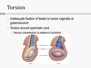
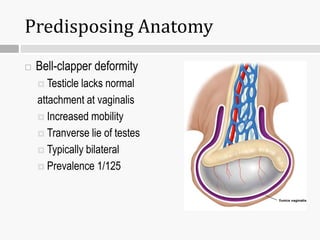
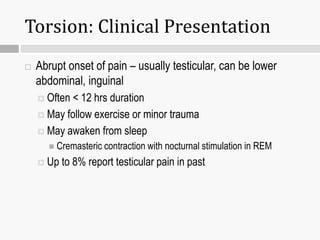
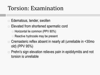
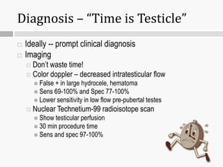
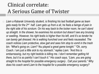
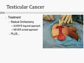
ACUTE SCROTAL PAIN
JAMES MWANGI K.
Embryology
 Descent of testes at 32-40 wks gestation
 Descends within processes vaginalis
 Outpouching of peritoneal cavity
 Tunica vaginalis is potential space that remains after
closure of process vaginalis
Anatomy
 Spermatic cord –testicular vessels, lymph, vas deferens
 Epididymis - sperm formed in testicle and undergo maturation, stored in
lower portion
 Vas Deferens – muscular action propels sperm up and out during
ejaculation
 Gubernaculum –
 fixation point for testicle to tunica vaginalis
 Tunica Vaginalis – potential space
 Encompasses anterior 2/3’s of testicle
 Tunica albuginea is inner layer opposing testis
Anatomy
Anterior
Posterior
Male External genitalia
 Scrotum
 Penis
Male internal genitalia
o Testes
o Epididymis
o Ductus deferens (Vas
deference)
o Ejaculatory duct
o Urethra.
o Spermatic cord
o Accessory organs are:-
seminal, prostate & bulbo-
urethral glands
Anatomy
Causes of Pain and Swelling
 Pain
 Testicular torsion
 Torsion of appendix testis
 Epididymitis
 Trauma
 Orchitis and Others
 Swelling
 Hydrocele
 Varicocele
 Spermatocele
 Tumor
Torsion
 Inadequate fixation of testes to tunica vaginalis at
gubernaculum
 Torsion around spermatic cord
 Venous compression to edema to ischemia
Epidemiology
 Accounts for 30% of all acute scrotal swelling
 Bimodal ages – neonatal (in utero) and pubertal ages
 65% occur in ages 12-18yo
 Incidence 1 in 4000 in males <25yo
 Increased incidence in puberty due to inc weight of testes
Predisposing Anatomy
 Bell-clapper deformity
 Testicle lacks normal
attachment at vaginalis
 Increased mobility
 Tranverse lie of testes
 Typically bilateral
 Prevalence 1/125
Torsion: Clinical Presentation
 Abrupt onset of pain – usually testicular, can be lower
abdominal, inguinal
 Often < 12 hrs duration
 May follow exercise or minor trauma
 May awaken from sleep
 Cremasteric contraction with nocturnal stimulation in REM
 Up to 8% report testicular pain in past
Torsion: Examination
 Edematous, tender, swollen
 Elevated from shortened spermatic cord
 Horizontal lie common (PPV 80%)
 Reactive hydrocele may be present
 Cremasteric reflex absent in nearly all (unreliable in <30mo
old) (PPV 95%)
 Prehn’s sign elevation relieves pain in epididymitis and not
torsion is unreliable
Diagnosis – “Time is Testicle”
 Ideally -- prompt clinical diagnosis
 Imaging
 Don’t waste time!
 Color doppler – decreased intratesticular flow
 False + in large hydrocele, hematoma
 Sens 69-100% and Spec 77-100%
 Lower sensitivity in low flow pre-pubertal testes
 Nuclear Technetium-99 radioisotope scan
 Show testicular perfusion
 30 min procedure time
 Sens and spec 97-100%
Management
 Detorsion within 6hr = 100% viability
 Within 12-24 hrs = 20% viability
 After 24 hrs = 0% viability
 Surgical detorsion and orchiopexy if viable
 Contralateral exploration and fixation if bell-clapper deformity
 Orchiectomy if non-viable testicle
 Never delay surgery on assumption of nonviability as
prolonged symptoms can represent periods of intermittent
torsion
Torsion: Special Considerations
 Adolescents may be embarrassed and not seek care until
late in course
 Torsion 10x more likely in undescended testicle
 Suspicious if empty scrotum, inguinal pain/swelling
Torsion of Appendix Testis
 Appendix testis
 Small vestigial structure,
remnant of Mullerium duct
 Pedunculated, 0.3cm long
 Other appendix structures
 Prepubertal estrogen may
enlarge appendix and cause
torsion
Torsion of Appendix Testis
 Peak age 3-13 yo (prepubertal)
 Sudden onset, pain less severe
 Classically, pain more often in abd or groin
 Non-tender testicle
 Tender mass at superior or inferior pole
 May be gangrenous, “blue-dot” (21% of cases)
 Normal cremasteric reflex, may have hydrocele
 Inc or normal flow by doppler U/S
Torsion of Appendix Testis
Blue dot of gangrenous
appendix testis
Torsion of Appendix Testis
 Management supportive
 analgesics, scrotal support to relieve swelling
 Surgery for persistent pain
 no need for contralateral exploration
 Liam a Kabarak University student, is finishing his last football game as team
gets ready for the 2nd half. Liam gets up from a sit, he feels a twinge of pain in
the right side of his scrotum. On his way back to the locker room, he can’t stand
up straight. In the shower, he examines his scrotum but doesn’t see any bruising
or swelling. However, his right testis is higher than his left, and it’s so tender he
can barely get dressed. He is walking hunched over and feels nauseated. The
coach notices Liam protective, slow gait and sees him stop to vomit in the trash
bin. “What’s going on, Liam? You played a great game tonight.” “Oh, sorry,
Coach. I am just a little sick to my stomach,” replies Liam. “And this is
embarrassing, but my right testicle is killing me. I don’t remember getting hit
there, but it is so painful I can’t touch it.” His coach says, “Liam, you are going
straight to the hospital for possible emergency surgery . Call your parents.” Why
does his coach send Liam to the hospital for a possible emergency surgery?
Clinical correlate:
A Serious Game of Twister
Epididymitis
 Inflammation of epididymis
 Subacute onset pain, swelling localized to epididymis,
duration of days
 With time swelling and pain less localized
 Testis has normal vertical lie
 Systemic signs of infection
 inc WBC and CRP, fever + in 95%
 Cremasteric reflex preserved
 Urinary complaints: discharge/dysuria PPV 80%
Epididymitis
 Scrotum has overlying erythema, edema in 60%
 Normal vertical
lie
Epididymitis
 Sexually active males
 Chlamydia > N. gonorrhea > E. coli
 Less commonly pseudomonas (elderly) and tuberculosis
(renal TB)
 Young boys, adolescents often post-infectious (adenovirus)
or anatomic
 Reflux of sterile urine through vas into epididymis
 50-75% of prepubertal boys have anatomic cause by imaging
Epididymitis Diagnosis
 Leukocytosis on UA in ~40% of patients
 PCR Chlamydia + in 50%, GC + in 20% of sexually active
 95% febrile at presentation
 Doppler scan show increased flow
 If hx consistent with STD, CDC recommends:
 Cx of urethral discharge, PCR for C and G
 Urine culture and UA
 Syphilis and HIV testing
Doppler Epididymitis
 Left Epididymitis
 Inc blood flow in
and around left testis
Epididymitis Treatment
 Sexually active treat with Ceftriaxone/Doxycycline or
Ofloxacin
 Pre-pubertal boys
 Treat for co-existing UTI if present
 Symptomatic tx with NASIDs, rest
 Referral all to GU for studies to rule out VUR, post urethral
valves, duplications
 Negative culture has 100% NPV for anomaly
Orchitis
 Inflammation/infection of testicle
 Swelling pain tenderness, erythema and shininess to overlying
skin
 Spread from epididymitis,
hematogenous, post-viral
 Viral: Mumps, coxsackie,
echovirus, parvovirus
 Bacterial: Brucellosis
Mumps Orchitis
 Extremely rare if vaccinated
 20-30% of pts with mumps, 70% unilateral, rare before
puberty
 Presents 4-6 days after mumps parotitis
 Impaired fertility in 15%, inc risk if bilateral
Trauma
 Result of testicular compression against the pubis bone,
from direct blow, or straddle injuries
 Extent depends on location of rupture
 Tunica albuginea ruptures (inner layer of tuncia vaginalis) allows
intratesticular hematoma to rupture into hematocele
 Rupture of tunica vaginalis allow blood to collect under scrotal
wall causing scrotal hematoma
 Doppler often sufficient to assess extent
 Surgery for uncertain dx, tunica albuginea rupture,
compromised doppler flow
Testicular Hematoma
 Blood as a filling
defect in testis
Other Causes of Pain
 Incarcerated inguinal hernia
 Henoch-Schonlein Purpura
 Vasculitis of testicular vessels
 Rarely presents with only scrotal pain
 Referred pain
 Retrocecal appendix, urolithiasis, lumbar/sacral nerve injury
 Non specific scrotal pain
 Minimal pain, nl exam – return immediately for inc
symptoms
Scrotal Swelling
 Hydrocele
 Varicocele
 Spermatocele
 Testicular Cancer
Approach to Scrotal Mass
Scrotal
Mass
Infectious Anatomic Malignancy
PAINFUL
• Epididymitis
• Orchitis
• Hydrocele
• Varicocele
• Spermatocele
• Torsion of Testis
• Torsion of Appendix Testis
• Testis Tumor
Hydrocele
 Fluid accumulation
in potential space of
tunica vaginalis
 May be primary from
patent PV or secondary
to torsion/epididymitis
Hydrocele
 Transilluminating
anterior cystic
mass
Hydrocele
 Mass increases in size during day or with crying and
decreases at night if communicating
 If non-communicating and <1 yo follow
 If communicating (enlarging), scrotum tense (may impair
blood flow) requires repair
 Unlikely to close spontaneously and predisposes to hernia
Varicocele
 Collection dilated veins in
pampiniform plexus
surrounding spermatic cord
 More common on left side
 R vein direct to IVC
 L vein acute angle to renal vein
 ~20% of all adolescent males
Varicocele
 Often asymptomatic or c/o dull ache/fullness upon standing
 Spermatic cord has ‘bag of worms’ appearance that
increased with standing/valsalva
 If prepubertal, rapidly enlarging, or persists in supine
position rule out IVC obstruction
 Most management conservatively
 Surgery if affected testis < unaffected testis volume
Spermatocele
 Painless sperm containing
cyst of testis, epipdidymis
 Distinct mass from testis
on exam
 Transilluminates
 Do not affect fertility
 Surgery for pain relief only
Acute Idiopathic Scrotal Edema
 Scrotal skin red and tender
 underlying testis normal
 no hydrocele
 Erythema extends off
scrotum onto perineum
 Empiric tx, cause unknown
 Antihistamine, steroids
 Resolves w/in 48-72hrs
Testicular Cancer
 Most common solid tumor in 15-30 yo males
 20% of all cancers in this group
 Painless mass
 Rapidly growing germ cell tumors may cause hemorrhage
and infarction
 Present as firm mass
 Typically do not transilluminate
 Diagnostic imaging with U/S initially
Testicular Cancer
 Typically occurs in young
healthy Men.
 Very good cure rates Even
for Metastatic Disease!
Testicular Cancer
Testis Cancer
Primary
Germ Cell
Tumors
Seminoma
Nonseminoma
Non-Germ Cell
Tumors
Secondary
Testicular Cancer
Testis Cancer
Primary
Germ Cell
Tumors
Seminoma
Nonseminoma
Non-Germ Cell
Tumors
Secondary
Germ Cell Testicular Cancer
 Seminoma
 Non-Seminoma
 Embryonal Carcinoma
 Teratoma
 Teratocarcinoma (Teratoma +Embryonal Carcinoma)
 Choriocarcinoma
 Yolk Sac Tumour (typically infants)
Testicular Cancer
Testis Cancer
Primary
Germ Cell
Tumors
Seminoma
Nonseminoma
Non-Germ Cell
Tumors
Secondary
Non-Germ Cell Testicular Cancer
 Leydig Cell Tumor
 Sertoli Cell Tumor
Testicular Cancer
Testis Cancer
Primary
Germ Cell
Tumors
Seminoma
Nonseminoma
Non-Germ Cell
Tumors
Secondary
Secondary Testicular Cancer
 Lymphoma
 Leukemia
Testicular Cancer
 Presentation
 Typically painless intratesticular mass discovered on self
examination
 Age 15-35
 Albeit some tumor subytpes cluster in infancy and some at later
age (60’s)
Testicular Cancer
 Investigations
 Labs
 B-HCG
 Produced by choriocarcinoma & in some Seminomas
 Alpha-fetoprotein
 Produced by Yolk Sac, Embryonal Carcinoma & Teratocarcinoma
 LDH
 Correlates with tumor volume
 Imaging
 Scrotal U/S
 CT Abdo and Pelvis: assess for retroperitoneal mets
 CXR
 +/- CT Head
Testicular Cancer
 Treatment:
 Radical Orchiectomy
 ALWAYS Inguinal approach
 NEVER scrotal approach
 PLUS…

scrotal conditions .pptx

  • 1.
  • 2.
    Embryology  Descent oftestes at 32-40 wks gestation  Descends within processes vaginalis  Outpouching of peritoneal cavity  Tunica vaginalis is potential space that remains after closure of process vaginalis
  • 3.
    Anatomy  Spermatic cord–testicular vessels, lymph, vas deferens  Epididymis - sperm formed in testicle and undergo maturation, stored in lower portion  Vas Deferens – muscular action propels sperm up and out during ejaculation  Gubernaculum –  fixation point for testicle to tunica vaginalis  Tunica Vaginalis – potential space  Encompasses anterior 2/3’s of testicle  Tunica albuginea is inner layer opposing testis
  • 4.
  • 5.
    Male External genitalia Scrotum  Penis Male internal genitalia o Testes o Epididymis o Ductus deferens (Vas deference) o Ejaculatory duct o Urethra. o Spermatic cord o Accessory organs are:- seminal, prostate & bulbo- urethral glands Anatomy
  • 6.
    Causes of Painand Swelling  Pain  Testicular torsion  Torsion of appendix testis  Epididymitis  Trauma  Orchitis and Others  Swelling  Hydrocele  Varicocele  Spermatocele  Tumor
  • 8.
    Torsion  Inadequate fixationof testes to tunica vaginalis at gubernaculum  Torsion around spermatic cord  Venous compression to edema to ischemia
  • 9.
    Epidemiology  Accounts for30% of all acute scrotal swelling  Bimodal ages – neonatal (in utero) and pubertal ages  65% occur in ages 12-18yo  Incidence 1 in 4000 in males <25yo  Increased incidence in puberty due to inc weight of testes
  • 10.
    Predisposing Anatomy  Bell-clapperdeformity  Testicle lacks normal attachment at vaginalis  Increased mobility  Tranverse lie of testes  Typically bilateral  Prevalence 1/125
  • 11.
    Torsion: Clinical Presentation Abrupt onset of pain – usually testicular, can be lower abdominal, inguinal  Often < 12 hrs duration  May follow exercise or minor trauma  May awaken from sleep  Cremasteric contraction with nocturnal stimulation in REM  Up to 8% report testicular pain in past
  • 12.
    Torsion: Examination  Edematous,tender, swollen  Elevated from shortened spermatic cord  Horizontal lie common (PPV 80%)  Reactive hydrocele may be present  Cremasteric reflex absent in nearly all (unreliable in <30mo old) (PPV 95%)  Prehn’s sign elevation relieves pain in epididymitis and not torsion is unreliable
  • 13.
    Diagnosis – “Timeis Testicle”  Ideally -- prompt clinical diagnosis  Imaging  Don’t waste time!  Color doppler – decreased intratesticular flow  False + in large hydrocele, hematoma  Sens 69-100% and Spec 77-100%  Lower sensitivity in low flow pre-pubertal testes  Nuclear Technetium-99 radioisotope scan  Show testicular perfusion  30 min procedure time  Sens and spec 97-100%
  • 14.
    Management  Detorsion within6hr = 100% viability  Within 12-24 hrs = 20% viability  After 24 hrs = 0% viability  Surgical detorsion and orchiopexy if viable  Contralateral exploration and fixation if bell-clapper deformity  Orchiectomy if non-viable testicle  Never delay surgery on assumption of nonviability as prolonged symptoms can represent periods of intermittent torsion
  • 15.
    Torsion: Special Considerations Adolescents may be embarrassed and not seek care until late in course  Torsion 10x more likely in undescended testicle  Suspicious if empty scrotum, inguinal pain/swelling
  • 16.
    Torsion of AppendixTestis  Appendix testis  Small vestigial structure, remnant of Mullerium duct  Pedunculated, 0.3cm long  Other appendix structures  Prepubertal estrogen may enlarge appendix and cause torsion
  • 17.
    Torsion of AppendixTestis  Peak age 3-13 yo (prepubertal)  Sudden onset, pain less severe  Classically, pain more often in abd or groin  Non-tender testicle  Tender mass at superior or inferior pole  May be gangrenous, “blue-dot” (21% of cases)  Normal cremasteric reflex, may have hydrocele  Inc or normal flow by doppler U/S
  • 18.
    Torsion of AppendixTestis Blue dot of gangrenous appendix testis
  • 19.
    Torsion of AppendixTestis  Management supportive  analgesics, scrotal support to relieve swelling  Surgery for persistent pain  no need for contralateral exploration
  • 20.
     Liam aKabarak University student, is finishing his last football game as team gets ready for the 2nd half. Liam gets up from a sit, he feels a twinge of pain in the right side of his scrotum. On his way back to the locker room, he can’t stand up straight. In the shower, he examines his scrotum but doesn’t see any bruising or swelling. However, his right testis is higher than his left, and it’s so tender he can barely get dressed. He is walking hunched over and feels nauseated. The coach notices Liam protective, slow gait and sees him stop to vomit in the trash bin. “What’s going on, Liam? You played a great game tonight.” “Oh, sorry, Coach. I am just a little sick to my stomach,” replies Liam. “And this is embarrassing, but my right testicle is killing me. I don’t remember getting hit there, but it is so painful I can’t touch it.” His coach says, “Liam, you are going straight to the hospital for possible emergency surgery . Call your parents.” Why does his coach send Liam to the hospital for a possible emergency surgery? Clinical correlate: A Serious Game of Twister
  • 21.
    Epididymitis  Inflammation ofepididymis  Subacute onset pain, swelling localized to epididymis, duration of days  With time swelling and pain less localized  Testis has normal vertical lie  Systemic signs of infection  inc WBC and CRP, fever + in 95%  Cremasteric reflex preserved  Urinary complaints: discharge/dysuria PPV 80%
  • 22.
    Epididymitis  Scrotum hasoverlying erythema, edema in 60%  Normal vertical lie
  • 23.
    Epididymitis  Sexually activemales  Chlamydia > N. gonorrhea > E. coli  Less commonly pseudomonas (elderly) and tuberculosis (renal TB)  Young boys, adolescents often post-infectious (adenovirus) or anatomic  Reflux of sterile urine through vas into epididymis  50-75% of prepubertal boys have anatomic cause by imaging
  • 24.
    Epididymitis Diagnosis  Leukocytosison UA in ~40% of patients  PCR Chlamydia + in 50%, GC + in 20% of sexually active  95% febrile at presentation  Doppler scan show increased flow  If hx consistent with STD, CDC recommends:  Cx of urethral discharge, PCR for C and G  Urine culture and UA  Syphilis and HIV testing
  • 25.
    Doppler Epididymitis  LeftEpididymitis  Inc blood flow in and around left testis
  • 26.
    Epididymitis Treatment  Sexuallyactive treat with Ceftriaxone/Doxycycline or Ofloxacin  Pre-pubertal boys  Treat for co-existing UTI if present  Symptomatic tx with NASIDs, rest  Referral all to GU for studies to rule out VUR, post urethral valves, duplications  Negative culture has 100% NPV for anomaly
  • 27.
    Orchitis  Inflammation/infection oftesticle  Swelling pain tenderness, erythema and shininess to overlying skin  Spread from epididymitis, hematogenous, post-viral  Viral: Mumps, coxsackie, echovirus, parvovirus  Bacterial: Brucellosis
  • 28.
    Mumps Orchitis  Extremelyrare if vaccinated  20-30% of pts with mumps, 70% unilateral, rare before puberty  Presents 4-6 days after mumps parotitis  Impaired fertility in 15%, inc risk if bilateral
  • 29.
    Trauma  Result oftesticular compression against the pubis bone, from direct blow, or straddle injuries  Extent depends on location of rupture  Tunica albuginea ruptures (inner layer of tuncia vaginalis) allows intratesticular hematoma to rupture into hematocele  Rupture of tunica vaginalis allow blood to collect under scrotal wall causing scrotal hematoma  Doppler often sufficient to assess extent  Surgery for uncertain dx, tunica albuginea rupture, compromised doppler flow
  • 30.
    Testicular Hematoma  Bloodas a filling defect in testis
  • 31.
    Other Causes ofPain  Incarcerated inguinal hernia  Henoch-Schonlein Purpura  Vasculitis of testicular vessels  Rarely presents with only scrotal pain  Referred pain  Retrocecal appendix, urolithiasis, lumbar/sacral nerve injury  Non specific scrotal pain  Minimal pain, nl exam – return immediately for inc symptoms
  • 32.
    Scrotal Swelling  Hydrocele Varicocele  Spermatocele  Testicular Cancer
  • 33.
    Approach to ScrotalMass Scrotal Mass Infectious Anatomic Malignancy PAINFUL • Epididymitis • Orchitis • Hydrocele • Varicocele • Spermatocele • Torsion of Testis • Torsion of Appendix Testis • Testis Tumor
  • 34.
    Hydrocele  Fluid accumulation inpotential space of tunica vaginalis  May be primary from patent PV or secondary to torsion/epididymitis
  • 35.
  • 36.
    Hydrocele  Mass increasesin size during day or with crying and decreases at night if communicating  If non-communicating and <1 yo follow  If communicating (enlarging), scrotum tense (may impair blood flow) requires repair  Unlikely to close spontaneously and predisposes to hernia
  • 37.
    Varicocele  Collection dilatedveins in pampiniform plexus surrounding spermatic cord  More common on left side  R vein direct to IVC  L vein acute angle to renal vein  ~20% of all adolescent males
  • 38.
    Varicocele  Often asymptomaticor c/o dull ache/fullness upon standing  Spermatic cord has ‘bag of worms’ appearance that increased with standing/valsalva  If prepubertal, rapidly enlarging, or persists in supine position rule out IVC obstruction  Most management conservatively  Surgery if affected testis < unaffected testis volume
  • 39.
    Spermatocele  Painless spermcontaining cyst of testis, epipdidymis  Distinct mass from testis on exam  Transilluminates  Do not affect fertility  Surgery for pain relief only
  • 40.
    Acute Idiopathic ScrotalEdema  Scrotal skin red and tender  underlying testis normal  no hydrocele  Erythema extends off scrotum onto perineum  Empiric tx, cause unknown  Antihistamine, steroids  Resolves w/in 48-72hrs
  • 41.
    Testicular Cancer  Mostcommon solid tumor in 15-30 yo males  20% of all cancers in this group  Painless mass  Rapidly growing germ cell tumors may cause hemorrhage and infarction  Present as firm mass  Typically do not transilluminate  Diagnostic imaging with U/S initially
  • 42.
    Testicular Cancer  Typicallyoccurs in young healthy Men.  Very good cure rates Even for Metastatic Disease!
  • 43.
    Testicular Cancer Testis Cancer Primary GermCell Tumors Seminoma Nonseminoma Non-Germ Cell Tumors Secondary
  • 44.
    Testicular Cancer Testis Cancer Primary GermCell Tumors Seminoma Nonseminoma Non-Germ Cell Tumors Secondary
  • 45.
    Germ Cell TesticularCancer  Seminoma  Non-Seminoma  Embryonal Carcinoma  Teratoma  Teratocarcinoma (Teratoma +Embryonal Carcinoma)  Choriocarcinoma  Yolk Sac Tumour (typically infants)
  • 46.
    Testicular Cancer Testis Cancer Primary GermCell Tumors Seminoma Nonseminoma Non-Germ Cell Tumors Secondary
  • 47.
    Non-Germ Cell TesticularCancer  Leydig Cell Tumor  Sertoli Cell Tumor
  • 48.
    Testicular Cancer Testis Cancer Primary GermCell Tumors Seminoma Nonseminoma Non-Germ Cell Tumors Secondary
  • 49.
    Secondary Testicular Cancer Lymphoma  Leukemia
  • 50.
    Testicular Cancer  Presentation Typically painless intratesticular mass discovered on self examination  Age 15-35  Albeit some tumor subytpes cluster in infancy and some at later age (60’s)
  • 51.
    Testicular Cancer  Investigations Labs  B-HCG  Produced by choriocarcinoma & in some Seminomas  Alpha-fetoprotein  Produced by Yolk Sac, Embryonal Carcinoma & Teratocarcinoma  LDH  Correlates with tumor volume  Imaging  Scrotal U/S  CT Abdo and Pelvis: assess for retroperitoneal mets  CXR  +/- CT Head
  • 52.
    Testicular Cancer  Treatment: Radical Orchiectomy  ALWAYS Inguinal approach  NEVER scrotal approach  PLUS…