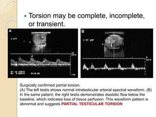
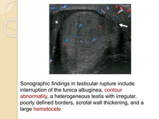
Acute Painful Scrotum
Dr. Tejas M. Tamhane.
Resident, Dept. of Radio-
Diagnosis.
Introduction
 High-frequency transducer
sonography using gray scale along
with pulsed and color Doppler is the
imaging modality of choice for
evaluating patients who present with
acute scrotal pain.
 This article is organized on the basis
of the pathophysiology of the disease
process with emphasis on color
Doppler when applicable.
Imaging Anatomy
The posterior surface of the tunica albuginea is reflected
into the interior of the gland, which forms the incomplete
septum known as the MEDIASTINUM of the testis.
Sonographically, the mediastinum of the testis is an
echogenic
Band.
Vascular supply
 Rt. & Lt. Testicular arteries- Testis.
 Deferential artery and Cremasteric
artery- epididymis, vas deferens, and
peritesticular tissue.
 Pudendal artery - Scrotal wall.
 Venous drainage – Pampiniform
Plexus.
 Four testicular appendages have been
described:- the appendix testis, the appendix
epididymis, the vas aberrans and the
paradidymis.
 They are remnants of embryologic ducts.
Pathology
Epididymo-orchitis :-
 Acute epididymo-orchitis or
epididymitis is the most common
cause of acute scrotum in adolescent
boys and adults.
 Sexually transmitted Chlamydia
trachomatis and Neisseria gonorrhea
are common pathogens in men
younger than 35 years.
 In prepubertal & >35yrs- Proteus &
Other causes –
 Sarcoidosis, brucellosis, tuberculosis,
cryptococcus and mumps.
 Amiodarone (Chemical epididymitis)
 Complications- chronic pain, infarction,
abscess, gangrene, infertility, atrophy
and pyocele.
USG
Markedly
enlarged
epididymis
(arrows) with
variable
echotexture.
Reactive hydrocele or pyocele with scrotal
wall thickening.
Diffuse testicular involvement is confirmed
by testicular enlargement and heterogenous
testicular Echotexture.
 The increased blood flow to the
epididymis and testis on color Doppler
examination is a well-established
criterion for the diagnosis of
epididymo-orchitis.
In normal- RI
rarely
< 0.5
Epididymorchitis
-
RI < 0.5 in 50%
cases.
Primary orchitis
 Mumps is the commonest cause of
orchitis without accompanying
epididymitis and is bilateral in 14% to
35% of cases.
 Enlarged and decreased
echogenecity.
 Because intratesticular venous flow is
difficult to detect in normal testes,
increased and easily detected venous
flow in the testes greatly suggests
Fournier’s Gangrene
 Urologic emergency.
 Diagnosis- primarily on clinical
examination rather than on imaging
studies.
 Fournier’s gangrene is a synergistic
polymicrobial necrotizing fascitis of the
perineum or perirectal or genital area
that predominantly affects the scrotum
in men and frequently extends to
involve the lower abdominal wall.
 Predisposing conditions include DM,
alcoholism, advanced age, and
immunodeficiency syndrome.
 Klebsiella, Proteus, Streptococcus,
Staphylococcus, Peptostreptococcus,
Escherichia coli and Clostridium
perfringens.
 Subcutaneous gas within the scrotal
wall is the sonographic hallmark of
Fournier’s gangrene.
Intratesticular abscess
 Usually secondary to
epididymoorchitis,but other causes
include mumps, trauma and testicular
infarction.
 The sonographic features include shaggy
irregular walls, an intratesticular location,
low-level internal echoes and occasional
hypervascular margins.
Surgically confirmed TESTICULAR ABSCESS.
Transverse
US of the testis (T) shows fluid-debris level (arrow)
consistent with intratesticular abscess that developed
secondary to epididymo-orchitis.
Testicular torsion
 Testicular torsion and epididymo-
orchitis commonly present with pain.
 Prehn test/maneuver.
 Sudden onset of pain followed by
nausea, vomiting and a low-grade
fever.
 Physical examination reveals a
swollen, tender and inflamed
hemiscrotum.
 The cremasteric reflex is usually
 Testicular torsion causes venous
engorgement that results in edema,
hemorrhage and subsequent arterial
compromise, which results in testicular
ischemia.
 The testicular salvage rate depends on
the degree of torsion and the duration
of ischemia. A nearly 100% salvage
rate exists within the first 6 hours after
the onset of symptoms, a 70% rate in 6
to 12 hours, and a 20% rate in 12 to 24
hours.
Surgically confirmed testicular torsion.
(A)Color Doppler US of the testis (T) demonstrates the absence of
intratesticular blood flow with peripheral hyperemia (arrows).
(B) Involvement of the epididymis in testicular torsion. There is no
blood flow within the testis (T) or epididymis (E).
Peripheral hyperemia is seen (arrow).
 Testicular swelling and decreased
echogenicity are the most commonly
encountered findings 4 to 6 hours after
the onset of torsion.
 Torsion may be complete, incomplete,
or transient.
Surgically confirmed partial torsion.
(A) The left testis shows normal intratesticular arterial spectral waveform. (B)
In the same patient, the right testis demonstrates diastolic flow below the
baseline, which indicates loss of tissue perfusion. This waveform pattern is
abnormal and suggests PARTIAL TESTICULAR TORSION
Varicocele
Idiopathic varicocele
 Abnormal dilatation of the veins of the
pampiniform plexus results in
varicocele,which is usually caused by
incompetent valves in the internal spermatic
vein.
 Patients with idiopathic varicoceles usually
present between the ages of 15 and 25
years.
 Varicocele is a clinical diagnosis, and
palpation reveals a scrotal mass that may
feel like a bag of worms with or without a
 Varicoceles are more common on the left side
for the following reasons:
(1) the left testicular vein is longer,
(2) the left testicular vein enters the left renal
vein at a right angle,
(3) in some men, the left testicular artery arches
over the left renal vein, thereby compressing it,
(4) the descending colon distended with feces
may compress the left testicular vein, and
(5) a ‘‘nutcracker’’ effect of compression of the
left renal vein may occur between the superior
mesenteric artery and the abdominal aorta.
The clinical gradation of
varicoceles
 Grade I - Not visible but palpable on
Valsalva’s maneuver.
 Grade II - Less visible but palpable
without Valsalva’s maneuver.
 Grade III - Always visually identifiable
and palpable without Valsalva’s
maneuver.
USG
varicocele consists of multiple, serpigenous, tubular
structures of varying sizes larger than 2 mm in diameter,
which are usually best visualized superior or
lateral to the testis.
sensitivity and specificity rates of varicocele detection
approach 100% with color Doppler sonography.
Secondary varicoceles
 Secondary varicoceles result from
increased pressure on the spermatic
vein produced by disease processes,
such as hydronephrosis, cirrhosis, or
abdominal neoplasm.
 Neoplasm is the most likely cause of
nondecompressible varicocele in men
over 40 years of age.
Intratesticular varicocele
 Patients with intratesticular varicocele may have
pain related to passive congestion of the testis,
which eventually stretches the tunica albuginea.
 Sonographic features include multiple
anechoic,serpigenous, tubular structures of
varying sizes within the testis.
Testicular trauma
 Testicular trauma typically results from
athletic injury, a motor vehicle accident, a
direct blow, straddle injury, or penetrating
gunshot trauma.
 direct blow to the testis with impingement
against the symphysis pubis or ischial ramus
is the most common mechanism of injury
from blunt trauma.
 Approximately 50 kg of pressure is necessary
to rupture the tunica albuginea during blunt
trauma.
 Testicular rupture is a surgical emergency
and more than 80% of ruptured testes can be
saved if surgery is performed within 72 hours
Sonographic findings in testicular rupture include
interruption of the tunica albuginea, contour
abnormality, a heterogeneous testis with irregular,
poorly defined borders, scrotal wall thickening, and a
large hematocele
Testicular tumors
 Testicular tumors sometimes can present with
acute pain. This presentation is usually
secondary to epididymo-orchitis or
hemorrhage within the tumor.
 Seminoma is the most common tumor to
masquerade as acute orchitis. It is presumed
to infiltrate and obstruct the seminiferous
tubules, and it results in orchitis.
 Gray scale findings of intratesticular tumors
are nonspecific and usually hypoechoic in
appearance.
 Hyperemia also can be seen in testicular
tumors.
Summary
 The ability of US to diagnose the pathogenesis of
the acute scrotum is unsurpassed by any other
imaging modality.
 It is the first imaging performed in patients with
acute scrotum.
 Knowledge of the normal and pathologic
sonographic appearance of the scrotum and
proper sonographic technique is essential for
accurate diagnosis of acute scrotum.
 High-frequency transducer sonography
combined with color flow Doppler sonography
provides the information essential to reach a
specific diagnosis in patients with testicular
torsion, epididymo-orchitis, and testicular trauma.
Thank You..!!

Acute painful scrotum

  • 1.
    Acute Painful Scrotum Dr.Tejas M. Tamhane. Resident, Dept. of Radio- Diagnosis.
  • 2.
    Introduction  High-frequency transducer sonographyusing gray scale along with pulsed and color Doppler is the imaging modality of choice for evaluating patients who present with acute scrotal pain.  This article is organized on the basis of the pathophysiology of the disease process with emphasis on color Doppler when applicable.
  • 3.
  • 4.
    The posterior surfaceof the tunica albuginea is reflected into the interior of the gland, which forms the incomplete septum known as the MEDIASTINUM of the testis. Sonographically, the mediastinum of the testis is an echogenic Band.
  • 5.
    Vascular supply  Rt.& Lt. Testicular arteries- Testis.  Deferential artery and Cremasteric artery- epididymis, vas deferens, and peritesticular tissue.  Pudendal artery - Scrotal wall.  Venous drainage – Pampiniform Plexus.
  • 6.
     Four testicularappendages have been described:- the appendix testis, the appendix epididymis, the vas aberrans and the paradidymis.  They are remnants of embryologic ducts.
  • 7.
    Pathology Epididymo-orchitis :-  Acuteepididymo-orchitis or epididymitis is the most common cause of acute scrotum in adolescent boys and adults.  Sexually transmitted Chlamydia trachomatis and Neisseria gonorrhea are common pathogens in men younger than 35 years.  In prepubertal & >35yrs- Proteus &
  • 8.
    Other causes – Sarcoidosis, brucellosis, tuberculosis, cryptococcus and mumps.  Amiodarone (Chemical epididymitis)  Complications- chronic pain, infarction, abscess, gangrene, infertility, atrophy and pyocele.
  • 9.
    USG Markedly enlarged epididymis (arrows) with variable echotexture. Reactive hydroceleor pyocele with scrotal wall thickening. Diffuse testicular involvement is confirmed by testicular enlargement and heterogenous testicular Echotexture.
  • 10.
     The increasedblood flow to the epididymis and testis on color Doppler examination is a well-established criterion for the diagnosis of epididymo-orchitis. In normal- RI rarely < 0.5 Epididymorchitis - RI < 0.5 in 50% cases.
  • 11.
    Primary orchitis  Mumpsis the commonest cause of orchitis without accompanying epididymitis and is bilateral in 14% to 35% of cases.  Enlarged and decreased echogenecity.  Because intratesticular venous flow is difficult to detect in normal testes, increased and easily detected venous flow in the testes greatly suggests
  • 12.
    Fournier’s Gangrene  Urologicemergency.  Diagnosis- primarily on clinical examination rather than on imaging studies.  Fournier’s gangrene is a synergistic polymicrobial necrotizing fascitis of the perineum or perirectal or genital area that predominantly affects the scrotum in men and frequently extends to involve the lower abdominal wall.
  • 13.
     Predisposing conditionsinclude DM, alcoholism, advanced age, and immunodeficiency syndrome.  Klebsiella, Proteus, Streptococcus, Staphylococcus, Peptostreptococcus, Escherichia coli and Clostridium perfringens.
  • 14.
     Subcutaneous gaswithin the scrotal wall is the sonographic hallmark of Fournier’s gangrene.
  • 15.
    Intratesticular abscess  Usuallysecondary to epididymoorchitis,but other causes include mumps, trauma and testicular infarction.  The sonographic features include shaggy irregular walls, an intratesticular location, low-level internal echoes and occasional hypervascular margins.
  • 16.
    Surgically confirmed TESTICULARABSCESS. Transverse US of the testis (T) shows fluid-debris level (arrow) consistent with intratesticular abscess that developed secondary to epididymo-orchitis.
  • 17.
    Testicular torsion  Testiculartorsion and epididymo- orchitis commonly present with pain.  Prehn test/maneuver.  Sudden onset of pain followed by nausea, vomiting and a low-grade fever.  Physical examination reveals a swollen, tender and inflamed hemiscrotum.  The cremasteric reflex is usually
  • 18.
     Testicular torsioncauses venous engorgement that results in edema, hemorrhage and subsequent arterial compromise, which results in testicular ischemia.  The testicular salvage rate depends on the degree of torsion and the duration of ischemia. A nearly 100% salvage rate exists within the first 6 hours after the onset of symptoms, a 70% rate in 6 to 12 hours, and a 20% rate in 12 to 24 hours.
  • 19.
    Surgically confirmed testiculartorsion. (A)Color Doppler US of the testis (T) demonstrates the absence of intratesticular blood flow with peripheral hyperemia (arrows). (B) Involvement of the epididymis in testicular torsion. There is no blood flow within the testis (T) or epididymis (E). Peripheral hyperemia is seen (arrow).
  • 20.
     Testicular swellingand decreased echogenicity are the most commonly encountered findings 4 to 6 hours after the onset of torsion.
  • 21.
     Torsion maybe complete, incomplete, or transient. Surgically confirmed partial torsion. (A) The left testis shows normal intratesticular arterial spectral waveform. (B) In the same patient, the right testis demonstrates diastolic flow below the baseline, which indicates loss of tissue perfusion. This waveform pattern is abnormal and suggests PARTIAL TESTICULAR TORSION
  • 22.
    Varicocele Idiopathic varicocele  Abnormaldilatation of the veins of the pampiniform plexus results in varicocele,which is usually caused by incompetent valves in the internal spermatic vein.  Patients with idiopathic varicoceles usually present between the ages of 15 and 25 years.  Varicocele is a clinical diagnosis, and palpation reveals a scrotal mass that may feel like a bag of worms with or without a
  • 23.
     Varicoceles aremore common on the left side for the following reasons: (1) the left testicular vein is longer, (2) the left testicular vein enters the left renal vein at a right angle, (3) in some men, the left testicular artery arches over the left renal vein, thereby compressing it, (4) the descending colon distended with feces may compress the left testicular vein, and (5) a ‘‘nutcracker’’ effect of compression of the left renal vein may occur between the superior mesenteric artery and the abdominal aorta.
  • 24.
    The clinical gradationof varicoceles  Grade I - Not visible but palpable on Valsalva’s maneuver.  Grade II - Less visible but palpable without Valsalva’s maneuver.  Grade III - Always visually identifiable and palpable without Valsalva’s maneuver.
  • 25.
    USG varicocele consists ofmultiple, serpigenous, tubular structures of varying sizes larger than 2 mm in diameter, which are usually best visualized superior or lateral to the testis. sensitivity and specificity rates of varicocele detection approach 100% with color Doppler sonography.
  • 26.
    Secondary varicoceles  Secondaryvaricoceles result from increased pressure on the spermatic vein produced by disease processes, such as hydronephrosis, cirrhosis, or abdominal neoplasm.  Neoplasm is the most likely cause of nondecompressible varicocele in men over 40 years of age.
  • 27.
    Intratesticular varicocele  Patientswith intratesticular varicocele may have pain related to passive congestion of the testis, which eventually stretches the tunica albuginea.  Sonographic features include multiple anechoic,serpigenous, tubular structures of varying sizes within the testis.
  • 28.
    Testicular trauma  Testiculartrauma typically results from athletic injury, a motor vehicle accident, a direct blow, straddle injury, or penetrating gunshot trauma.  direct blow to the testis with impingement against the symphysis pubis or ischial ramus is the most common mechanism of injury from blunt trauma.  Approximately 50 kg of pressure is necessary to rupture the tunica albuginea during blunt trauma.  Testicular rupture is a surgical emergency and more than 80% of ruptured testes can be saved if surgery is performed within 72 hours
  • 29.
    Sonographic findings intesticular rupture include interruption of the tunica albuginea, contour abnormality, a heterogeneous testis with irregular, poorly defined borders, scrotal wall thickening, and a large hematocele
  • 30.
    Testicular tumors  Testiculartumors sometimes can present with acute pain. This presentation is usually secondary to epididymo-orchitis or hemorrhage within the tumor.  Seminoma is the most common tumor to masquerade as acute orchitis. It is presumed to infiltrate and obstruct the seminiferous tubules, and it results in orchitis.  Gray scale findings of intratesticular tumors are nonspecific and usually hypoechoic in appearance.  Hyperemia also can be seen in testicular tumors.
  • 31.
    Summary  The abilityof US to diagnose the pathogenesis of the acute scrotum is unsurpassed by any other imaging modality.  It is the first imaging performed in patients with acute scrotum.  Knowledge of the normal and pathologic sonographic appearance of the scrotum and proper sonographic technique is essential for accurate diagnosis of acute scrotum.  High-frequency transducer sonography combined with color flow Doppler sonography provides the information essential to reach a specific diagnosis in patients with testicular torsion, epididymo-orchitis, and testicular trauma.
  • 32.