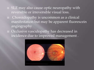
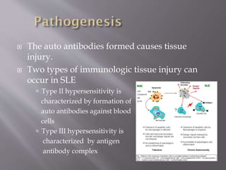
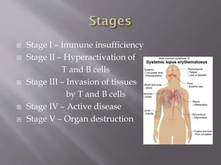
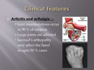
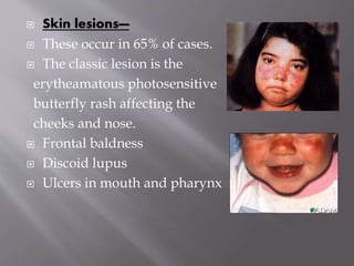
2. Classification
1. Systemic form – Acute and chronic
inflammatory lesions widely scattered in the
body
2. Discoid form – Chronic and localized skin
lesions involving the bridge of nose and
adjacent cheeks
3. 1) Antinuclear antibodies (ANA) are the
antibodies against common nuclear antigen
that includes DNA as well as RNA
2) Antibodies to double stranded DNA most specific
for SLE
3) Anti smith Antibodies act against smith antigens
which is part of ribonucleoproteins
4) Other non specific antibodies – Anti RNP, Anti
Histone, Antiphospholipid
4. The source of these autoantibodies as well
hyper gammaglobulinaemia seen in SLE is the
polyclonal activation of B cells brought about
by following derangements
1. Immunologic factors–B cell or T cell defect
2. Genetic factors-Class II HLA gene defect
3. Other factors-Drugs, Viral infections,
Hormones
5. The auto antibodies formed causes tissue
injury.
Two types of immunologic tissue injury can
occur in SLE
Type II hypersensitivity is
characterized by formation of
auto antibodies against blood
cells
Type III hypersensitivity is
characterized by antigen
antibody complex
6. Stage I – Immune insufficiency
Stage II – Hyperactivation of
T and B cells
Stage III – Invasion of tissues
by T and B cells
Stage IV – Active disease
Stage V – Organ destruction
7. Arthritis and arthalgia ---
Joint manifestations occur
in 90 % of subjects
Large joints are affected
Jaccoud’s arthopathy
may affect the hand
in upto 50 % cases
8. Skin lesions—
These occur in 65% of cases.
The classic lesion is the
erytheamatous photosensitive
butterfly rash affecting the
cheeks and nose.
Frontal baldness
Discoid lupus
Ulcers in mouth and pharynx
9. Cardiovascular lesions
Develop in 25 – 40% cases
Cardiac lesions include
pericarditis, pericardial
effusion, myocarditis etc
Libman sacks endocarditis
is occasionally seen
Vasculitis leads to
Raynaud’s phenomenon,
necrotic ulcers of the finger pulp
10. Respiratory system
30 % cases
Common lesions are dry
pleurisy, pleural effusion ,
fibrosing alveolitis and lupus
pneumonitis
Kidney
60 % cases
Renal involvement includes
Nephrotic syndrome ,
haemturia, Acute nephritic
syndrome
11. Nervous system
Affected in 40 % cases
Neurological manifestation
usually is a late manifestation
Lesions are caused due to
vasculitis
Includes stroke, psychosis,
convulsions, cranial nerve palsies,
peripheral neuropathy.
Muscular system
Myalgia, Muscle wasting,
12. Eye
Occulr involvement in SLE
may arise from embolic lesions
of libman sacks endocarditis or
vascular occlusions resulting
from the hypercoagubility
associated with lupus
anticoagulants
Retinal vascular disease
including retinal arterial or
venous occlusion and Retinal
haemorroage or retinal oedema
may be seen
13. SLE may also cause optic neuropathy with
reversible or irreversible visual loss.
Choroidopathy is uncommon as a clinical
manifestation but may be apparent fluorescein
angiography
Occlusive vasculopathy has decreased in
incidence due to improved management .
14. There is no cure for SLE. The goal of treatment
is to control symptoms.
Mild disease may be treated with: ----
Nonsteroidal anti-inflammatory medications
(NSAIDs) treat arthritis and pleurisy
Corticosteroid creams to treat skin rashes
An antimalarial drug (hydroxychloroquine) and
low-dose corticosteroids for skin and arthritis
symptoms
Intravenous Immunoglobulins
15. Severe or life-threatening symptoms (such
as hemolytic anemia, extensive heart or lung
involvement, kidney disease, or central
nervous system involvement) often require
more aggressive treatment. Treatment for
more severe lupus may include :-
High-dose corticosteroids or medications to
decrease the immune system response
Cytotoxic drugs