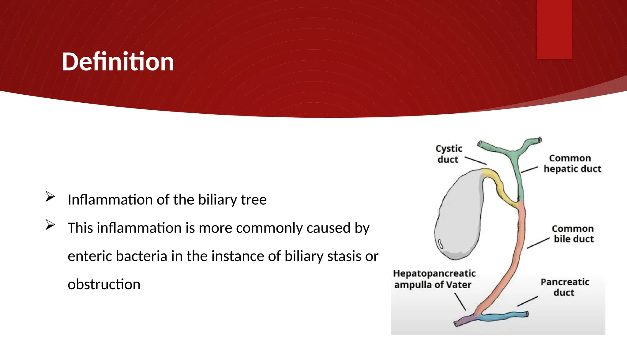
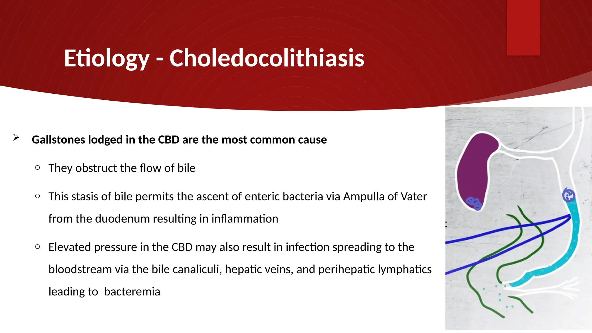
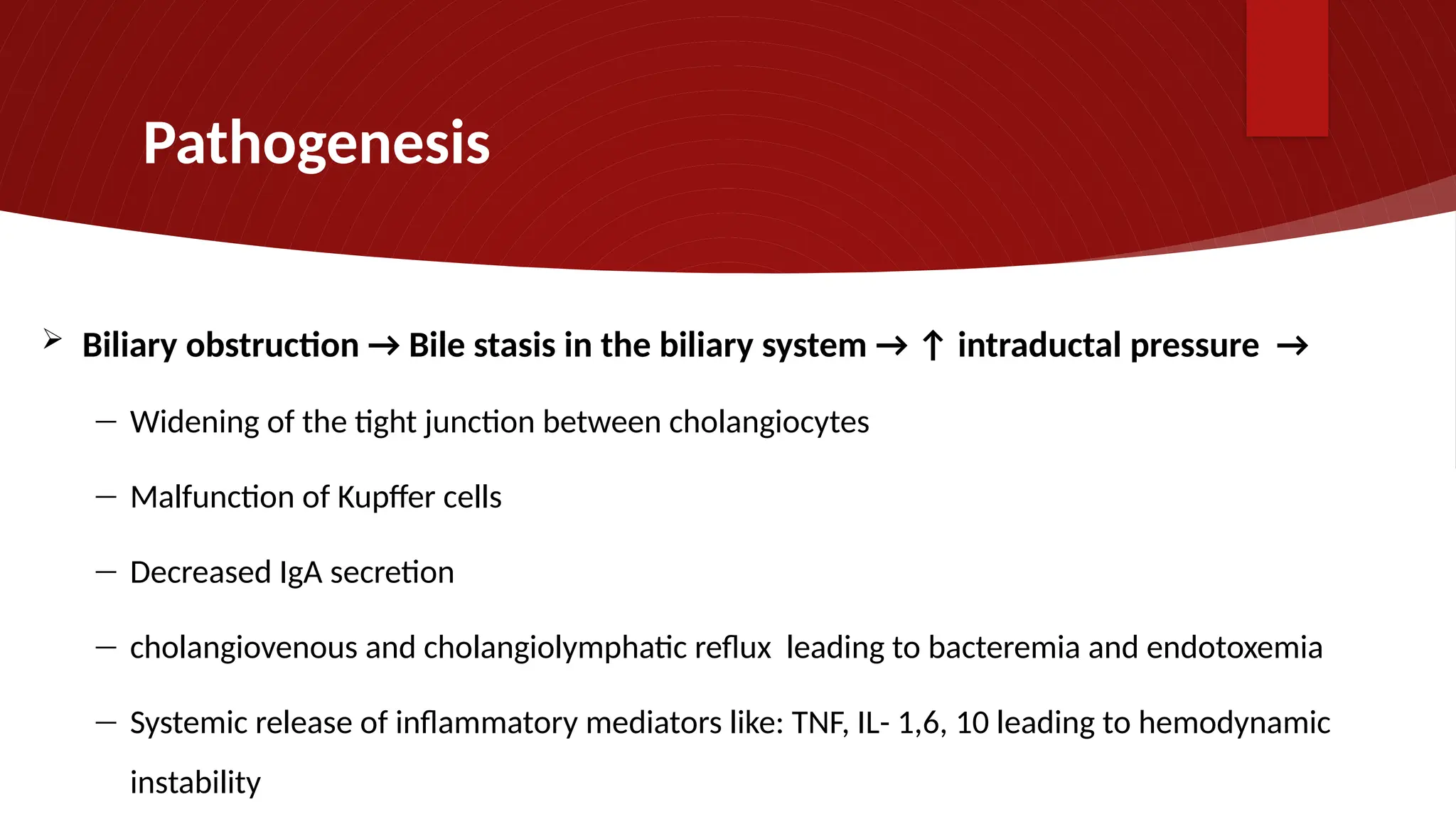
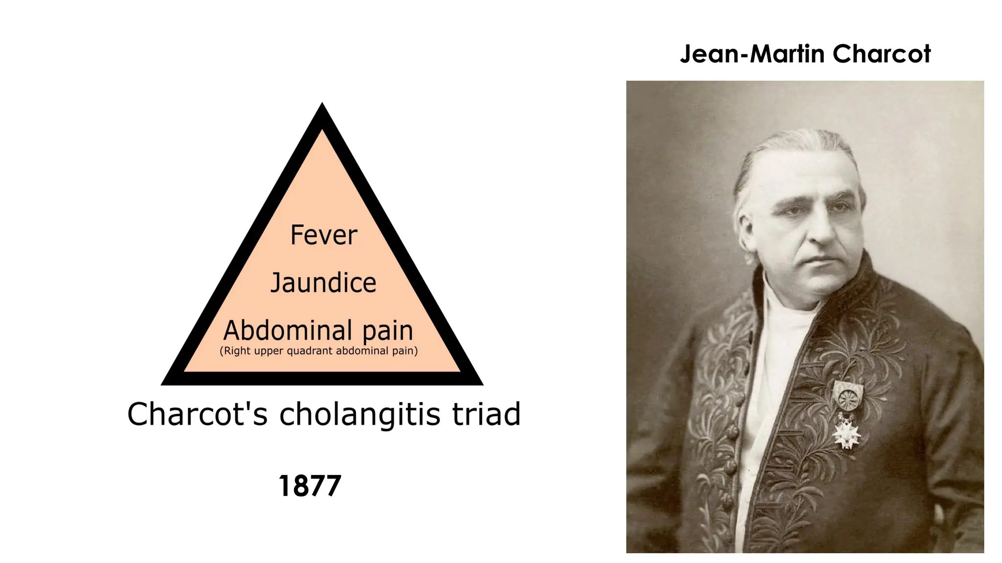
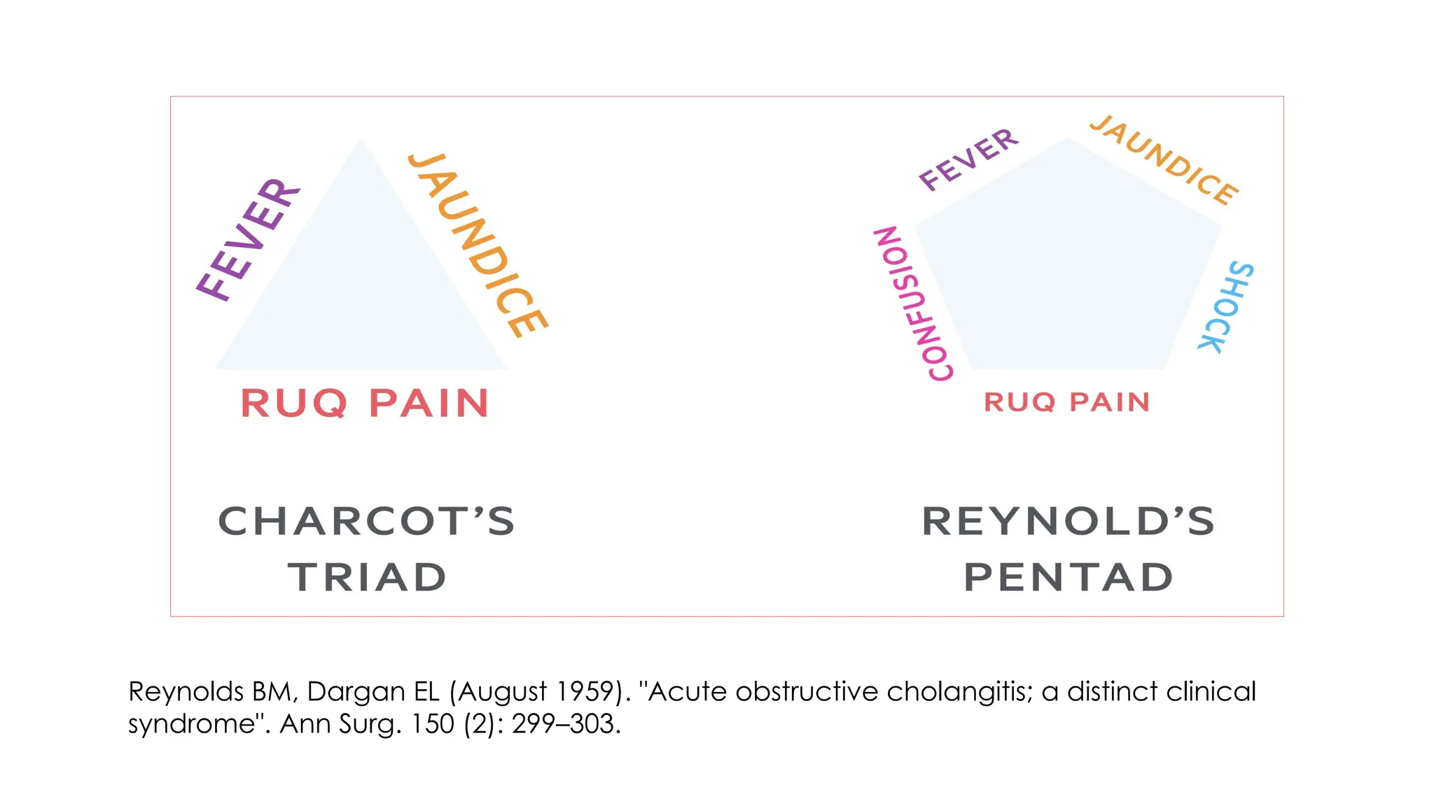
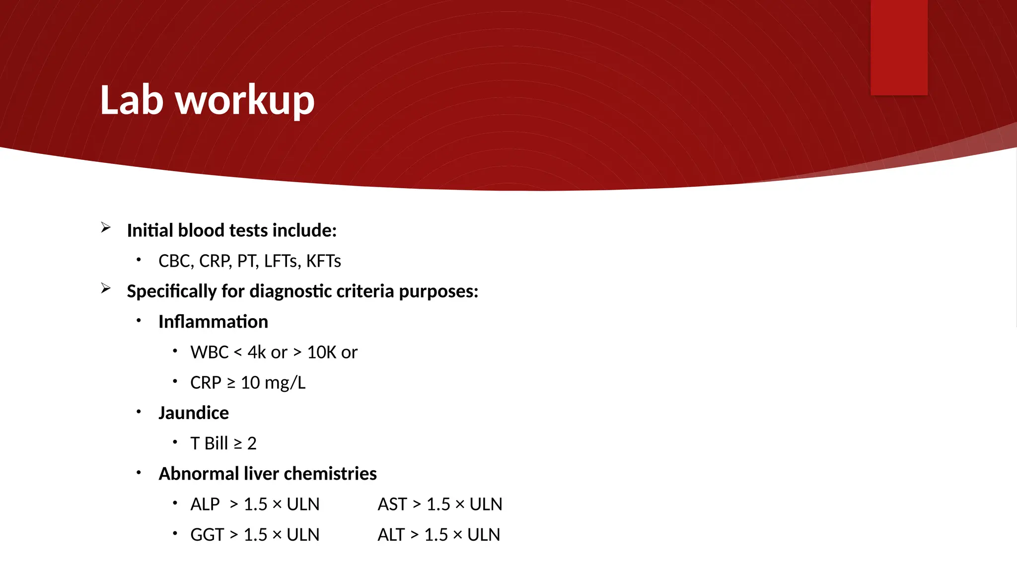
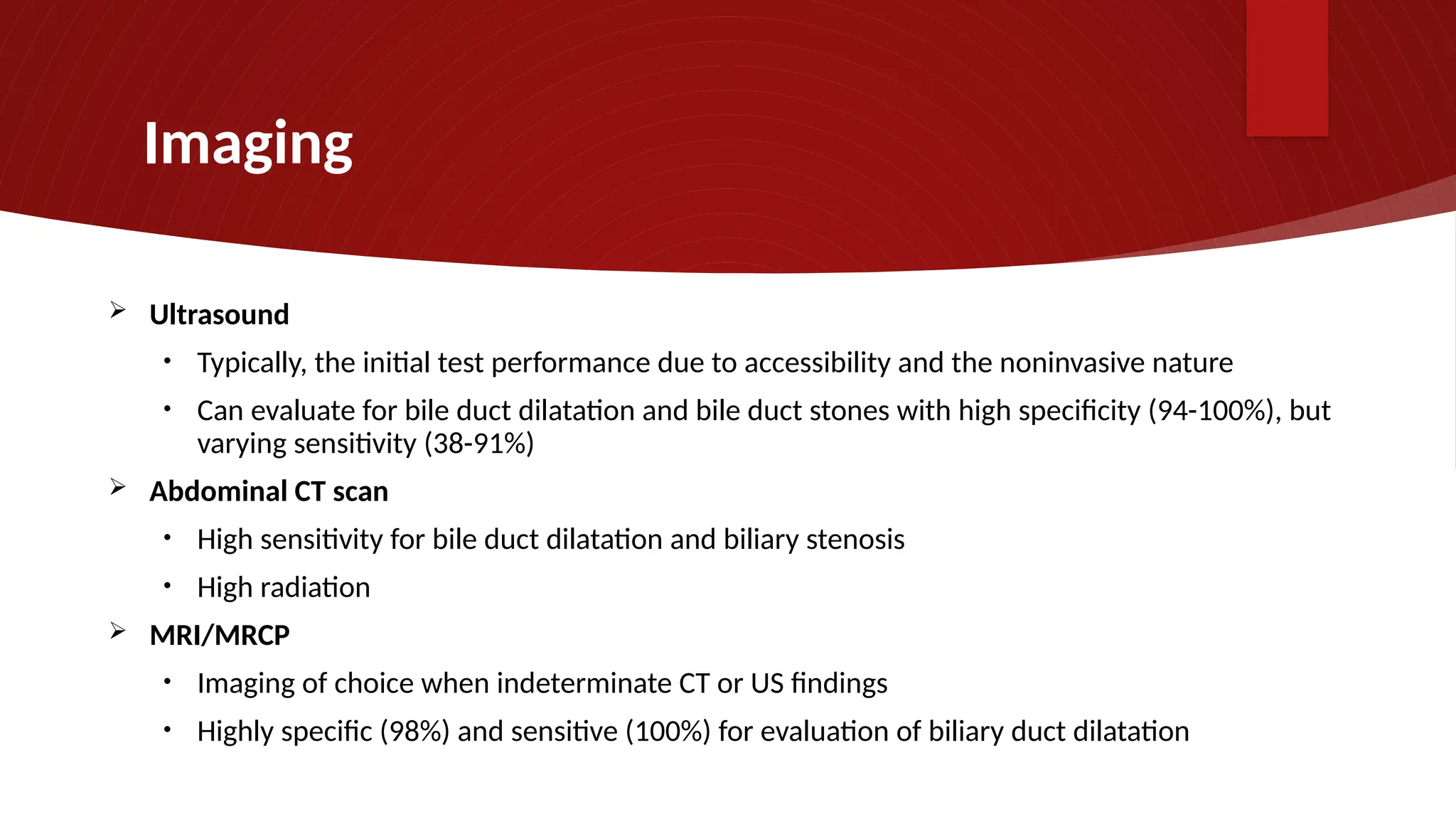
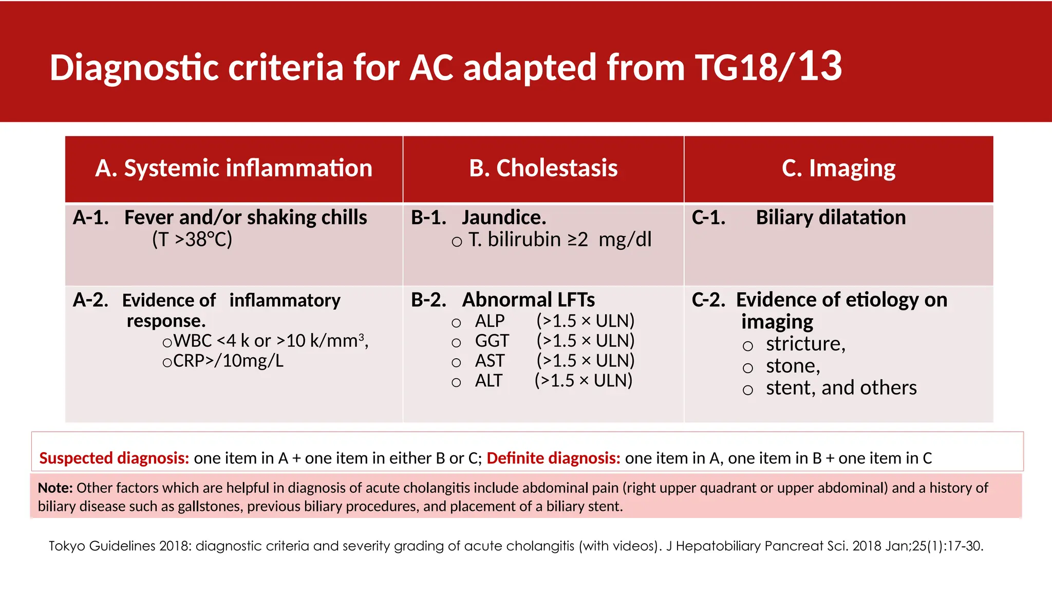
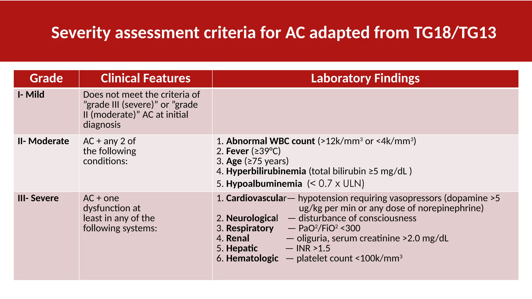
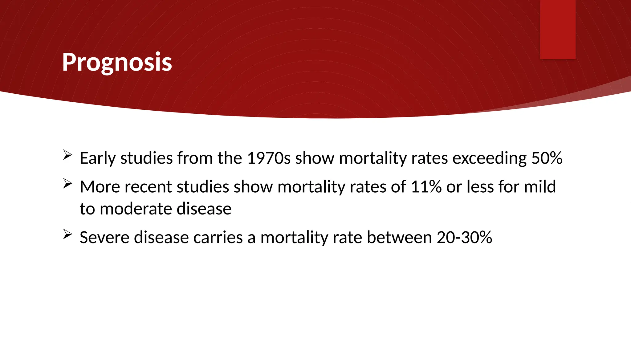
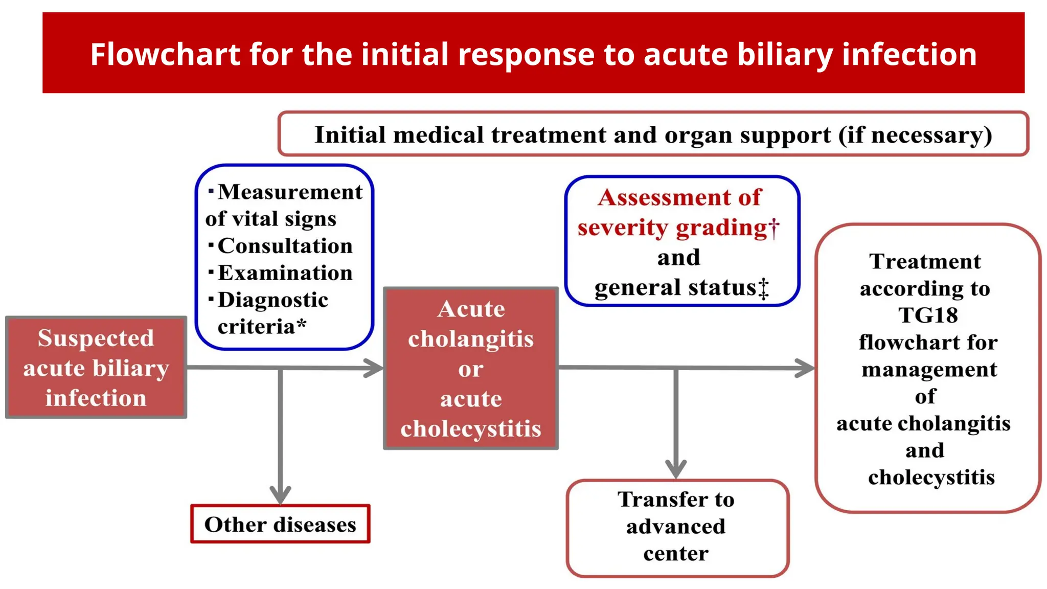
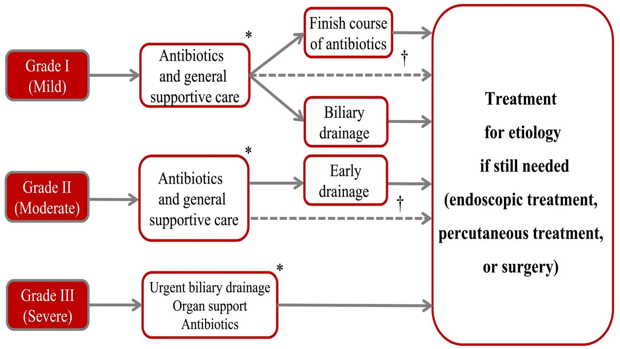
Acute Cholangitis
Diagnostic Criteria & Severity Assessment
Dr Monkez M Yousif MD, AGAF
Professor of Internal Medicine
Zagazig University
2023
Definition
 Inflammation of the biliary tree
 This inflammation is more commonly caused by
enteric bacteria in the instance of biliary stasis or
obstruction
Epidemiology
 Acute cholangitis is not uncommon
• Occurs in patients with gallstones (0.3-1.6%)
• 12% of these cases fit the Tokyo guidelines for severe disease
 Median age of presentation is between 50-60 y
Etiology
Etiology - Choledocolithiasis
 Gallstones lodged in the CBD are the most common cause
o They obstruct the flow of bile
o This stasis of bile permits the ascent of enteric bacteria via Ampulla of Vater
from the duodenum resulting in inflammation
o Elevated pressure in the CBD may also result in infection spreading to the
bloodstream via the bile canaliculi, hepatic veins, and perihepatic lymphatics
leading to bacteremia
Etiology - ERCP
 ERCP can cause cholangitis where endoscopes and accessories introduce infection
into the biliary tree if not adequately sterilized
 Risk factors for ERCP infection include:
o Incomplete drainage of the biliary tree
o Presence of jaundice
o Placement of a stent in malignant bile duct strictures (such as those caused by pancreatic cancer)
Etiology – Obstructive tumors
Carcinoma of head of pancreas causing
narrowing of CBD
Etiology – Obstructive tumors
 Choloangiocarcinoma – these most commonly present in the
seventh decade of life
 The position of these tumors can vary
 There are 3 main classifications based on the site of these tumors
― Intrahepatic tumors of the left and right hepatic ducts
― Extrahepatic tumors
o Hilar (occur near the junction of Rt and Lt hepatic ducts
o Distal (can present from the ampulla of Vater upwards)
Etiology – Obstructive tumors
 Porta hepatis mass
― The porta hepatis is a groove on the inferior surface of the
liver where various structures pass-through
― A wide array of conditions can cause a mass to develop at
this point, impeding bile flow
― This could include for example,
o thrombosis,
o stenosis or aneurysm of the portal triad vessels, or
o even a rare mass from nerves that pass through a
neurofibrosarcoma
Etiology – Benign Bile Strictures
 The majority of these strictures (80%) are iatrogenic,
caused by procedures such as ERCP
 Other causes of benign biliary strictures include:
― Primary sclerosing cholangitis
― Pancreatitis
Etiology – Malignant Bile Strictures
 Malignant bile strictures are strictures secondary to malignancy:
― Choloangiocarcinoma
― Pancreatic carcinoma
― Ampullary carcinoma
― Hepatocellular carcinoma
― Lymphoma
― Metastasis
Etiology – Mirizzi Syndrome
 This is where the CBD is obstructed by
a large gallstone in the cystic duct
Pathogenesis
 Normally, there are different defensive mechanisms to prevent cholangitis.
1. The bile salts have bacteriostatic activity
2. The biliary epithelium secretes IgA and mucous which probably act as anti-adherent
factors.
3. Kupffer cells on the biliary epithelium and the tight junction between the
cholangiocytes prevent translocation of bacteria from the hepatobiliary system into the
portal venous system.
4. Normal bile flow flushes out any bacteria into the duodenum.
5. The sphincter of Oddi also prevents any migration of bacteria from the duodenum into
the biliary system.
Pathogenesis
 Biliary obstruction → Bile stasis in the biliary system → ↑ intraductal pressure →
― Widening of the tight junction between cholangiocytes
― Malfunction of Kupffer cells
― Decreased IgA secretion
― cholangiovenous and cholangiolymphatic reflux leading to bacteremia and endotoxemia
― Systemic release of inflammatory mediators like: TNF, IL- 1,6, 10 leading to hemodynamic
instability
Diagnosis
Jean-Martin Charcot
1877
Reynolds BM, Dargan EL (August 1959). "Acute obstructive cholangitis; a distinct clinical
syndrome". Ann Surg. 150 (2): 299–303.
Lab workup
 Initial blood tests include:
• CBC, CRP, PT, LFTs, KFTs
 Specifically for diagnostic criteria purposes:
• Inflammation
• WBC < 4k or > 10K or
• CRP ≥ 10 mg/L
• Jaundice
• T Bill ≥ 2
• Abnormal liver chemistries
• ALP > 1.5 × ULN AST > 1.5 × ULN
• GGT > 1.5 × ULN ALT > 1.5 × ULN
Imaging
 Ultrasound
• Typically, the initial test performance due to accessibility and the noninvasive nature
• Can evaluate for bile duct dilatation and bile duct stones with high specificity (94-100%), but
varying sensitivity (38-91%)
 Abdominal CT scan
• High sensitivity for bile duct dilatation and biliary stenosis
• High radiation
 MRI/MRCP
• Imaging of choice when indeterminate CT or US findings
• Highly specific (98%) and sensitive (100%) for evaluation of biliary duct dilatation
Endoscopic Ultrasound (EUS)
• Best imaging test
• Only takes 5 minutes
• Can perform ERCP at the
same time
• Invasive
Diagnostic criteria for AC adapted from TG18/13
A. Systemic inflammation B. Cholestasis C. Imaging
A-1. Fever and/or shaking chills
(T >38°C)
B-1. Jaundice.
o T. bilirubin ≥2 mg/dl
C-1. Biliary dilatation
A-2. Evidence of inflammatory
response.
oWBC <4 k or >10 k/mm3
,
oCRP>/10mg/L
B-2. Abnormal LFTs
o ALP (>1.5 × ULN)
o GGT (>1.5 × ULN)
o AST (>1.5 × ULN)
o ALT (>1.5 × ULN)
C-2. Evidence of etiology on
imaging
o stricture,
o stone,
o stent, and others
Suspected diagnosis: one item in A + one item in either B or C; Definite diagnosis: one item in A, one item in B + one item in C
Tokyo Guidelines 2018: diagnostic criteria and severity grading of acute cholangitis (with videos). J Hepatobiliary Pancreat Sci. 2018 Jan;25(1):17-30.
Note: Other factors which are helpful in diagnosis of acute cholangitis include abdominal pain (right upper quadrant or upper abdominal) and a history of
biliary disease such as gallstones, previous biliary procedures, and placement of a biliary stent.
Severity assessment criteria for AC adapted from TG18/TG13
Grade Clinical Features Laboratory Findings
I- Mild Does not meet the criteria of
”grade III (severe)” or ”grade
II (moderate)” AC at initial
diagnosis
II- Moderate AC + any 2 of
the following
conditions:
1. Abnormal WBC count (>12k/mm3
or <4k/mm3
)
2. Fever (≥39°C)
3. Age (≥75 years)
4. Hyperbilirubinemia (total bilirubin ≥5 mg/dL )
5. Hypoalbuminemia (< 0.7 x ULN)
III- Severe AC + one
dysfunction at
least in any of the
following systems:
1. Cardiovascular— hypotension requiring vasopressors (dopamine >5
ug/kg per min or any dose of norepinephrine)
2. Neurological — disturbance of consciousness
3. Respiratory — PaO2
/FiO2
<300
4. Renal — oliguria, serum creatinine >2.0 mg/dL
5. Hepatic — INR >1.5
6. Hematologic — platelet count <100k/mm3
Differential Diagnosis
 Acute cholecystitis
 Biliary leak
 Acute pancreatitis
 Liver abscess
 Acute hepatitis
Triage Disposition
 Mild AC
• Regular floor
• Will need access to endoscopic surfaces for biliary drainage if no response to
treatment within 24 hours or choledocholithiasis suspected
 Moderate AC
• Regular floor or step down
• Will need access to endoscopic surfaces for biliary drainage
 Severe AC
• ICU level care
• Emergent biliary drainage
Overall Treatment Approach
 Disease severity may help guide the treatment plan
 Mortality can exceed 50% if cholangitis is not treated
 General management approach
• Supportive treatment and hemodynamic stabilization
• Antibiotic therapy
• Biliary decompression
Supportive Therapy
 Volume resuscitation with IV fluids
 Analgesics for pain control
 Antiemetics
 Correction of electrolyte disturbances
 Treatment of end-organ dysfunction
• Pressors for septic shock
• Intubation for respiratory support
 NPO for potential biliary drainage procedures
 Discontinuation and reversal of anticoagulation
Antibiotic Therapy
 Antibiotic selection should target the most common intra-
abdominal bacteria
 Initial choice can be piperacillin-tazobactam, cephalosporin plus
metronidazole, or ampicillin-sulbactam depending on severity
 Tailor antibiotic selection to culture and susceptibility results
 Duration of antibiotic treatment is usually 7 days after source
control is achieved and longer if bacteremia is present
Isolated microorganisms from
bile cultures
Proportions of isolated
organisms (%)
Gram-negative organisms
o Escherichia coli
o Klebsiella spp.
o Pseudomonas spp.
o Enterobacter spp.
Gram-positive organisms
o Enterococcus spp.
o Streptococcus spp.
o Staphylococcus spp.
Anaerobes
31–44
9–20
0.5–19
5–9
3–34
2–10
0 in bile but 3.6 in blood
culture
4–20
Common microorganisms
isolated from bile cultures
among patients with acute
biliary infections
(endorsed from the TG13)
Endoscopic Intervention
 ERCP is the primary modality of choice
• Stone Extraction
• Endoscopic drainage with or without biliary
stent
Percutaneous Transhepatic Biliary
Drainage
o Performed when endoscopic
drainage is unavailable,
unsuccessful, or the patient is too
unstable for anesthesia
o Can be done by IR via intrahepatic
approach into the bile duct
Alternative Intervention
 EUS-guided biliary drainage
• in patients with moderate-severe cholangitis
 Surgical Drainage
• Rarely used, when all other methods of source control
have failed
Timing of Endoscopic Intervention
 Early biliary drainage via ERCP within 24 h has been
associated with decreased hospital stay and recurrent
cholangitis, though no significant difference in mortality if
done within 72 h
Prognosis
 Early studies from the 1970s show mortality rates exceeding 50%
 More recent studies show mortality rates of 11% or less for mild
to moderate disease
 Severe disease carries a mortality rate between 20-30%
Flowchart for the initial response to acute biliary infection
Take-Home Messages
 AC is an infection of the biliary tree
 Diagnosis is based on evidence of systemic inflammation, cholestasis, and
imaging findings of biliary obstruction
 Mainstays of treatment are end-organ support, IV antibiotics, and biliary
decompression
 Disease severity can be mild, moderate, or severe, which can help inform the
disposition and timing of biliary drainage
 Mortality rates are decreasing with improvements in patient care and biliary
drainage techniques
Monkez Yousif

Acute Cholangitis: Diagnostic Criteria and Management.pptx

  • 1.
    Acute Cholangitis Diagnostic Criteria& Severity Assessment Dr Monkez M Yousif MD, AGAF Professor of Internal Medicine Zagazig University 2023
  • 2.
    Definition  Inflammation ofthe biliary tree  This inflammation is more commonly caused by enteric bacteria in the instance of biliary stasis or obstruction
  • 3.
    Epidemiology  Acute cholangitisis not uncommon • Occurs in patients with gallstones (0.3-1.6%) • 12% of these cases fit the Tokyo guidelines for severe disease  Median age of presentation is between 50-60 y
  • 4.
  • 5.
    Etiology - Choledocolithiasis Gallstones lodged in the CBD are the most common cause o They obstruct the flow of bile o This stasis of bile permits the ascent of enteric bacteria via Ampulla of Vater from the duodenum resulting in inflammation o Elevated pressure in the CBD may also result in infection spreading to the bloodstream via the bile canaliculi, hepatic veins, and perihepatic lymphatics leading to bacteremia
  • 6.
    Etiology - ERCP ERCP can cause cholangitis where endoscopes and accessories introduce infection into the biliary tree if not adequately sterilized  Risk factors for ERCP infection include: o Incomplete drainage of the biliary tree o Presence of jaundice o Placement of a stent in malignant bile duct strictures (such as those caused by pancreatic cancer)
  • 7.
    Etiology – Obstructivetumors Carcinoma of head of pancreas causing narrowing of CBD
  • 8.
    Etiology – Obstructivetumors  Choloangiocarcinoma – these most commonly present in the seventh decade of life  The position of these tumors can vary  There are 3 main classifications based on the site of these tumors ― Intrahepatic tumors of the left and right hepatic ducts ― Extrahepatic tumors o Hilar (occur near the junction of Rt and Lt hepatic ducts o Distal (can present from the ampulla of Vater upwards)
  • 9.
    Etiology – Obstructivetumors  Porta hepatis mass ― The porta hepatis is a groove on the inferior surface of the liver where various structures pass-through ― A wide array of conditions can cause a mass to develop at this point, impeding bile flow ― This could include for example, o thrombosis, o stenosis or aneurysm of the portal triad vessels, or o even a rare mass from nerves that pass through a neurofibrosarcoma
  • 10.
    Etiology – BenignBile Strictures  The majority of these strictures (80%) are iatrogenic, caused by procedures such as ERCP  Other causes of benign biliary strictures include: ― Primary sclerosing cholangitis ― Pancreatitis
  • 11.
    Etiology – MalignantBile Strictures  Malignant bile strictures are strictures secondary to malignancy: ― Choloangiocarcinoma ― Pancreatic carcinoma ― Ampullary carcinoma ― Hepatocellular carcinoma ― Lymphoma ― Metastasis
  • 12.
    Etiology – MirizziSyndrome  This is where the CBD is obstructed by a large gallstone in the cystic duct
  • 13.
    Pathogenesis  Normally, thereare different defensive mechanisms to prevent cholangitis. 1. The bile salts have bacteriostatic activity 2. The biliary epithelium secretes IgA and mucous which probably act as anti-adherent factors. 3. Kupffer cells on the biliary epithelium and the tight junction between the cholangiocytes prevent translocation of bacteria from the hepatobiliary system into the portal venous system. 4. Normal bile flow flushes out any bacteria into the duodenum. 5. The sphincter of Oddi also prevents any migration of bacteria from the duodenum into the biliary system.
  • 14.
    Pathogenesis  Biliary obstruction→ Bile stasis in the biliary system → ↑ intraductal pressure → ― Widening of the tight junction between cholangiocytes ― Malfunction of Kupffer cells ― Decreased IgA secretion ― cholangiovenous and cholangiolymphatic reflux leading to bacteremia and endotoxemia ― Systemic release of inflammatory mediators like: TNF, IL- 1,6, 10 leading to hemodynamic instability
  • 15.
  • 16.
  • 17.
    Reynolds BM, DarganEL (August 1959). "Acute obstructive cholangitis; a distinct clinical syndrome". Ann Surg. 150 (2): 299–303.
  • 18.
    Lab workup  Initialblood tests include: • CBC, CRP, PT, LFTs, KFTs  Specifically for diagnostic criteria purposes: • Inflammation • WBC < 4k or > 10K or • CRP ≥ 10 mg/L • Jaundice • T Bill ≥ 2 • Abnormal liver chemistries • ALP > 1.5 × ULN AST > 1.5 × ULN • GGT > 1.5 × ULN ALT > 1.5 × ULN
  • 19.
    Imaging  Ultrasound • Typically,the initial test performance due to accessibility and the noninvasive nature • Can evaluate for bile duct dilatation and bile duct stones with high specificity (94-100%), but varying sensitivity (38-91%)  Abdominal CT scan • High sensitivity for bile duct dilatation and biliary stenosis • High radiation  MRI/MRCP • Imaging of choice when indeterminate CT or US findings • Highly specific (98%) and sensitive (100%) for evaluation of biliary duct dilatation
  • 20.
    Endoscopic Ultrasound (EUS) •Best imaging test • Only takes 5 minutes • Can perform ERCP at the same time • Invasive
  • 21.
    Diagnostic criteria forAC adapted from TG18/13 A. Systemic inflammation B. Cholestasis C. Imaging A-1. Fever and/or shaking chills (T >38°C) B-1. Jaundice. o T. bilirubin ≥2 mg/dl C-1. Biliary dilatation A-2. Evidence of inflammatory response. oWBC <4 k or >10 k/mm3 , oCRP>/10mg/L B-2. Abnormal LFTs o ALP (>1.5 × ULN) o GGT (>1.5 × ULN) o AST (>1.5 × ULN) o ALT (>1.5 × ULN) C-2. Evidence of etiology on imaging o stricture, o stone, o stent, and others Suspected diagnosis: one item in A + one item in either B or C; Definite diagnosis: one item in A, one item in B + one item in C Tokyo Guidelines 2018: diagnostic criteria and severity grading of acute cholangitis (with videos). J Hepatobiliary Pancreat Sci. 2018 Jan;25(1):17-30. Note: Other factors which are helpful in diagnosis of acute cholangitis include abdominal pain (right upper quadrant or upper abdominal) and a history of biliary disease such as gallstones, previous biliary procedures, and placement of a biliary stent.
  • 22.
    Severity assessment criteriafor AC adapted from TG18/TG13 Grade Clinical Features Laboratory Findings I- Mild Does not meet the criteria of ”grade III (severe)” or ”grade II (moderate)” AC at initial diagnosis II- Moderate AC + any 2 of the following conditions: 1. Abnormal WBC count (>12k/mm3 or <4k/mm3 ) 2. Fever (≥39°C) 3. Age (≥75 years) 4. Hyperbilirubinemia (total bilirubin ≥5 mg/dL ) 5. Hypoalbuminemia (< 0.7 x ULN) III- Severe AC + one dysfunction at least in any of the following systems: 1. Cardiovascular— hypotension requiring vasopressors (dopamine >5 ug/kg per min or any dose of norepinephrine) 2. Neurological — disturbance of consciousness 3. Respiratory — PaO2 /FiO2 <300 4. Renal — oliguria, serum creatinine >2.0 mg/dL 5. Hepatic — INR >1.5 6. Hematologic — platelet count <100k/mm3
  • 23.
    Differential Diagnosis  Acutecholecystitis  Biliary leak  Acute pancreatitis  Liver abscess  Acute hepatitis
  • 24.
    Triage Disposition  MildAC • Regular floor • Will need access to endoscopic surfaces for biliary drainage if no response to treatment within 24 hours or choledocholithiasis suspected  Moderate AC • Regular floor or step down • Will need access to endoscopic surfaces for biliary drainage  Severe AC • ICU level care • Emergent biliary drainage
  • 25.
    Overall Treatment Approach Disease severity may help guide the treatment plan  Mortality can exceed 50% if cholangitis is not treated  General management approach • Supportive treatment and hemodynamic stabilization • Antibiotic therapy • Biliary decompression
  • 26.
    Supportive Therapy  Volumeresuscitation with IV fluids  Analgesics for pain control  Antiemetics  Correction of electrolyte disturbances  Treatment of end-organ dysfunction • Pressors for septic shock • Intubation for respiratory support  NPO for potential biliary drainage procedures  Discontinuation and reversal of anticoagulation
  • 27.
    Antibiotic Therapy  Antibioticselection should target the most common intra- abdominal bacteria  Initial choice can be piperacillin-tazobactam, cephalosporin plus metronidazole, or ampicillin-sulbactam depending on severity  Tailor antibiotic selection to culture and susceptibility results  Duration of antibiotic treatment is usually 7 days after source control is achieved and longer if bacteremia is present
  • 28.
    Isolated microorganisms from bilecultures Proportions of isolated organisms (%) Gram-negative organisms o Escherichia coli o Klebsiella spp. o Pseudomonas spp. o Enterobacter spp. Gram-positive organisms o Enterococcus spp. o Streptococcus spp. o Staphylococcus spp. Anaerobes 31–44 9–20 0.5–19 5–9 3–34 2–10 0 in bile but 3.6 in blood culture 4–20 Common microorganisms isolated from bile cultures among patients with acute biliary infections (endorsed from the TG13)
  • 29.
    Endoscopic Intervention  ERCPis the primary modality of choice • Stone Extraction • Endoscopic drainage with or without biliary stent
  • 30.
    Percutaneous Transhepatic Biliary Drainage oPerformed when endoscopic drainage is unavailable, unsuccessful, or the patient is too unstable for anesthesia o Can be done by IR via intrahepatic approach into the bile duct
  • 31.
    Alternative Intervention  EUS-guidedbiliary drainage • in patients with moderate-severe cholangitis  Surgical Drainage • Rarely used, when all other methods of source control have failed
  • 32.
    Timing of EndoscopicIntervention  Early biliary drainage via ERCP within 24 h has been associated with decreased hospital stay and recurrent cholangitis, though no significant difference in mortality if done within 72 h
  • 33.
    Prognosis  Early studiesfrom the 1970s show mortality rates exceeding 50%  More recent studies show mortality rates of 11% or less for mild to moderate disease  Severe disease carries a mortality rate between 20-30%
  • 34.
    Flowchart for theinitial response to acute biliary infection
  • 36.
    Take-Home Messages  ACis an infection of the biliary tree  Diagnosis is based on evidence of systemic inflammation, cholestasis, and imaging findings of biliary obstruction  Mainstays of treatment are end-organ support, IV antibiotics, and biliary decompression  Disease severity can be mild, moderate, or severe, which can help inform the disposition and timing of biliary drainage  Mortality rates are decreasing with improvements in patient care and biliary drainage techniques
  • 37.

Editor's Notes

  • #13 Bacteria can invade the biliary tract by ascending from the duodenum and via the hematogenous route from the hepatic portal venous blood. The sphincter of Oddi, situated at the junction of the biliary tract and the upper gastrointestinal tract, forms an effective mechanical barrier to duodenal reflux and ascending bacterial infection. Kupffer cells and the tight junctions between hepatocytes help prevent bacteria and toxic metabolites from entering the hepatobiliary system from the portal circulation. The continuous flushing action of bile and the bacteriostatic effects of bile salts keeps the biliary tract sterile under normal conditions. Secretory immunoglobulin A (slgA), the predominant immunoglobulin in the bile, and mucus excreted by the biliary epithelium probably function as antiadherence factors, preventing microbial colonization.
  • #14 When barrier mechanisms break down, as in surgical or endoscopic sphincterotomy and with insertion of biliary stents, pathogenic bacteria enter the biliary system at high concentrations and take up residence on any foreign bodies. Intrabiliary pressure is a key factor in the development of cholangitis. Chronic biliary obstruction raises the intrabiliary pressure. This adversely influences the defensive mechanisms such as the tight junctions, Kupffer cell functions, bile flow, and slgA production in the system, resulting in a higher incidence of septicemia and endotoxemia in these patients.
  • #15 The diagnosis of AC is made based on clinical presentation, laboratory investigations, and imaging studies
  • #21 Since the 19th century, Charcot’s triad of abdominal pain, fever, and jaundice has been the most widespread system to identify patients with cholangitis. Reynolds and Dargan identified clinical features of severely ill patients requiring emergent surgical decompression. Both clinical investigators operated in a system in which reliable noninvasive imaging studies and biochemical laboratory tests were not readily available. Therefore, several observational studies demonstrated that the performance of both criteria is highly variable. For example, in a systematic review, Rumsey et al reported a median sensitivity of 43.6% and specificity of 91% for Charcot’s triad. The more recent Tokyo criteria versions in 2013 and 2018 eliminated abdominal pain and history of biliary disease because these had suboptimal specificity. Specifically, they found that more than a third of patients with cholecystitis were incorrectly diagnosed with cholangitis. The diagnostic performance of TG18 in identifying definite cholangitis recently showed an 82% sensitivity but had low specificity at 60%.
  • #31 IR = Interventional Radiologist
  • #34 The initial management of patients with suspected acute biliary infection starts with the measurement of vital signs to assess whether or not the situation is urgent. If the case is judged to be urgent, initial medical treatment should be started immediately including respiratory/circulatory management if required, without waiting for the definitive diagnosis. A detailed examination (consultation and physical examination) is then performed, after which blood tests and diagnostic imaging are performed; on the basis of the results, a definitive diagnosis is made following the diagnostic criteria for acute cholangitis and cholecystitis. Once the diagnosis has been confirmed, initial medical treatment should be started immediately, the severity should be assessed according to the severity grading criteria for acute cholangitis/cholecystitis, and the patient's general status should be evaluated. The Charlson comorbidity index (CCI) (case series, CS) and the American Society of Anesthesiologists (ASA) Physical Status (PS) classification are useful for the evaluation of general status. After the severity assessment, a treatment strategy should be decided on the basis of the flowchart for the management of acute cholangitis or acute cholecystitis, and treatment should immediately be provided. The Charlson Comorbidity Index was first developed in 1987 by Mary Charlson and colleagues as a weighted index to predict risk of death within 1 year of hospitalization for patients with specific comorbid conditions. The American Society of Anesthesiologists (ASA) physical status classification system came about to offer perioperative clinicians a simple categorization of a patient's physiological status to help predict operative risk. ASA 1: A normal healthy patient. ASA 2: A patient with mild systemic disease. ASA 3: A patient with a severe systemic disease that is not life-threatening. ASA 4: A patient with a severe systemic disease that is a constant threat to life. ASA 5: A moribund patient who is not expected to survive without the operation. ASA 6: A brain-dead patient whose organs are being removed with the intention of transplanting them into another patient.
  • #35 Mild acute cholangitis is defined as cholangitis that does not meet the TG18 severity assessment criteria for moderate or severe cholangitis below. In most cases initial treatment including antibiotics is sufficient, and most patients do not require biliary drainage. However, biliary drainage should be considered if a patient does not respond to initial treatment. EST and subsequent choledocholithotomy may be performed at the same time as biliary drainage. Postoperative cholangitis usually improves with antibiotic treatment alone, and biliary drainage is not usually required. Moderate acute cholangitis is cholangitis that is not severe but requires early biliary drainage. Early endoscopic or percutaneous transhepatic biliary drainage is indicated. If the underlying etiology requires treatment, this should be provided after the patient's general condition has improved and EST and subsequent choledocholithotomy may be performed together with biliary drainage. Severe acute cholangitis is cholangitis with sepsis-induced organ damage. As the patient's condition may deteriorate rapidly, a swift response is essential including appropriate respiratory/circulatory management (tracheal intubation followed by artificial ventilation and the use of hypertensive agents). Endoscopic or percutaneous transhepatic biliary drainage should be performed as soon as possible after the patient's condition has been improved by initial treatment and respiratory/circulatory management. If treatment for the underlying etiology is required, this should be provided after the patient's general status has improved