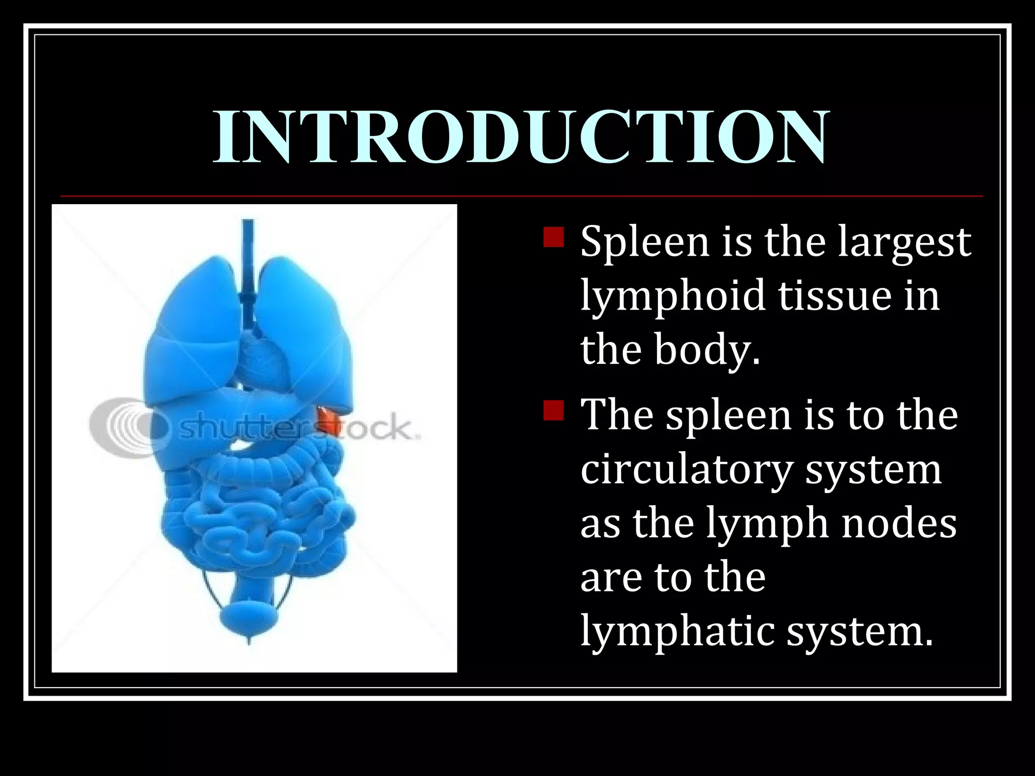
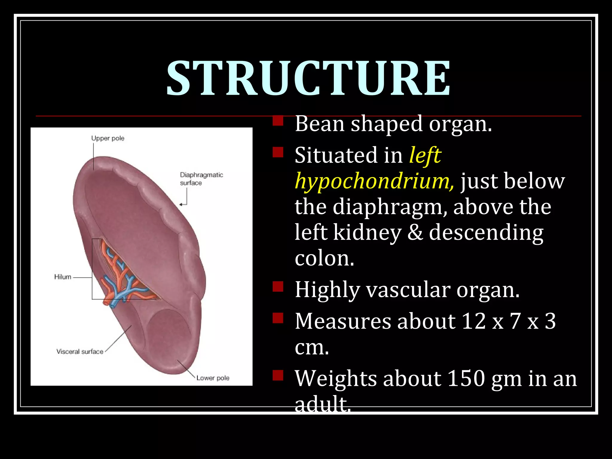
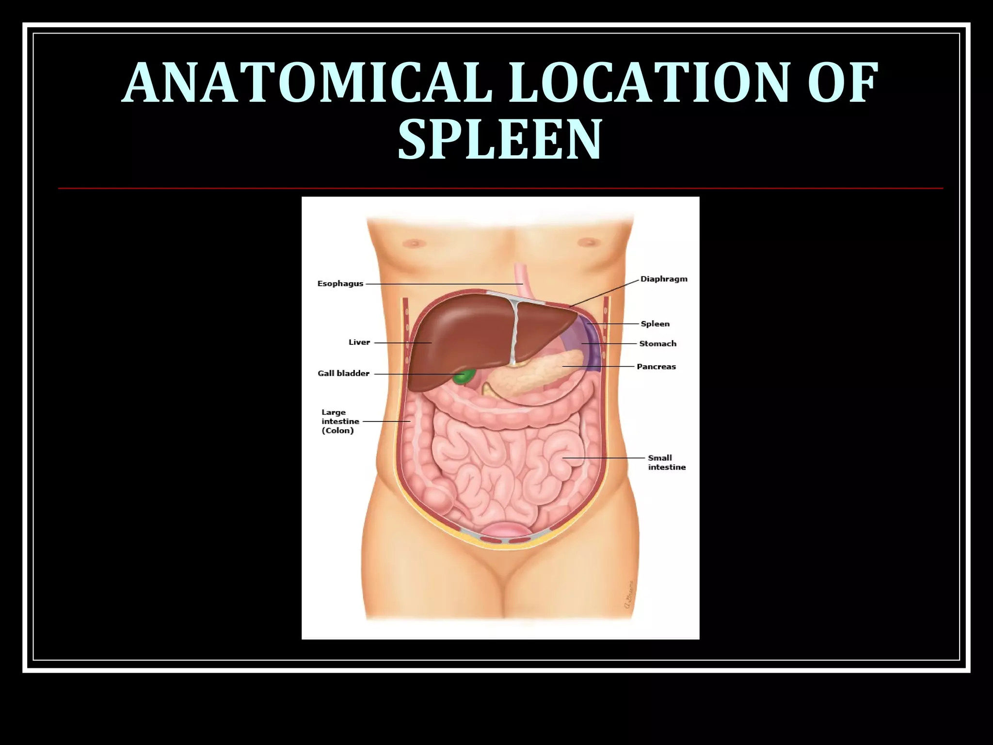
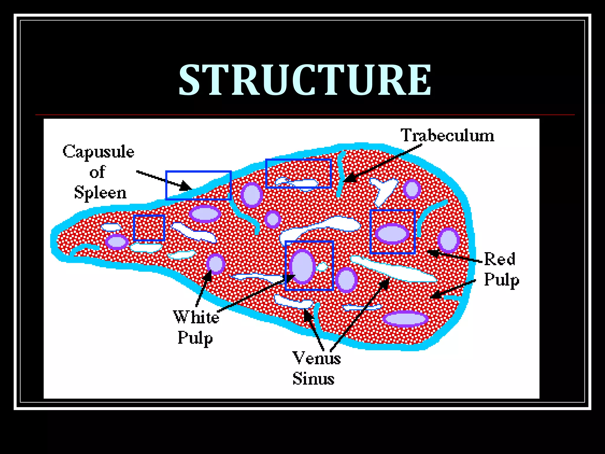
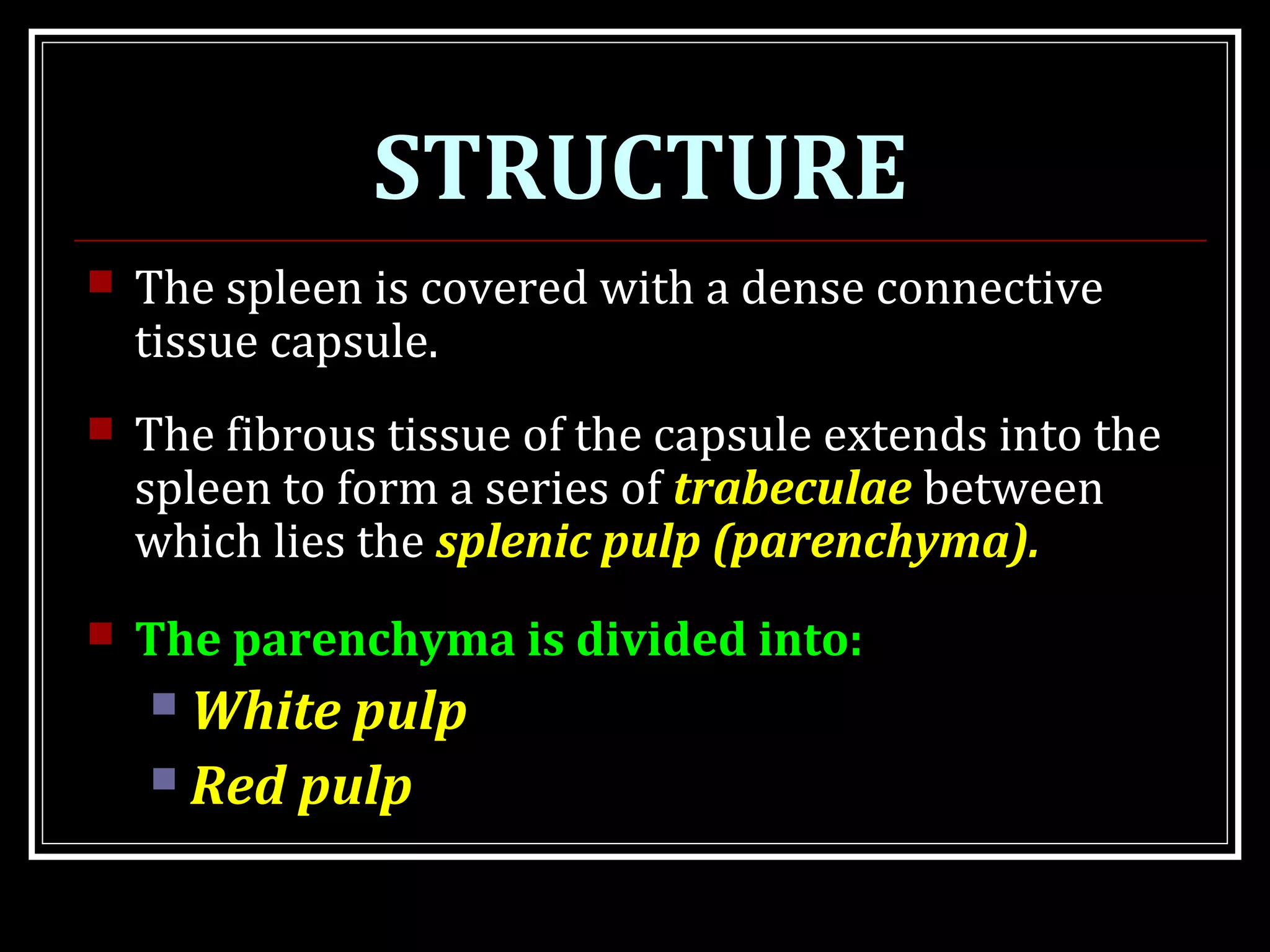
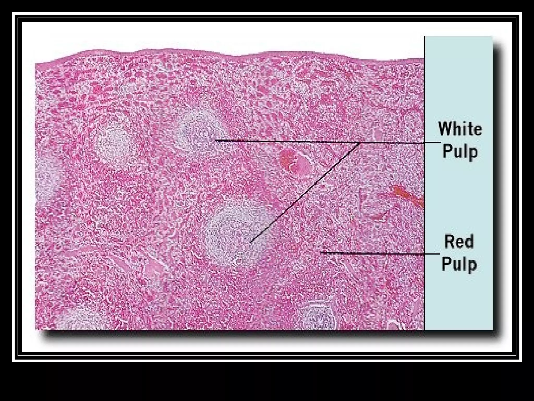
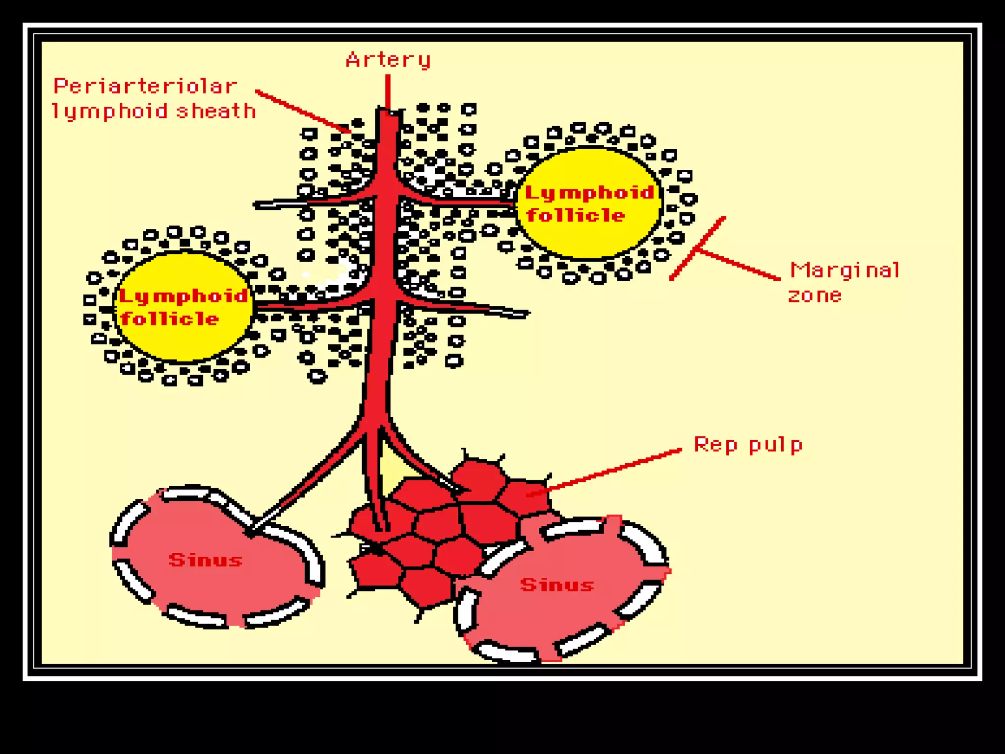
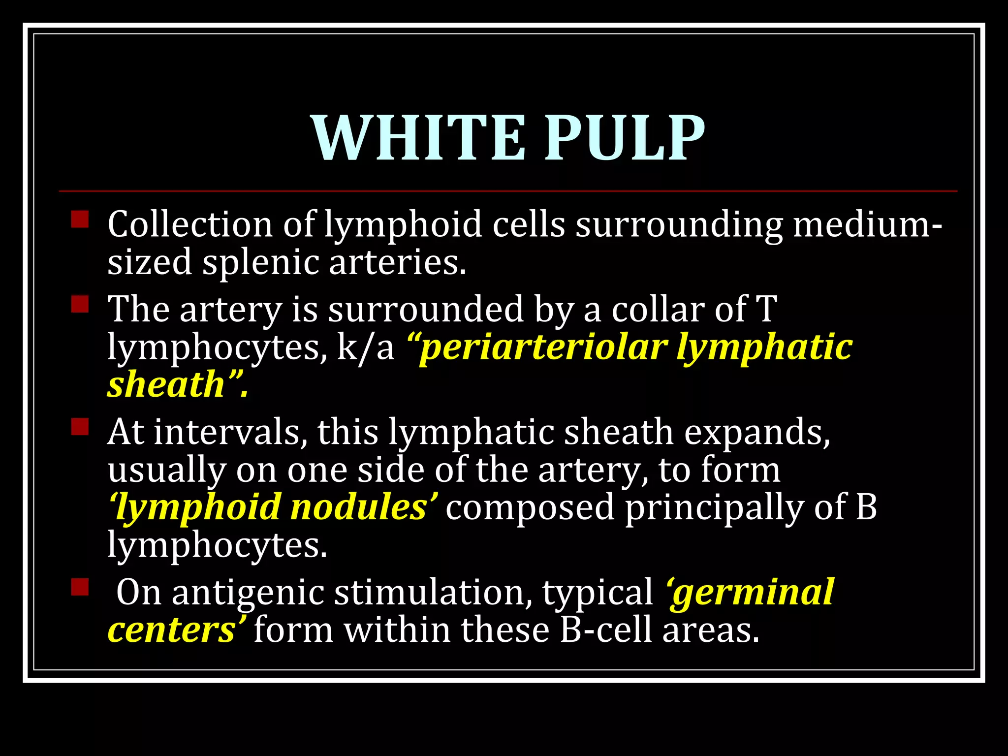
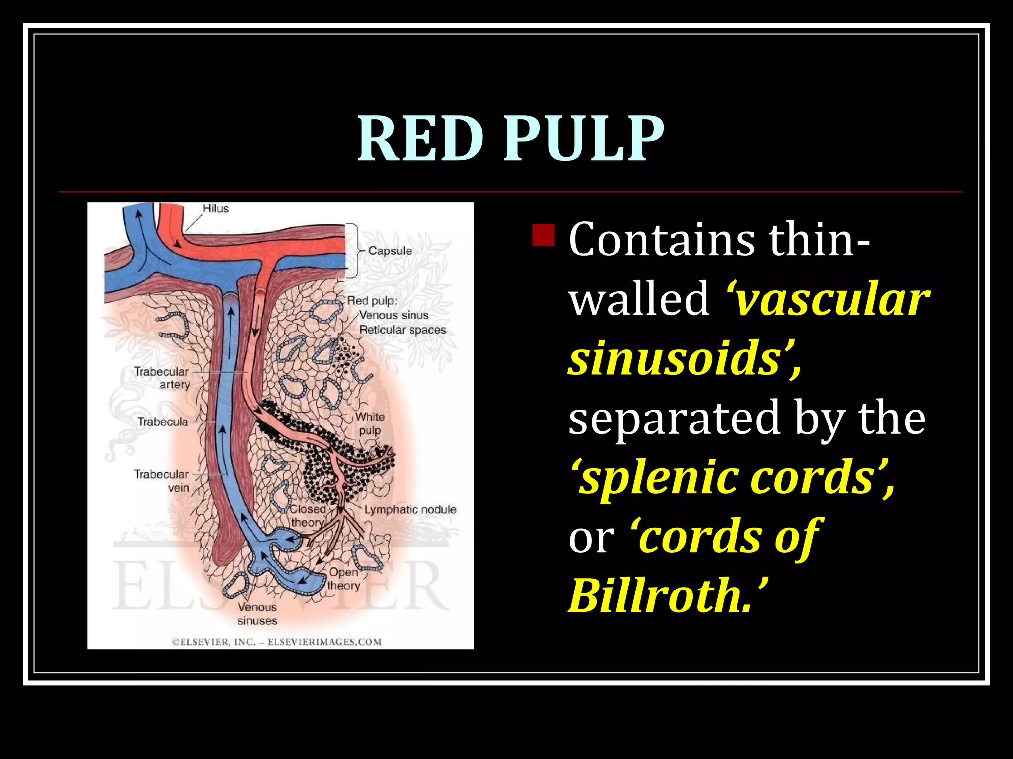
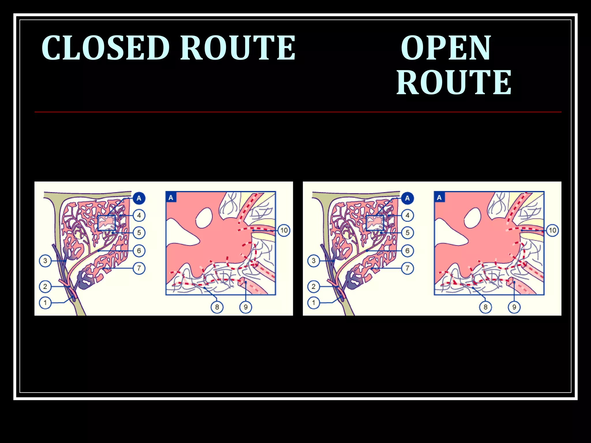
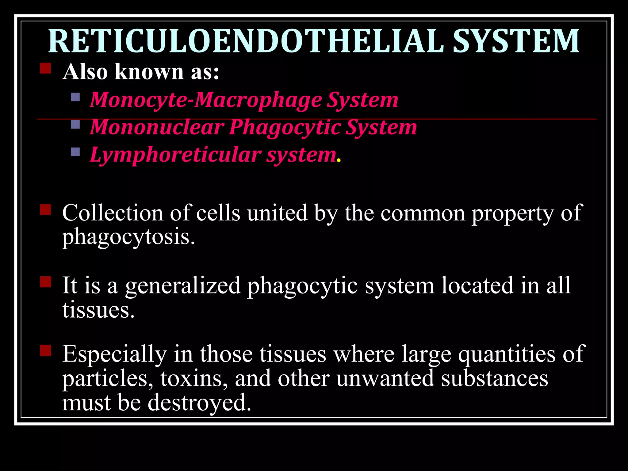
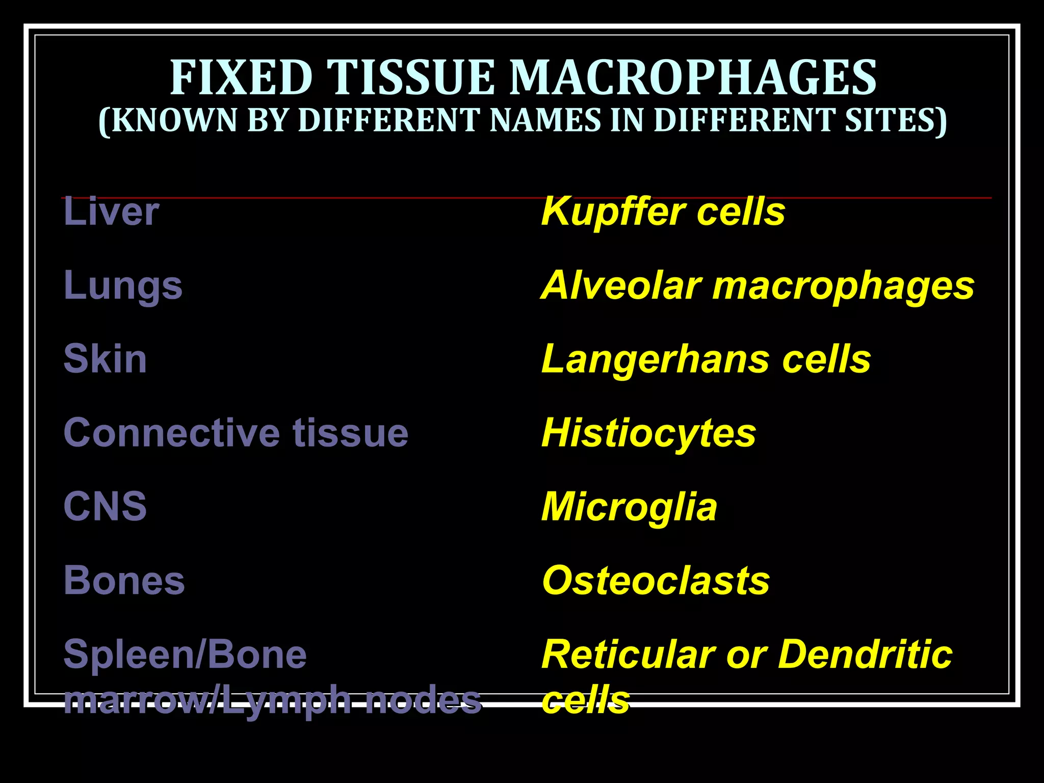
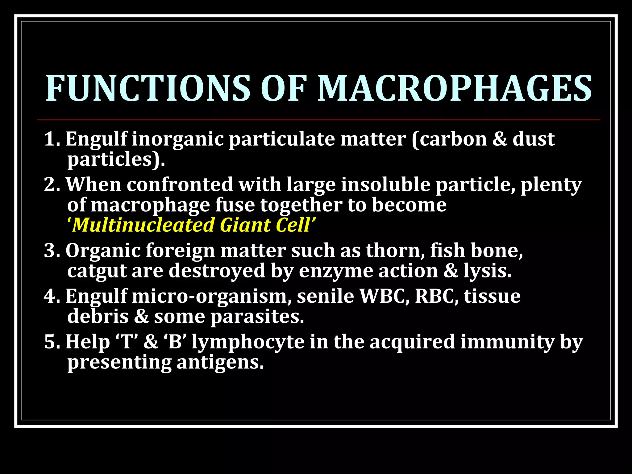
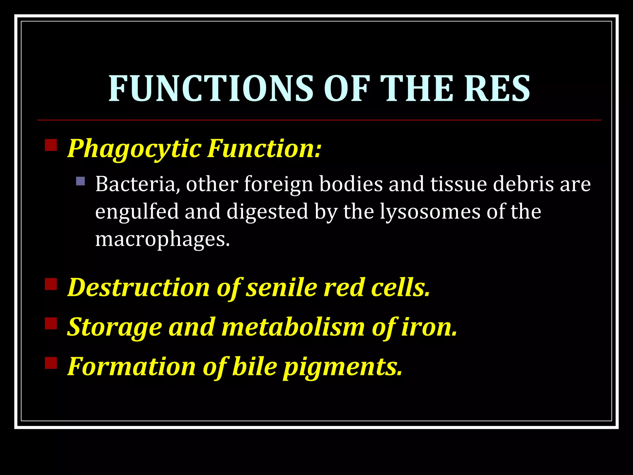
The document discusses the spleen and reticuloendothelial system (RES). It describes the spleen's structure as being divided into red and white pulp. Red pulp contains sinusoids and cords that filter blood, while white pulp contains lymphatic nodules. The spleen plays roles in filtering blood, forming red blood cells, mounting an immune response, and storing iron. The RES consists of tissue-based macrophages that phagocytose pathogens and debris. It is a generalized innate defense system located throughout the body.
For More Medicine Free PPT - http://playnever.blogspot.com/
For Health benefits and medicine videos Subscribe youtube channel - https://www.youtube.com/playlist?list=PLKg-H-sMh9G01zEg4YpndngXODW2bq92w
This was done as a Student presentation on the kidney.
Here following topics are covered.
Macroscopic structure of the urinary system
Microscopic anatomy of the urinary system
Functions of the nephron
Renal blood supply
Kidneys and blood pressure regulation
Structure of ureters and urinary bladder to perform its function
Renal failure
synovial joint, definition of synovial joint, diarthrodial joints, components of synovial joint, types of synovial joints, hinge joint with examples, pivot joint with examples, condyloid joint with examples, saddle joint with examples, ball and socket joint with examples, gliding joint with examples, features of synovial joint, synovial membrane, synovial fluid, components of synovial membrane, meniscus, true and accessory ligament of synovial joint, bursae, blood supply of synovial joint, innervation of synovial joint
Each kidney contains over 1 million tiny structures called nephrons. Each nephron has a glomerulus, the site of blood filtration. The glomerulus is a network of capillaries surrounded by a cuplike structure, the glomerular capsule (or Bowman’s capsule). As blood flows through the glomerulus, blood pressure pushes water and solutes from the capillaries into the capsule through a filtration membrane. This glomerular filtration begins the urine formation process.Inside the glomerulus, blood pressure pushes fluid from capillaries into the glomerular capsule through a specialized layer of cells. This layer, the filtration membrane, allows water and small solutes to pass but blocks blood cells and large proteins. Those components remain in the bloodstream. The filtrate (the fluid that has passed through the membrane) flows from the glomerular capsule further into the nephron.The glomerulus filters water and small solutes out of the bloodstream. The resulting filtrate contains waste, but also other substances the body needs: essential ions, glucose, amino acids, and smaller proteins. When the filtrate exits the glomerulus, it flows into a duct in the nephron called the renal tubule. As it moves, the needed substances and some water are reabsorbed through the tube wall into adjacent capillaries. This reabsorption of vital nutrients from the filtrate is the second step in urine creation.The filtrate absorbed in the glomerulus flows through the renal tubule, where nutrients and water are reabsorbed into capillaries. At the same time, waste ions and hydrogen ions pass from the capillaries into the renal tubule. This process is called secretion. The secreted ions combine with the remaining filtrate and become urine. The urine flows out of the nephron tubule into a collecting duct. It passes out of the kidney through the renal pelvis, into the ureter, and down to the bladder.The nephrons of the kidneys process blood and create urine through a process of filtration, reabsorption, and secretion. Urine is about 95% water and 5% waste products. Nitrogenous wastes excreted in urine include urea, creatinine, ammonia, and uric acid. Ions such as sodium, potassium, hydrogen, and calcium are also excreted
Flu Vaccine Alert in Bangalore Karnatakaaddon Scans
As flu season approaches, health officials in Bangalore, Karnataka, are urging residents to get their flu vaccinations. The seasonal flu, while common, can lead to severe health complications, particularly for vulnerable populations such as young children, the elderly, and those with underlying health conditions.
Dr. Vidisha Kumari, a leading epidemiologist in Bangalore, emphasizes the importance of getting vaccinated. "The flu vaccine is our best defense against the influenza virus. It not only protects individuals but also helps prevent the spread of the virus in our communities," he says.
This year, the flu season is expected to coincide with a potential increase in other respiratory illnesses. The Karnataka Health Department has launched an awareness campaign highlighting the significance of flu vaccinations. They have set up multiple vaccination centers across Bangalore, making it convenient for residents to receive their shots.
To encourage widespread vaccination, the government is also collaborating with local schools, workplaces, and community centers to facilitate vaccination drives. Special attention is being given to ensuring that the vaccine is accessible to all, including marginalized communities who may have limited access to healthcare.
Residents are reminded that the flu vaccine is safe and effective. Common side effects are mild and may include soreness at the injection site, mild fever, or muscle aches. These side effects are generally short-lived and far less severe than the flu itself.
Healthcare providers are also stressing the importance of continuing COVID-19 precautions. Wearing masks, practicing good hand hygiene, and maintaining social distancing are still crucial, especially in crowded places.
Protect yourself and your loved ones by getting vaccinated. Together, we can help keep Bangalore healthy and safe this flu season. For more information on vaccination centers and schedules, residents can visit the Karnataka Health Department’s official website or follow their social media pages.
Stay informed, stay safe, and get your flu shot today!
TEST BANK for Operations Management, 14th Edition by William J. Stevenson, Ve...kevinkariuki227
TEST BANK for Operations Management, 14th Edition by William J. Stevenson, Verified Chapters 1 - 19, Complete Newest Version.pdf
Lung Cancer: Artificial Intelligence, Synergetics, Complex System Analysis, S...Oleg Kshivets
RESULTS: Overall life span (LS) was 2252.1±1742.5 days and cumulative 5-year survival (5YS) reached 73.2%, 10 years – 64.8%, 20 years – 42.5%. 513 LCP lived more than 5 years (LS=3124.6±1525.6 days), 148 LCP – more than 10 years (LS=5054.4±1504.1 days).199 LCP died because of LC (LS=562.7±374.5 days). 5YS of LCP after bi/lobectomies was significantly superior in comparison with LCP after pneumonectomies (78.1% vs.63.7%, P=0.00001 by log-rank test). AT significantly improved 5YS (66.3% vs. 34.8%) (P=0.00000 by log-rank test) only for LCP with N1-2. Cox modeling displayed that 5YS of LCP significantly depended on: phase transition (PT) early-invasive LC in terms of synergetics, PT N0—N12, cell ratio factors (ratio between cancer cells- CC and blood cells subpopulations), G1-3, histology, glucose, AT, blood cell circuit, prothrombin index, heparin tolerance, recalcification time (P=0.000-0.038). Neural networks, genetic algorithm selection and bootstrap simulation revealed relationships between 5YS and PT early-invasive LC (rank=1), PT N0—N12 (rank=2), thrombocytes/CC (3), erythrocytes/CC (4), eosinophils/CC (5), healthy cells/CC (6), lymphocytes/CC (7), segmented neutrophils/CC (8), stick neutrophils/CC (9), monocytes/CC (10); leucocytes/CC (11). Correct prediction of 5YS was 100% by neural networks computing (area under ROC curve=1.0; error=0.0).
CONCLUSIONS: 5YS of LCP after radical procedures significantly depended on: 1) PT early-invasive cancer; 2) PT N0--N12; 3) cell ratio factors; 4) blood cell circuit; 5) biochemical factors; 6) hemostasis system; 7) AT; 8) LC characteristics; 9) LC cell dynamics; 10) surgery type: lobectomy/pneumonectomy; 11) anthropometric data. Optimal diagnosis and treatment strategies for LC are: 1) screening and early detection of LC; 2) availability of experienced thoracic surgeons because of complexity of radical procedures; 3) aggressive en block surgery and adequate lymph node dissection for completeness; 4) precise prediction; 5) adjuvant chemoimmunoradiotherapy for LCP with unfavorable prognosis.
Ethanol (CH3CH2OH), or beverage alcohol, is a two-carbon alcohol
that is rapidly distributed in the body and brain. Ethanol alters many
neurochemical systems and has rewarding and addictive properties. It
is the oldest recreational drug and likely contributes to more morbidity,
mortality, and public health costs than all illicit drugs combined. The
5th edition of the Diagnostic and Statistical Manual of Mental Disorders
(DSM-5) integrates alcohol abuse and alcohol dependence into a single
disorder called alcohol use disorder (AUD), with mild, moderate,
and severe subclassifications (American Psychiatric Association, 2013).
In the DSM-5, all types of substance abuse and dependence have been
combined into a single substance use disorder (SUD) on a continuum
from mild to severe. A diagnosis of AUD requires that at least two of
the 11 DSM-5 behaviors be present within a 12-month period (mild
AUD: 2–3 criteria; moderate AUD: 4–5 criteria; severe AUD: 6–11 criteria).
The four main behavioral effects of AUD are impaired control over
drinking, negative social consequences, risky use, and altered physiological
effects (tolerance, withdrawal). This chapter presents an overview
of the prevalence and harmful consequences of AUD in the U.S.,
the systemic nature of the disease, neurocircuitry and stages of AUD,
comorbidities, fetal alcohol spectrum disorders, genetic risk factors, and
pharmacotherapies for AUD.
Prix Galien International 2024 Forum ProgramLevi Shapiro
June 20, 2024, Prix Galien International and Jerusalem Ethics Forum in ROME. Detailed agenda including panels:
- ADVANCES IN CARDIOLOGY: A NEW PARADIGM IS COMING
- WOMEN’S HEALTH: FERTILITY PRESERVATION
- WHAT’S NEW IN THE TREATMENT OF INFECTIOUS,
ONCOLOGICAL AND INFLAMMATORY SKIN DISEASES?
- ARTIFICIAL INTELLIGENCE AND ETHICS
- GENE THERAPY
- BEYOND BORDERS: GLOBAL INITIATIVES FOR DEMOCRATIZING LIFE SCIENCE TECHNOLOGIES AND PROMOTING ACCESS TO HEALTHCARE
- ETHICAL CHALLENGES IN LIFE SCIENCES
- Prix Galien International Awards Ceremony
Couples presenting to the infertility clinic- Do they really have infertility...Sujoy Dasgupta
Dr Sujoy Dasgupta presented the study on "Couples presenting to the infertility clinic- Do they really have infertility? – The unexplored stories of non-consummation" in the 13th Congress of the Asia Pacific Initiative on Reproduction (ASPIRE 2024) at Manila on 24 May, 2024.
These lecture slides, by Dr Sidra Arshad, offer a quick overview of physiological basis of a normal electrocardiogram.
Learning objectives:
1. Define an electrocardiogram (ECG) and electrocardiography
2. Describe how dipoles generated by the heart produce the waveforms of the ECG
3. Describe the components of a normal electrocardiogram of a typical bipolar leads (limb II)
4. Differentiate between intervals and segments
5. Enlist some common indications for obtaining an ECG
Study Resources:
1. Chapter 11, Guyton and Hall Textbook of Medical Physiology, 14th edition
2. Chapter 9, Human Physiology - From Cells to Systems, Lauralee Sherwood, 9th edition
3. Chapter 29, Ganong’s Review of Medical Physiology, 26th edition
4. Electrocardiogram, StatPearls - https://www.ncbi.nlm.nih.gov/books/NBK549803/
5. ECG in Medical Practice by ABM Abdullah, 4th edition
6. ECG Basics, http://www.nataliescasebook.com/tag/e-c-g-basics
Anti ulcer drugs and their Advance pharmacology ||
Anti-ulcer drugs are medications used to prevent and treat ulcers in the stomach and upper part of the small intestine (duodenal ulcers). These ulcers are often caused by an imbalance between stomach acid and the mucosal lining, which protects the stomach lining.
||Scope: Overview of various classes of anti-ulcer drugs, their mechanisms of action, indications, side effects, and clinical considerations.
micro teaching on communication m.sc nursing.pdfAnurag Sharma
Microteaching is a unique model of practice teaching. It is a viable instrument for the. desired change in the teaching behavior or the behavior potential which, in specified types of real. classroom situations, tends to facilitate the achievement of specified types of objectives.
2. OBJECTIVES
At the end of the lecture you should know:
The functional anatomy of the spleen.
The histological structure of spleen (red & white
pulp)
How to draw the schematic diagram of histological
structure.
Functions of spleen.
Definition & causes for splenomegaly &
hypersplenism.
Definition, components & function of RES.
3. INTRODUCTION
Spleen is the largest
lymphoid tissue in
the body.
The spleen is to the
circulatory system
as the lymph nodes
are to the
lymphatic system.
4. STRUCTURE
Bean shaped organ.
Situated in left
hypochondrium, just below
the diaphragm, above the
left kidney & descending
colon.
Highly vascular organ.
Measures about 12 x 7 x 3
cm.
Weights about 150 gm in an
adult.
8. STRUCTURE
The spleen is covered with a dense connective
tissue capsule.
The fibrous tissue of the capsule extends into the
spleen to form a series of trabeculae between
which lies the splenic pulp (parenchyma).
The parenchyma is divided into:
White pulp
Red pulp
9.
10.
11. WHITE PULP
Collection of lymphoid cells surrounding medium-
sized splenic arteries.
The artery is surrounded by a collar of T
lymphocytes, k/a “periarteriolar lymphatic
sheath”.
At intervals, this lymphatic sheath expands,
usually on one side of the artery, to form
‘lymphoid nodules’ composed principally of B
lymphocytes.
On antigenic stimulation, typical ‘germinal
centers’ form within these B-cell areas.
12.
13. RED PULP
Contains thin-
walled ‘vascular
sinusoids’,
separated by the
‘splenic cords’,
or ‘cords of
Billroth.’
14. RED PULP
The endothelial lining
of the sinusoid is of
the open or
discontinuous type,
providing passage of
blood cells between
the sinusoids and
cords.
15.
16. RED PULP
Blood reaching the capillaries in red pulp may
take one of two routes:
Open route: filters into the splenic cords.
Closed route: passes directly into
sinusoides.
The splenic cords are lined by macrophages
and function as seive through which blood in
red pulp filters into sinusoides.
19. FUNCTIONS OF SPLEEN
Formation of RBC
Removal of unwanted elements from
the blood.
Reservoir function
Role in defense mechanism
Storage and metabolism of iron
20. FORMATION OF RBC
During fetal life (3-
5m IUL) spleen
along with liver act
as an important
hemopoietic organ.
Lymphocytes are
also formed in the
white pulp.
21. REMOVAL OF UNWANTED
ELEMENTS FROM THE BLOOD.
By splenic phagocytes in the cord.
Removes:
Old effaced RBCs
Damaged RBCs & leucocytes
Abnormal RBCs (e.g. Hereditary spherocytes)
Bacteria, cell debris etc.
RBCs undergo extreme deformation during
passage from the cords into the sinusoids.
↓ RBC elasticity RBC entrapped in cords
phagocytosed by cordal macrophages.
22. RESERVOIR FUNCTION
More important in animals with contractile
spleen (e.g. Dog). Human spleen is non-
contractile.
It contains about 30-40 ml of RBCs. Also
contains 30-40% of total platelet mass in the
body.
Enlarged spleen can trap 80-90% of platelets
and can cause thrombocytopenia.
23. ROLE IN DEFENSE MECHANISM
Macrophages of spleen
engulf bacteria and other
infectious agents.
Lymphoid cells of the
spleen react against
infections by formation of
antibodies.
Contains about 25% of T
& 15% of B lymphocytes
26. HYPERSPLENISM
(INCREASED ACTIVITY OF SPLEEN IS KNOWN AS
HYPERSPELNISM)
Characterized by:
splenomegaly
cytopenia(s)
normal or hyperplastic bone marrow
responds to splenectomy.
All possible causes of splenomegaly cause
hypersplenism.
27. RETICULOENDOTHELIAL SYSTEM
Also known as:
Monocyte-Macrophage System
Mononuclear Phagocytic System
Lymphoreticular system.
Collection of cells united by the common property of
phagocytosis.
It is a generalized phagocytic system located in all
tissues.
Especially in those tissues where large quantities of
particles, toxins, and other unwanted substances
must be destroyed.
28. RETICULOENDOTHELIAL
SYSTEM
System of cells which have highly
phagocytic properties.
RES consists of:
Monocytes
Mobile (wandering) tissue
macrophages
Fixed tissue macrophages.
29. MONOCYTES
Largest leucocytes.
Immature cells present in blood,
with little ability to fight infectious
agent.
After 72 hours they enter the tissues
to become ‘tissue macrophages’.
In the tissue they swell to become
large in size and cytoplasm is filled
with lysosomes.
30. FUNCTIONS OF MONOCYTES
1. Enter tissue and form tissue macrophages – act as
scavengers.
2. Phagocyte several bacteria. (up to 100)
3. Engulf large particulate matter, dead tissue cells and
senile cells.
4. Along with macrophage involved in phagocytosis &
destruction of necrotic material.
5. Co-operate with B & T lymphocyte in both Humoral
& Cellular immunity.
31. WANDERING TISSUE MACROPHAGES
Monocytes leaving the blood become
activated and differentiate into
macrophages.
Those that have recently left the blood are
sometimes referred to as wandering
macrophages.
Monocyte changes during maturation:
A. Increase in cell size
B. Number and complexity of intracellular
organelles increase, i.e., Golgi, mitochondria,
lysosomes
C. Increase in intracellular digestive enzymes
32. FIXED TISSUE MACROPHAGES
(KNOWN BY DIFFERENT NAMES IN DIFFERENT SITES)
LiverLiver Kupffer cellsKupffer cells
LungsLungs Alveolar macrophagesAlveolar macrophages
SkinSkin Langerhans cellsLangerhans cells
Connective tissueConnective tissue HistiocytesHistiocytes
CNSCNS MicrogliaMicroglia
BonesBones OsteoclastsOsteoclasts
Spleen/BoneSpleen/Bone
marrow/Lymph nodesmarrow/Lymph nodes
Reticular or DendriticReticular or Dendritic
cellscells
34. FUNCTIONS OF MACROPHAGES
1. Engulf inorganic particulate matter (carbon & dust
particles).
2. When confronted with large insoluble particle, plenty
of macrophage fuse together to become
‘Multinucleated Giant Cell’
3. Organic foreign matter such as thorn, fish bone,
catgut are destroyed by enzyme action & lysis.
4. Engulf micro-organism, senile WBC, RBC, tissue
debris & some parasites.
5. Help ‘T’ & ‘B’ lymphocyte in the acquired immunity by
presenting antigens.
35. FUNCTIONS OF THE RES
Phagocytic Function:
Bacteria, other foreign bodies and tissue debris are
engulfed and digested by the lysosomes of the
macrophages.
Destruction of senile red cells.
Storage and metabolism of iron.
Formation of bile pigments.
36. SUMMARY
In this lecture we have seen that:
Spleen is divided into red & white pulp and
plays an important role in defense
mechanism.
RES is an innate defense mechanism of the
body and is located in all the tissues of the
body.