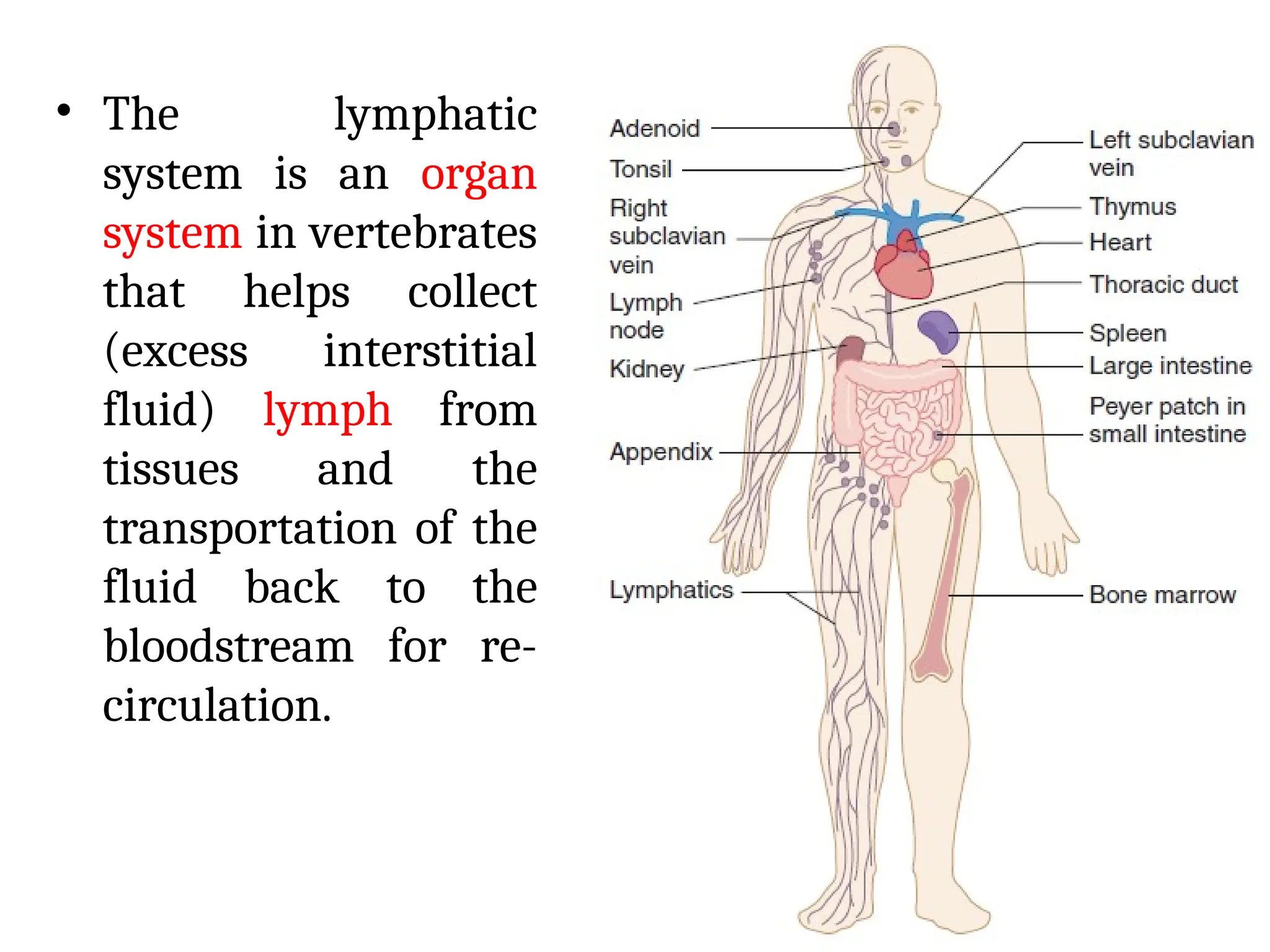
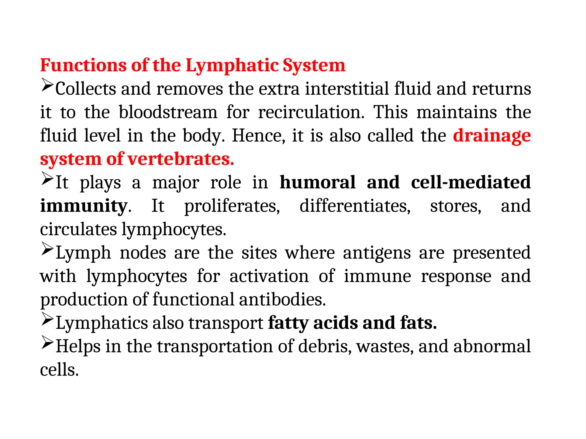
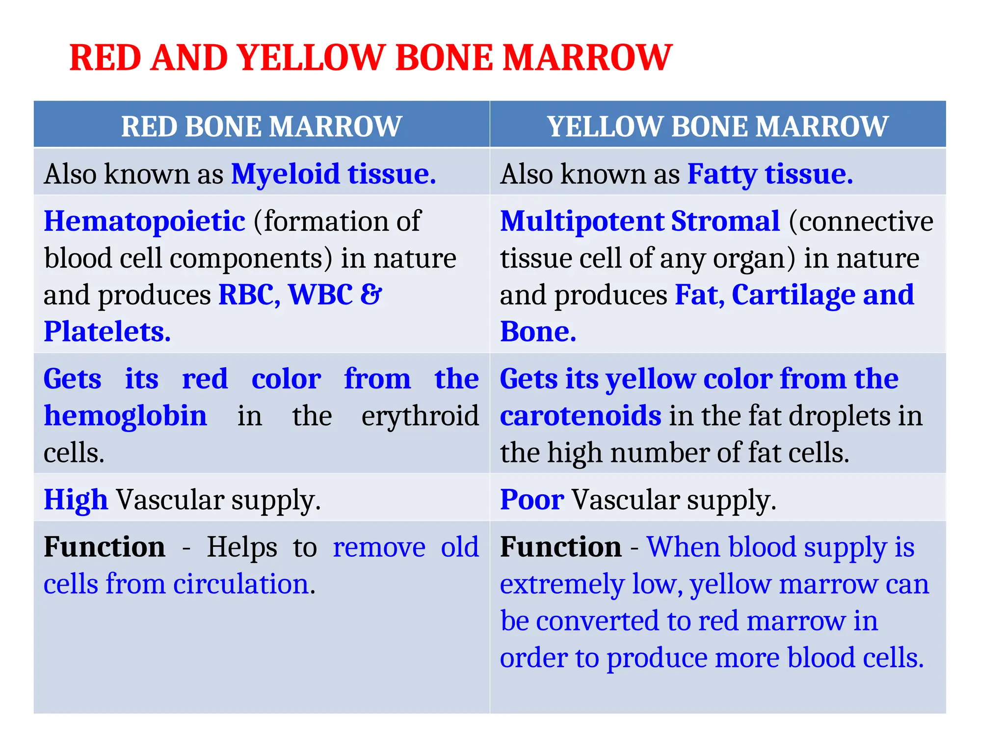
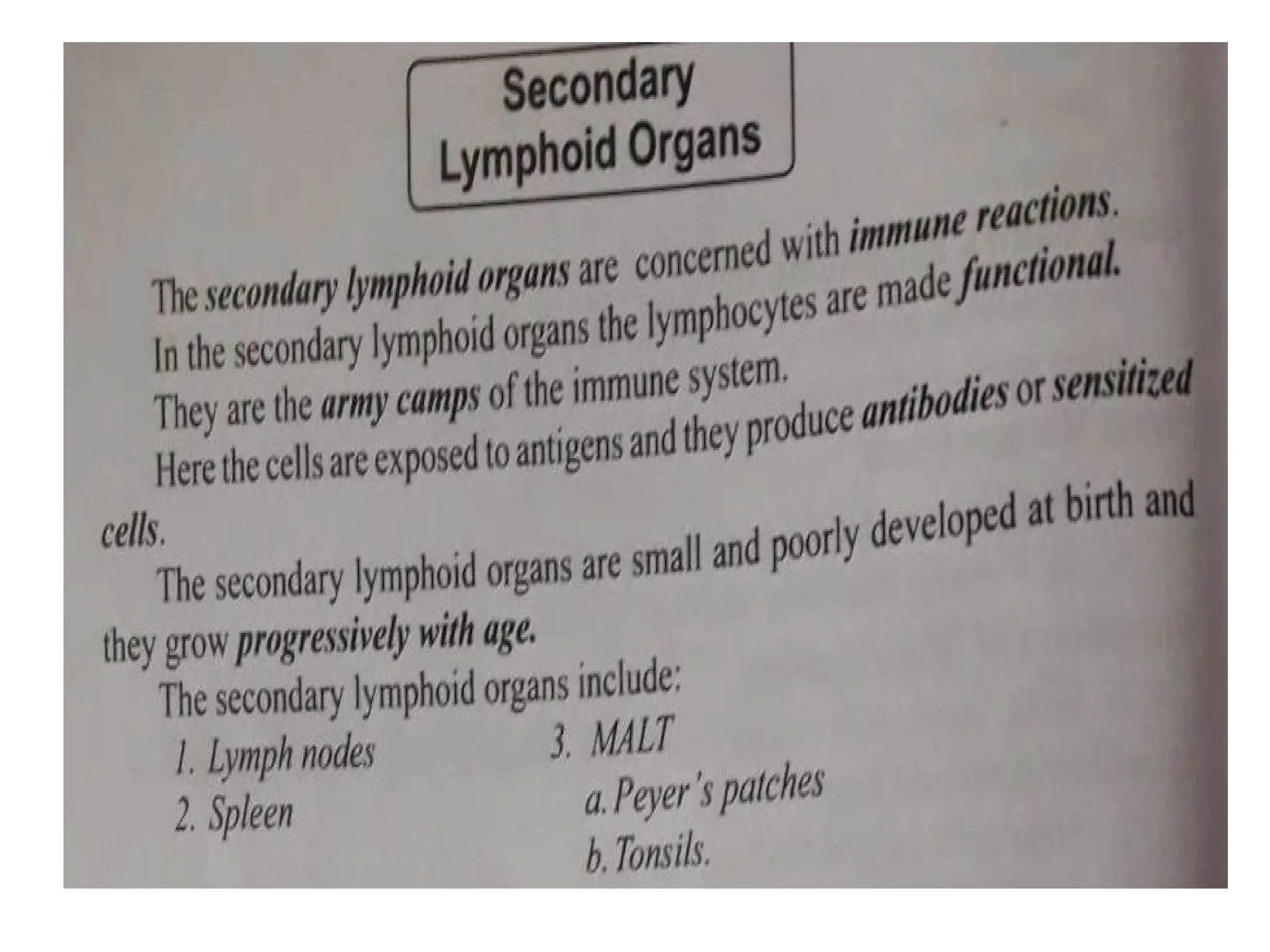
Lymphatic system
• The lymphatic
system is an organ
system in vertebrates
that helps collect
(excess interstitial
fluid) lymph from
tissues and the
transportation of the
fluid back to the
bloodstream for re-
circulation.
• Lymphatic System is composed of lymphoid organs,
lymph nodes, lymphatic vessels, and circulating clear
fluid called Lymph.
• The Lymphatic system was first described in the seventeenth
century independently by Olaus Rudbeck and Thomas
Bartholin.
• The Immune system consists of Immune cells that
continuously circulate between the Blood and Lymphoid
organs.
• Lymphoid organs are classified into 2 types. They are:
(1) Primary Lymphoid Organs-includes lymphoid organs
where the lymphocytes are produced, maintained, and
matured.
(2) Secondary Lymphoid Organs-includes lymphoid organs
where the lymphocytes undergo further maturation and
contact the bloodstream with the lymph.
Functions of the Lymphatic System
Collects and removes the extra interstitial fluid and returns
it to the bloodstream for recirculation. This maintains the
fluid level in the body. Hence, it is also called the drainage
system of vertebrates.
It plays a major role in humoral and cell-mediated
immunity. It proliferates, differentiates, stores, and
circulates lymphocytes.
Lymph nodes are the sites where antigens are presented
with lymphocytes for activation of immune response and
production of functional antibodies.
Lymphatics also transport fatty acids and fats.
Helps in the transportation of debris, wastes, and abnormal
cells.
PRIMARY & SECONDARY LYMPHOID ORGANS
PRIMARY LYMPHOID ORGANS SECONDARY LYMPHOID
ORGANS
Central lymphoid organs. Peripheral lymphoid organs.
Primary lymphoid organs are the
sites where Leukocytes (WBCs)
are generated. It includes: (i)
Bone marrow and (ii) Thymus.
Secondary lymphoid organs are
the sites where Adaptive
immune responses (Acquired
immunity) are initiated. It
includes: (i) Spleen, (ii) Lymph
Node and (iii) MALT.
The Lymphoid stem cells
proliferate (rapid increase),
differentiate and mature.
The Lymphoid stem cells becomes
functional.
Contains either B – cell or T – cell. Contains both B – cell or T – cell.
Antigens cannot enter in. Antigens enter in and stimulates
the Lymphoid cells.
PRIMARY & SECONDARY LYMPHOID ORGANS
PRIMARY LYMPHOID ORGANS SECONDARY LYMPHOID
ORGANS
Site of Maturation for T & B cells. Site of Cell function for T & B cells.
Develop before Secondary
lymphoid organs during Ontogeny
(Organization and development of
organs).
Develop after Primary lymphoid
organs during Ontogeny.
Differentiation of Lymphocytes
(Type of WBCs, E.g., T cells, B
cells & NK cells) is taken place in
Primary lymphoid organs.
Interaction of immune cells
with each other and antigen
processing are taken place in
Secondary lymphoid organs.
Decrease in size and strength
with age.
Increase in size with age.
THYMUS
• Growth and maturation of T - lymphocytes takes
place in Thymus only.
• It is large at the time of birth (70 g) but with age, the
size keep on reducing and becomes very small by
attaining puberty (3 g).
• It is a flat, bilobed organ situated above the heart.
• Each Lobe is surrounded by a Capsule and is divided
into Lobules, which are separated from each other by
strands of Connective tissue called Trabeculae.
• Each lobule is organized into 2 compartments: the outer
compartment, or cortex, is densely packed with
immature T cells, called Thymocytes, whereas the
inner compartment, or medulla, is sparsely populated
with Thymocytes.
• Both the cortex and medulla of the thymus are
crisscrossed (moved or travel around) by a three -
dimensional stromal - cell network composed of
Epithelial cells, Dendritic cells and Macrophages,
which make up the framework of the organ and
contribute to the growth and maturation of
Thymocytes.
• Some thymic epithelial cells in the outer cortex, called
Nurse cells, have long membrane extensions that
surround as many as 50 Thymocytes, forming large
multicellular complexes.
• Hassall corpuscles are a characteristic morphologic
feature located within the medullary region of the
thymus.
FUNCTIONS OF THYMUS
• The main function of the Thymus is to release
Thymosin hormone that will stimulate the
maturation of T - cells.
• Failure of Thymus development shows dramatic
decrease in circulating Lymphocytes of the T-cell
lineage and absence of Cell - mediated immunity.
(i.e. it brings about cell mediated immunity).
• Aging is accompanied by a decline in Thymic
function.
• It brings about graft rejection.
BONE MARROW
• Bone marrow is the soft, flexible connective
tissue present within the bone cavities.
• In humans and Mice, bone marrow is the site of B - cell
origin and development.
• Bone marrow forms around 4 % of total body weight.
• There are two categories of bone marrow tissue: Red
marrow and Yellow marrow. From birth to early
adolescence, the majority of our bone marrow is red
marrow. As we grow and mature, increasing amounts of
red marrow is replaced by yellow marrow.
• Bone marrow can generate 200 billions of new blood
cells every day.
RED AND YELLOW BONE MARROW
RED BONE MARROW YELLOW BONE MARROW
Also known as Myeloid tissue. Also known as Fatty tissue.
Hematopoietic (formation of
blood cell components) in nature
and produces RBC, WBC &
Platelets.
Multipotent Stromal (connective
tissue cell of any organ) in nature
and produces Fat, Cartilage and
Bone.
Gets its red color from the
hemoglobin in the erythroid
cells.
Gets its yellow color from the
carotenoids in the fat droplets in
the high number of fat cells.
High Vascular supply. Poor Vascular supply.
Function - Helps to remove old
cells from circulation.
Function - When blood supply is
extremely low, yellow marrow can
be converted to red marrow in
order to produce more blood cells.
FUNCTIONS OF BONE MARROW
• Bone marrow is the site of B - cell origin and
development-site of antibody synthesis.
• A bone marrow transplant can save the lives of people
battling leukemia, lymphoma and other blood
cancers.
• Bone marrow generates RBCs which carry oxygen to
the tissues.
• Bone marrow generates Platelets or Thrombocytes
help prevent bleeding and aid in clotting of blood.
• Granulocytes (Neutrophils, Basophils & Eosinophils) and
Macrophages fight against microbial infections. They
also remove dead cells and remodel tissue and bones.
• Stroma-Cytokines and growth factors are produced.
LYMPH NODE
• Lymph nodes are a group of small, bean-shaped
organs (2.6 cm in length) found mainly in the
neck and trunk of the human body.
• They play vital roles in the filtration of antigens
and debris from Lymph (circulating colourless
watery fluid) and in the generation of immune
responses to pathogens.
• Lymph nodes are often removed from cancer
patients as their filtration function catches tumor
cells metastasized (spread to other sites in the body)
from primary tumors.
STRUCTURE OF LYMPH NODE
• The Capsule is made of Collagen and has a sub-
capsular Sinus.
• The Lymph flows into the Sinus carrying Lymphocytes,
Antigen processing macrophages and Dendritic
cells to the node Cortex, Paracortex and Medulla.
• Morphologically, Lymph node can be divided into 3
roughly concentric regions: (1) Cortex, (2) Paracortex
and (3) Medulla.
• The outermost layer, Cortex contains Lymphocytes
(mostly B - cells), Macrophages and Follicular
dendritic cells arranged in Primary follicles.
• The Primary follicles enlarge into Secondary follicles,
each containing a Germinal center.
• Beneath the cortex is the Paracortex, which is
populated largely by T - lymphocytes and also
contains Interdigitating dendritic cells thought to
have migrated from tissues to the node.
• The innermost layer of a lymph node, the Medulla is
more sparsely populated with Lymphoid-lineage
cells of those present, many are Plasma cells
actively secreting antibody molecules.
• The Medulla in the core of the lymph node mainly
processes T - lymphocytes.
FUNCTIONS OF LYMPH NODE
• Drainage of fluid from blood stream into the tissues.
• Filtration of the lymph at the lymph nodes.
• Filtering blood.
• Raise an immune reaction and fight against
microbial infections.
• Important centre for phagocytosis.
SPLEEN
• The Spleen is an organ found in virtually
all Vertebrates.
• The spleen is an organ located in the left side of
the Abdominal cavity under the Diaphragm, the
muscular partition between the Abdomen and the
Chest.
• Similar to a Lymph node, it acts primarily as a blood
filter.
• Old RBCs are recycled in the Spleen.
• Platelets and WBCs are stored in Spleen.
• The spleen also helps to fight against certain kinds of
bacteria that cause Pneumonia and Meningitis.
STRUCTURE OF SPLEEN
• The spleen varies in size and shape between
people, but it’s commonly Ovoid shaped and
Reddish brown in colour.
• The spleen, in healthy adult humans, is
approximately 7 cm (2.8 in) to 14 cm (5.5 in) in
length. It usually weighs between 150 g and 200 g.
• The spleen is surrounded by a Capsule that extends
a number of projections (Trabeculae) into the
interior to form a compartmentalized structure.
• The compartments are of two types, the (i) Red
pulp & (ii) White pulp, which are separated by a
diffuse marginal zone.
• The splenic Red pulp consists of a network of
sinusoids populated by Macrophages and
numerous RBCs and few Lymphocytes. It is the
site where old and defective red blood cells are
destroyed and removed.
• The splenic White pulp surrounds the branches of
the Splenic artery, forming a Periarteriolar
lymphoid sheath (PALS) populated mainly by T -
lymphocytes.
• It is a site where lymphocytes are activated by the
antigens present in the blood resulting in the
activation of the humoral and cell-mediated
immune response.
• Primary lymphoid follicles - Rich in B - cells and
some of them contain Germinal centers.
• The Marginal zone, located peripheral to the PALS,
is populated by Lymphocytes and Macrophages.
• In the marginal zone, antigen is trapped by
Interdigitating dendritic cells, which carry it to
the PALS.
• The initial activation of B and T - cells takes place
in the T- cell - rich PALS.
• Like Lymph node, the Primary follicles enlarge into
Secondary follicles, each containing a Germinal
center.
FUNCTIONS OF SPLEEN
• Similar to a Lymph node, Spleen acts primarily as a
blood filter.
• Old RBCs are recycled in the Spleen.
• Platelets and WBCs are stored in Spleen.
• The spleen also helps to fight against certain
kinds of bacteria that cause Pneumonia and
Meningitis.
SPLENOMEGALY
• Splenomegaly – Enlargement of Spleen.
• A spleen weight of 400 - 500 g indicates
Splenomegaly, while a weight of more than 1000 g is
Labelled Massive Splenomegaly.
• Causes - Viral infections, Bacterial infections
(Syphilis), Parasitic infections (Malaria), Cirrhosis
(Chronic liver damage), Hemolytic anaemia, Blood
cancers, Metabolic disorders, Inflammatory disease
(Rheumatoid arthritis) and Pressure on veins.
• Symptoms – Enlarged abdomen, Painful abdomen
and shoulder, Weakness, Feeling stomach full
without eating, Anaemia, Fatigue, Frequent
infections and Easy bleeding.
MALT
• The mucous membranes lining the digestive, respiratory,
and urogenital systems have a combined surface area of
about 400 m2
and are the major sites of entry for most
pathogens.
• These vulnerable membrane surfaces are defended by a
group of organized lymphoid tissues mentioned earlier
and known collectively as Mucosal-associated lymphoid
tissue (MALT).
• MALT can be further classified as Gut-associated
lymphoid tissue (GALT) or Bronchus-associated
lymphoid tissue (BALT).
• The Tonsils, Appendix and Peyer’s patches are good
examples of lymphoid tissue found in the mucosal layer
of the alimentary canal (GALT).
Tonsils
• Tonsils are collections of Lymphoid tissue facing into the Aero
digestive tract.
• The Tonsils play a role in protecting the body against
Respiratory and Gastrointestinal infections.
• Each tonsil consists of a network of crypts (pits) that store
cells used to fight infection.
• The tonsils contain B & T- cells, that fights against infections.
• Tonsils also produce Antibodies against Polio, Streptococcal
pneumonia, Influenza, and numerous infections.
• Tonsillitis occurs when bacterial or viral organisms cause
inflammation of the Tonsillar tissue. This results in fever,
difficulty swallowing, sore throat, ear pain, loss of voice and
throat tenderness.
Appendix
• Appendix - Blind - ended tube connected to the Cecum.
• The Cecum is a pouch like structure of the Colon, located
at the junction of the Small and the Large intestines.
• The human appendix averages 9 cm in length but can
range from 2 to 20 cm. The diameter of the appendix is
usually between 7 and 8 mm.
• Appendicitis – Inflammation of Appendix.
• Functions of Appendix - 1) Maintaining Gut flora, 2)
Important component of Mucosal immune function and
3) Storehouse for good bacteria, “rebooting” the digestive
system after diarrheal illnesses.
• Surgical removal of the appendix causes no observable
health problems.
Peyer’s Patches
• Peyer’s patches are small masses of lymphatic tissue
found throughout the Ileum region of the Small
intestine.
• Peyer’s patches are roughly egg-shaped lymphatic tissue
nodules that are similar to lymph nodes in structure,
except that they are not surrounded by a connective
tissue capsule.
• Important part of the immune system by monitoring
intestinal bacteria populations and preventing the
growth of pathogenic bacteria in the intestines.
• Peyer’s patches also playing an important role in traping
antigens and destroying them.
Primary and secondary lymphoid organs.ppt

Primary and secondary lymphoid organs.ppt

  • 1.
  • 2.
    • The lymphatic systemis an organ system in vertebrates that helps collect (excess interstitial fluid) lymph from tissues and the transportation of the fluid back to the bloodstream for re- circulation.
  • 3.
    • Lymphatic Systemis composed of lymphoid organs, lymph nodes, lymphatic vessels, and circulating clear fluid called Lymph. • The Lymphatic system was first described in the seventeenth century independently by Olaus Rudbeck and Thomas Bartholin. • The Immune system consists of Immune cells that continuously circulate between the Blood and Lymphoid organs. • Lymphoid organs are classified into 2 types. They are: (1) Primary Lymphoid Organs-includes lymphoid organs where the lymphocytes are produced, maintained, and matured. (2) Secondary Lymphoid Organs-includes lymphoid organs where the lymphocytes undergo further maturation and contact the bloodstream with the lymph.
  • 4.
    Functions of theLymphatic System Collects and removes the extra interstitial fluid and returns it to the bloodstream for recirculation. This maintains the fluid level in the body. Hence, it is also called the drainage system of vertebrates. It plays a major role in humoral and cell-mediated immunity. It proliferates, differentiates, stores, and circulates lymphocytes. Lymph nodes are the sites where antigens are presented with lymphocytes for activation of immune response and production of functional antibodies. Lymphatics also transport fatty acids and fats. Helps in the transportation of debris, wastes, and abnormal cells.
  • 5.
    PRIMARY & SECONDARYLYMPHOID ORGANS PRIMARY LYMPHOID ORGANS SECONDARY LYMPHOID ORGANS Central lymphoid organs. Peripheral lymphoid organs. Primary lymphoid organs are the sites where Leukocytes (WBCs) are generated. It includes: (i) Bone marrow and (ii) Thymus. Secondary lymphoid organs are the sites where Adaptive immune responses (Acquired immunity) are initiated. It includes: (i) Spleen, (ii) Lymph Node and (iii) MALT. The Lymphoid stem cells proliferate (rapid increase), differentiate and mature. The Lymphoid stem cells becomes functional. Contains either B – cell or T – cell. Contains both B – cell or T – cell. Antigens cannot enter in. Antigens enter in and stimulates the Lymphoid cells.
  • 6.
    PRIMARY & SECONDARYLYMPHOID ORGANS PRIMARY LYMPHOID ORGANS SECONDARY LYMPHOID ORGANS Site of Maturation for T & B cells. Site of Cell function for T & B cells. Develop before Secondary lymphoid organs during Ontogeny (Organization and development of organs). Develop after Primary lymphoid organs during Ontogeny. Differentiation of Lymphocytes (Type of WBCs, E.g., T cells, B cells & NK cells) is taken place in Primary lymphoid organs. Interaction of immune cells with each other and antigen processing are taken place in Secondary lymphoid organs. Decrease in size and strength with age. Increase in size with age.
  • 8.
  • 9.
    • Growth andmaturation of T - lymphocytes takes place in Thymus only. • It is large at the time of birth (70 g) but with age, the size keep on reducing and becomes very small by attaining puberty (3 g). • It is a flat, bilobed organ situated above the heart. • Each Lobe is surrounded by a Capsule and is divided into Lobules, which are separated from each other by strands of Connective tissue called Trabeculae. • Each lobule is organized into 2 compartments: the outer compartment, or cortex, is densely packed with immature T cells, called Thymocytes, whereas the inner compartment, or medulla, is sparsely populated with Thymocytes.
  • 10.
    • Both thecortex and medulla of the thymus are crisscrossed (moved or travel around) by a three - dimensional stromal - cell network composed of Epithelial cells, Dendritic cells and Macrophages, which make up the framework of the organ and contribute to the growth and maturation of Thymocytes. • Some thymic epithelial cells in the outer cortex, called Nurse cells, have long membrane extensions that surround as many as 50 Thymocytes, forming large multicellular complexes. • Hassall corpuscles are a characteristic morphologic feature located within the medullary region of the thymus.
  • 11.
    FUNCTIONS OF THYMUS •The main function of the Thymus is to release Thymosin hormone that will stimulate the maturation of T - cells. • Failure of Thymus development shows dramatic decrease in circulating Lymphocytes of the T-cell lineage and absence of Cell - mediated immunity. (i.e. it brings about cell mediated immunity). • Aging is accompanied by a decline in Thymic function. • It brings about graft rejection.
  • 12.
    BONE MARROW • Bonemarrow is the soft, flexible connective tissue present within the bone cavities. • In humans and Mice, bone marrow is the site of B - cell origin and development. • Bone marrow forms around 4 % of total body weight. • There are two categories of bone marrow tissue: Red marrow and Yellow marrow. From birth to early adolescence, the majority of our bone marrow is red marrow. As we grow and mature, increasing amounts of red marrow is replaced by yellow marrow. • Bone marrow can generate 200 billions of new blood cells every day.
  • 14.
    RED AND YELLOWBONE MARROW RED BONE MARROW YELLOW BONE MARROW Also known as Myeloid tissue. Also known as Fatty tissue. Hematopoietic (formation of blood cell components) in nature and produces RBC, WBC & Platelets. Multipotent Stromal (connective tissue cell of any organ) in nature and produces Fat, Cartilage and Bone. Gets its red color from the hemoglobin in the erythroid cells. Gets its yellow color from the carotenoids in the fat droplets in the high number of fat cells. High Vascular supply. Poor Vascular supply. Function - Helps to remove old cells from circulation. Function - When blood supply is extremely low, yellow marrow can be converted to red marrow in order to produce more blood cells.
  • 15.
    FUNCTIONS OF BONEMARROW • Bone marrow is the site of B - cell origin and development-site of antibody synthesis. • A bone marrow transplant can save the lives of people battling leukemia, lymphoma and other blood cancers. • Bone marrow generates RBCs which carry oxygen to the tissues. • Bone marrow generates Platelets or Thrombocytes help prevent bleeding and aid in clotting of blood. • Granulocytes (Neutrophils, Basophils & Eosinophils) and Macrophages fight against microbial infections. They also remove dead cells and remodel tissue and bones. • Stroma-Cytokines and growth factors are produced.
  • 17.
    LYMPH NODE • Lymphnodes are a group of small, bean-shaped organs (2.6 cm in length) found mainly in the neck and trunk of the human body. • They play vital roles in the filtration of antigens and debris from Lymph (circulating colourless watery fluid) and in the generation of immune responses to pathogens. • Lymph nodes are often removed from cancer patients as their filtration function catches tumor cells metastasized (spread to other sites in the body) from primary tumors.
  • 19.
    STRUCTURE OF LYMPHNODE • The Capsule is made of Collagen and has a sub- capsular Sinus. • The Lymph flows into the Sinus carrying Lymphocytes, Antigen processing macrophages and Dendritic cells to the node Cortex, Paracortex and Medulla. • Morphologically, Lymph node can be divided into 3 roughly concentric regions: (1) Cortex, (2) Paracortex and (3) Medulla. • The outermost layer, Cortex contains Lymphocytes (mostly B - cells), Macrophages and Follicular dendritic cells arranged in Primary follicles. • The Primary follicles enlarge into Secondary follicles, each containing a Germinal center.
  • 20.
    • Beneath thecortex is the Paracortex, which is populated largely by T - lymphocytes and also contains Interdigitating dendritic cells thought to have migrated from tissues to the node. • The innermost layer of a lymph node, the Medulla is more sparsely populated with Lymphoid-lineage cells of those present, many are Plasma cells actively secreting antibody molecules. • The Medulla in the core of the lymph node mainly processes T - lymphocytes.
  • 21.
    FUNCTIONS OF LYMPHNODE • Drainage of fluid from blood stream into the tissues. • Filtration of the lymph at the lymph nodes. • Filtering blood. • Raise an immune reaction and fight against microbial infections. • Important centre for phagocytosis.
  • 22.
    SPLEEN • The Spleenis an organ found in virtually all Vertebrates. • The spleen is an organ located in the left side of the Abdominal cavity under the Diaphragm, the muscular partition between the Abdomen and the Chest. • Similar to a Lymph node, it acts primarily as a blood filter. • Old RBCs are recycled in the Spleen. • Platelets and WBCs are stored in Spleen. • The spleen also helps to fight against certain kinds of bacteria that cause Pneumonia and Meningitis.
  • 24.
    STRUCTURE OF SPLEEN •The spleen varies in size and shape between people, but it’s commonly Ovoid shaped and Reddish brown in colour. • The spleen, in healthy adult humans, is approximately 7 cm (2.8 in) to 14 cm (5.5 in) in length. It usually weighs between 150 g and 200 g. • The spleen is surrounded by a Capsule that extends a number of projections (Trabeculae) into the interior to form a compartmentalized structure. • The compartments are of two types, the (i) Red pulp & (ii) White pulp, which are separated by a diffuse marginal zone.
  • 25.
    • The splenicRed pulp consists of a network of sinusoids populated by Macrophages and numerous RBCs and few Lymphocytes. It is the site where old and defective red blood cells are destroyed and removed. • The splenic White pulp surrounds the branches of the Splenic artery, forming a Periarteriolar lymphoid sheath (PALS) populated mainly by T - lymphocytes. • It is a site where lymphocytes are activated by the antigens present in the blood resulting in the activation of the humoral and cell-mediated immune response.
  • 26.
    • Primary lymphoidfollicles - Rich in B - cells and some of them contain Germinal centers. • The Marginal zone, located peripheral to the PALS, is populated by Lymphocytes and Macrophages. • In the marginal zone, antigen is trapped by Interdigitating dendritic cells, which carry it to the PALS. • The initial activation of B and T - cells takes place in the T- cell - rich PALS. • Like Lymph node, the Primary follicles enlarge into Secondary follicles, each containing a Germinal center.
  • 27.
    FUNCTIONS OF SPLEEN •Similar to a Lymph node, Spleen acts primarily as a blood filter. • Old RBCs are recycled in the Spleen. • Platelets and WBCs are stored in Spleen. • The spleen also helps to fight against certain kinds of bacteria that cause Pneumonia and Meningitis.
  • 28.
    SPLENOMEGALY • Splenomegaly –Enlargement of Spleen. • A spleen weight of 400 - 500 g indicates Splenomegaly, while a weight of more than 1000 g is Labelled Massive Splenomegaly. • Causes - Viral infections, Bacterial infections (Syphilis), Parasitic infections (Malaria), Cirrhosis (Chronic liver damage), Hemolytic anaemia, Blood cancers, Metabolic disorders, Inflammatory disease (Rheumatoid arthritis) and Pressure on veins. • Symptoms – Enlarged abdomen, Painful abdomen and shoulder, Weakness, Feeling stomach full without eating, Anaemia, Fatigue, Frequent infections and Easy bleeding.
  • 29.
    MALT • The mucousmembranes lining the digestive, respiratory, and urogenital systems have a combined surface area of about 400 m2 and are the major sites of entry for most pathogens. • These vulnerable membrane surfaces are defended by a group of organized lymphoid tissues mentioned earlier and known collectively as Mucosal-associated lymphoid tissue (MALT). • MALT can be further classified as Gut-associated lymphoid tissue (GALT) or Bronchus-associated lymphoid tissue (BALT). • The Tonsils, Appendix and Peyer’s patches are good examples of lymphoid tissue found in the mucosal layer of the alimentary canal (GALT).
  • 30.
    Tonsils • Tonsils arecollections of Lymphoid tissue facing into the Aero digestive tract. • The Tonsils play a role in protecting the body against Respiratory and Gastrointestinal infections. • Each tonsil consists of a network of crypts (pits) that store cells used to fight infection. • The tonsils contain B & T- cells, that fights against infections. • Tonsils also produce Antibodies against Polio, Streptococcal pneumonia, Influenza, and numerous infections. • Tonsillitis occurs when bacterial or viral organisms cause inflammation of the Tonsillar tissue. This results in fever, difficulty swallowing, sore throat, ear pain, loss of voice and throat tenderness.
  • 33.
    Appendix • Appendix -Blind - ended tube connected to the Cecum. • The Cecum is a pouch like structure of the Colon, located at the junction of the Small and the Large intestines. • The human appendix averages 9 cm in length but can range from 2 to 20 cm. The diameter of the appendix is usually between 7 and 8 mm. • Appendicitis – Inflammation of Appendix. • Functions of Appendix - 1) Maintaining Gut flora, 2) Important component of Mucosal immune function and 3) Storehouse for good bacteria, “rebooting” the digestive system after diarrheal illnesses. • Surgical removal of the appendix causes no observable health problems.
  • 35.
    Peyer’s Patches • Peyer’spatches are small masses of lymphatic tissue found throughout the Ileum region of the Small intestine. • Peyer’s patches are roughly egg-shaped lymphatic tissue nodules that are similar to lymph nodes in structure, except that they are not surrounded by a connective tissue capsule. • Important part of the immune system by monitoring intestinal bacteria populations and preventing the growth of pathogenic bacteria in the intestines. • Peyer’s patches also playing an important role in traping antigens and destroying them.