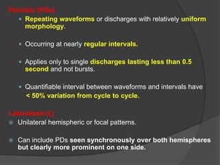
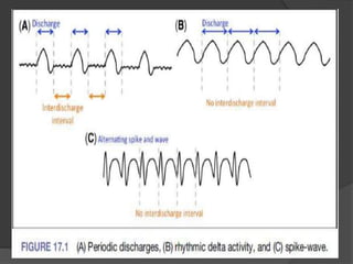
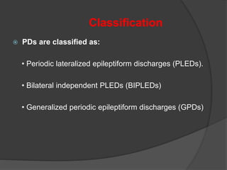
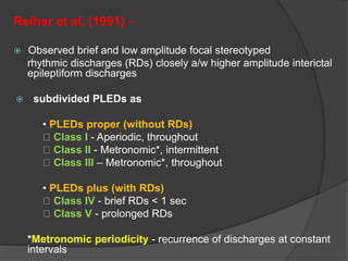
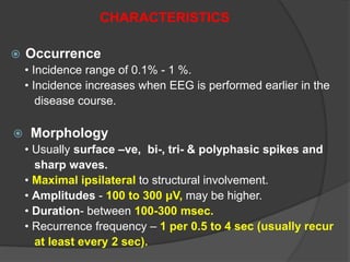
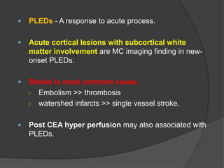
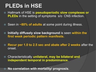
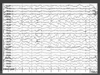
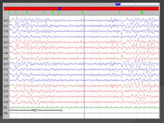
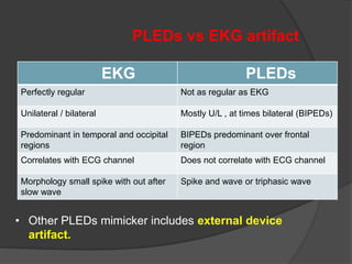
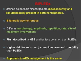
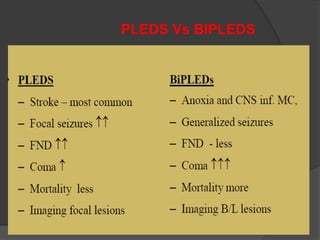
Periodic Lateralized Epileptiform
Discharges (PLEDs)
Periodic (PDs):
 Repeating waveforms or discharges with relatively uniform
morphology.
 Occurring at nearly regular intervals.
 Applies only to single discharges lasting less than 0.5
second and not bursts.
 Quantifiable interval between waveforms and intervals have
< 50% variation from cycle to cycle.
Lateralized (L)
 Unilateral hemispheric or focal patterns.
 Can include PDs seen synchronously over both hemispheres
but clearly more prominent on one side.
Classification
 PDs are classified as:
• Periodic lateralized epileptiform discharges (PLEDs).
• Bilateral independent PLEDs (BIPLEDs)
• Generalized periodic epileptiform discharges (GPDs)
Reiher et al. (1991) –
 Observed brief and low amplitude focal stereotyped
rhythmic discharges (RDs) closely a/w higher amplitude interictal
epileptiform discharges
 subdivided PLEDs as
• PLEDs proper (without RDs)
Class I - Aperiodic, throughout
Class II - Metronomic*, intermittent
Class III – Metronomic*, throughout
• PLEDs plus (with RDs)
Class IV - brief RDs < 1 sec
Class V - prolonged RDs
*Metronomic periodicity - recurrence of discharges at constant
intervals
CHARACTERISTICS
 Occurrence
• Incidence range of 0.1% - 1 %.
• Incidence increases when EEG is performed earlier in the
disease course.
 Morphology
• Usually surface –ve, bi-, tri- & polyphasic spikes and
sharp waves.
• Maximal ipsilateral to structural involvement.
• Amplitudes - 100 to 300 μV, may be higher.
• Duration- between 100-300 msec.
• Recurrence frequency – 1 per 0.5 to 4 sec (usually recur
at least every 2 sec).
ETIOLOGY
 Cortical strokes - most common cause (50%)
 Cerebral infections(20%)
- HSE most common
- Other viral infections include influenza B.
 Tumors
 Prion diseases (CJD)
• Trauma - Extra-axial hematoma, SAH
• Anoxic encephalopathy.
• Rare causes:
– Alzheimer's disease
– Mitochondrial disease
– MS
– Intoxication with baclofen, lithium, levodopa, ifosfamide.
– Trauma with out subsequent hemorrhage.
 PLEDs - A response to acute process.
 Acute cortical lesions with subcortical white
matter involvement are MC imaging finding in new-
onset PLEDs.
 Stroke is most common cause.
○ Embolism >> thrombosis
○ watershed infarcts >> single vessel stroke.
 Post CEA hyper perfusion may also associated with
PLEDs.
PLEDs in HSE
 Hallmark of HSE is pseudoperiodic slow complexes or
PLEDs in the setting of symptoms s/o CNS infection.
 Seen in ~80% of adults at some point during illness.
 Initially diffusely slow background is seen within the
first week periodic pattern manifests.
 Recur per 1.0 to 2.5 sec and abate after 2 weeks after the
onset.
 Characteristically unilateral, may be bilateral and
independent temporal in predominance.
 No correlation with mortality/ prognosis.
PLEDs in CJD
 Pseudoperiodic , biphasic or triphasic sharply contoured
wave forms with diffuse slow background.
 67 – 100% CJD will have PLEDs and appear in within 3
months of onset.
 Repeat with a period of 0.5 to 2.0 sec and shorten with
disease progress.
 Rarely unilateral, typically anterior predominant.
 Present only during wakefulness and frequently time
locked to myoclonic jerks.
SSPE
 The typical EEG pattern is usually seen in myoclonic phase
and is virtually diagnostic.
 Periodic complexes consisting of bilaterally symmetrical,
synchronous, high voltage (200–500 mv), polyphasic,
stereotyped delta waves.
 Waveforms remain identical in any given lead.
 These periodic complexes repeat at fairly regular 4–10
second intervals and have 1:1 relationship with
myoclonic jerks.
 Shortening of interval between periodic complexes with
progression of the disease.
 The periodic complexes of SSPE first appear during
sleep, when they are not accompanied by myoclonic
spasms.
 Late in the course of disease, the EEG may become
increasingly disorganised and show high amplitudes
and random dysrhythmic slowing.
 In terminal stages the amplitude of waveforms may fall.
Other types of EEG abnormalities include:
Type II pattern:
 Characterized by periodic giant delta waves intermixed
with rapid spikes as fast activity.
 In this pattern of periodic complexes background is
usually slow.
Type III pattern:
 Characterized by long spike-wave discharges
interrupted by giant delta waves.
Prognosis: Type II > Type I > Type III
THEORIES OF PLEDs NEUROPHYSIOLOGY
Pohlmann et al (1996)
○ PLEDs are EEG signature of a dynamic patho-
physiological state in which unstable neurobiological
processes create an ictal-interictal continuum.
○ An unstable brain state related to the combination of
one or more of seizures, structural injury and
metabolic derangement.
○ No single common unifying mechanism.
Lee 1988, Handforth 1994
 PET and SPECT show hyper metabolism and hyper
perfusion in PLED foci, respectively.
 But these reflect increased neuronal activity rather than
seizure.
Kalamangalam (2015)
 Synchronization of pre-existing local field potentials,
through enhancement of excitatory neurotransmission
and inactivation of inhibitory neurotransmission
provoked by the PLED associated disease process.
PLEDs vs EKG artifact
EKG PLEDs
Perfectly regular Not as regular as EKG
Unilateral / bilateral Mostly U/L , at times bilateral (BIPEDs)
Predominant in temporal and occipital
regions
BIPEDs predominant over frontal
region
Correlates with ECG channel Does not correlate with ECG channel
Morphology small spike with out after
slow wave
Spike and wave or triphasic wave
• Other PLEDs mimicker includes external device
artifact.
Relation of PLEDs to seizures
 Most clinicians, do not consider PLEDs to be “ictal”.
 PLEDs can exist in patients who never develop either
clinical or electrographic seizure.
 However, they highly associated with clinical seizures and
may lie somewhere along an “ictal-interictal continuum”.
 Seizure occur in up to 80% of patients with PLEDs and
focal motor seizure MC.
 No significant association between seizures and etiology.
 No significant difference in degree of functional outcome
between patients with or without PLED associated seizures.
Prognosis
 Prognosis depends on the underlying etiology.
 The worst prognosis noted for acute severe stroke.
 Mortality was unchanged with or without treatment of
patients with PLEDs on cEEG.
 PLEDs without structural lesion can be ictal, interictal or
postictal finding on EEG, resulted in a higher mortality rate.
Treatment
 No standard management for diagnosis, prevention and Rx of
seizures associated to PLEDs.
 Strongly consider treatment if:
 Presence of myoclonic or clonic movements, nystagmus or
rhythmic blinking time locked to appearance of PDs
(i.e, ictal PDs)
 Decline in clinical state that coincides with onset of PD
 History of any of the following:
○ Epilepsy or recent clinical seizure/SE
○ Acute structural lesion a/w high risk of seizures
(SAH,ICH,TBI).
BIPLEDs
 Defined as periodic discharges are independently and
simultaneously present in both hemispheres.
 Bilaterally asynchronous
 Differ in morphology, amplitude, repetition, rate, site of
maximum involvement
 First described in HSE and far less common than PLEDs.
 Higher risk for seizures, ↓ consciousness and mortality
than PLEDs.
 Approach to AED management is the same.
PLEDS Vs BIPLEDS
BIPLEDs
Ipsilateral Independent PLEDs (IpsiIPs)
• Rare subtype
• Ipsilateral but independent in temporal & topographical
relationship.
Multifocal PLEDS
• 3 or more independent foci of PLEDs over both hemispheres.
• 3 foci are also called TriPLEDs.
• Reflect severe brain dysfunction
• Significant mortality rate.
Periodic lateralized epileptiform discharges

Periodic lateralized epileptiform discharges

  • 1.
  • 2.
    Periodic (PDs):  Repeatingwaveforms or discharges with relatively uniform morphology.  Occurring at nearly regular intervals.  Applies only to single discharges lasting less than 0.5 second and not bursts.  Quantifiable interval between waveforms and intervals have < 50% variation from cycle to cycle. Lateralized (L)  Unilateral hemispheric or focal patterns.  Can include PDs seen synchronously over both hemispheres but clearly more prominent on one side.
  • 4.
    Classification  PDs areclassified as: • Periodic lateralized epileptiform discharges (PLEDs). • Bilateral independent PLEDs (BIPLEDs) • Generalized periodic epileptiform discharges (GPDs)
  • 5.
    Reiher et al.(1991) –  Observed brief and low amplitude focal stereotyped rhythmic discharges (RDs) closely a/w higher amplitude interictal epileptiform discharges  subdivided PLEDs as • PLEDs proper (without RDs) Class I - Aperiodic, throughout Class II - Metronomic*, intermittent Class III – Metronomic*, throughout • PLEDs plus (with RDs) Class IV - brief RDs < 1 sec Class V - prolonged RDs *Metronomic periodicity - recurrence of discharges at constant intervals
  • 6.
    CHARACTERISTICS  Occurrence • Incidencerange of 0.1% - 1 %. • Incidence increases when EEG is performed earlier in the disease course.  Morphology • Usually surface –ve, bi-, tri- & polyphasic spikes and sharp waves. • Maximal ipsilateral to structural involvement. • Amplitudes - 100 to 300 μV, may be higher. • Duration- between 100-300 msec. • Recurrence frequency – 1 per 0.5 to 4 sec (usually recur at least every 2 sec).
  • 7.
    ETIOLOGY  Cortical strokes- most common cause (50%)  Cerebral infections(20%) - HSE most common - Other viral infections include influenza B.  Tumors  Prion diseases (CJD) • Trauma - Extra-axial hematoma, SAH • Anoxic encephalopathy.
  • 8.
    • Rare causes: –Alzheimer's disease – Mitochondrial disease – MS – Intoxication with baclofen, lithium, levodopa, ifosfamide. – Trauma with out subsequent hemorrhage.
  • 9.
     PLEDs -A response to acute process.  Acute cortical lesions with subcortical white matter involvement are MC imaging finding in new- onset PLEDs.  Stroke is most common cause. ○ Embolism >> thrombosis ○ watershed infarcts >> single vessel stroke.  Post CEA hyper perfusion may also associated with PLEDs.
  • 10.
    PLEDs in HSE Hallmark of HSE is pseudoperiodic slow complexes or PLEDs in the setting of symptoms s/o CNS infection.  Seen in ~80% of adults at some point during illness.  Initially diffusely slow background is seen within the first week periodic pattern manifests.  Recur per 1.0 to 2.5 sec and abate after 2 weeks after the onset.  Characteristically unilateral, may be bilateral and independent temporal in predominance.  No correlation with mortality/ prognosis.
  • 12.
    PLEDs in CJD Pseudoperiodic , biphasic or triphasic sharply contoured wave forms with diffuse slow background.  67 – 100% CJD will have PLEDs and appear in within 3 months of onset.  Repeat with a period of 0.5 to 2.0 sec and shorten with disease progress.  Rarely unilateral, typically anterior predominant.  Present only during wakefulness and frequently time locked to myoclonic jerks.
  • 14.
    SSPE  The typicalEEG pattern is usually seen in myoclonic phase and is virtually diagnostic.  Periodic complexes consisting of bilaterally symmetrical, synchronous, high voltage (200–500 mv), polyphasic, stereotyped delta waves.  Waveforms remain identical in any given lead.  These periodic complexes repeat at fairly regular 4–10 second intervals and have 1:1 relationship with myoclonic jerks.
  • 15.
     Shortening ofinterval between periodic complexes with progression of the disease.  The periodic complexes of SSPE first appear during sleep, when they are not accompanied by myoclonic spasms.  Late in the course of disease, the EEG may become increasingly disorganised and show high amplitudes and random dysrhythmic slowing.  In terminal stages the amplitude of waveforms may fall.
  • 17.
    Other types ofEEG abnormalities include: Type II pattern:  Characterized by periodic giant delta waves intermixed with rapid spikes as fast activity.  In this pattern of periodic complexes background is usually slow. Type III pattern:  Characterized by long spike-wave discharges interrupted by giant delta waves. Prognosis: Type II > Type I > Type III
  • 19.
    THEORIES OF PLEDsNEUROPHYSIOLOGY Pohlmann et al (1996) ○ PLEDs are EEG signature of a dynamic patho- physiological state in which unstable neurobiological processes create an ictal-interictal continuum. ○ An unstable brain state related to the combination of one or more of seizures, structural injury and metabolic derangement. ○ No single common unifying mechanism.
  • 20.
    Lee 1988, Handforth1994  PET and SPECT show hyper metabolism and hyper perfusion in PLED foci, respectively.  But these reflect increased neuronal activity rather than seizure. Kalamangalam (2015)  Synchronization of pre-existing local field potentials, through enhancement of excitatory neurotransmission and inactivation of inhibitory neurotransmission provoked by the PLED associated disease process.
  • 21.
    PLEDs vs EKGartifact EKG PLEDs Perfectly regular Not as regular as EKG Unilateral / bilateral Mostly U/L , at times bilateral (BIPEDs) Predominant in temporal and occipital regions BIPEDs predominant over frontal region Correlates with ECG channel Does not correlate with ECG channel Morphology small spike with out after slow wave Spike and wave or triphasic wave • Other PLEDs mimicker includes external device artifact.
  • 22.
    Relation of PLEDsto seizures  Most clinicians, do not consider PLEDs to be “ictal”.  PLEDs can exist in patients who never develop either clinical or electrographic seizure.  However, they highly associated with clinical seizures and may lie somewhere along an “ictal-interictal continuum”.  Seizure occur in up to 80% of patients with PLEDs and focal motor seizure MC.  No significant association between seizures and etiology.  No significant difference in degree of functional outcome between patients with or without PLED associated seizures.
  • 23.
    Prognosis  Prognosis dependson the underlying etiology.  The worst prognosis noted for acute severe stroke.  Mortality was unchanged with or without treatment of patients with PLEDs on cEEG.  PLEDs without structural lesion can be ictal, interictal or postictal finding on EEG, resulted in a higher mortality rate.
  • 24.
    Treatment  No standardmanagement for diagnosis, prevention and Rx of seizures associated to PLEDs.  Strongly consider treatment if:  Presence of myoclonic or clonic movements, nystagmus or rhythmic blinking time locked to appearance of PDs (i.e, ictal PDs)  Decline in clinical state that coincides with onset of PD  History of any of the following: ○ Epilepsy or recent clinical seizure/SE ○ Acute structural lesion a/w high risk of seizures (SAH,ICH,TBI).
  • 25.
    BIPLEDs  Defined asperiodic discharges are independently and simultaneously present in both hemispheres.  Bilaterally asynchronous  Differ in morphology, amplitude, repetition, rate, site of maximum involvement  First described in HSE and far less common than PLEDs.  Higher risk for seizures, ↓ consciousness and mortality than PLEDs.  Approach to AED management is the same.
  • 26.
  • 27.
  • 28.
    Ipsilateral Independent PLEDs(IpsiIPs) • Rare subtype • Ipsilateral but independent in temporal & topographical relationship. Multifocal PLEDS • 3 or more independent foci of PLEDs over both hemispheres. • 3 foci are also called TriPLEDs. • Reflect severe brain dysfunction • Significant mortality rate.