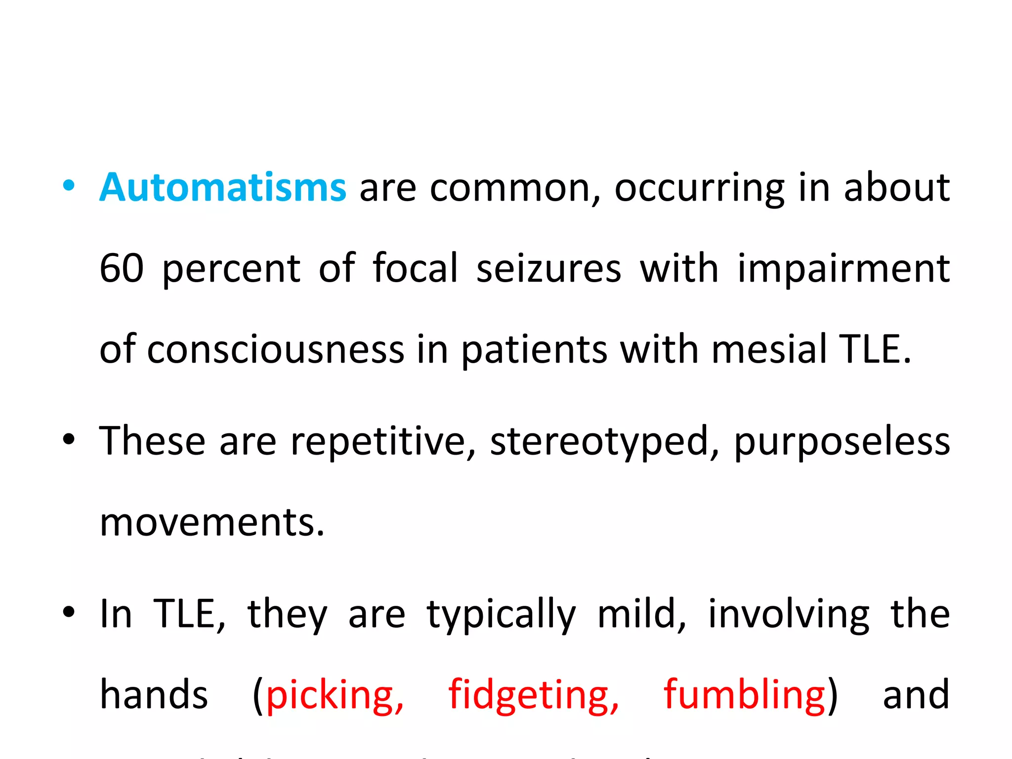
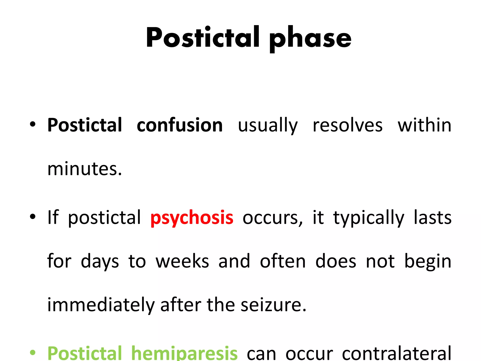
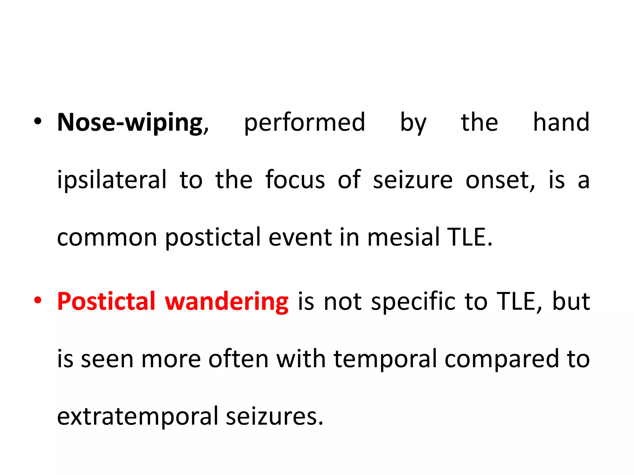
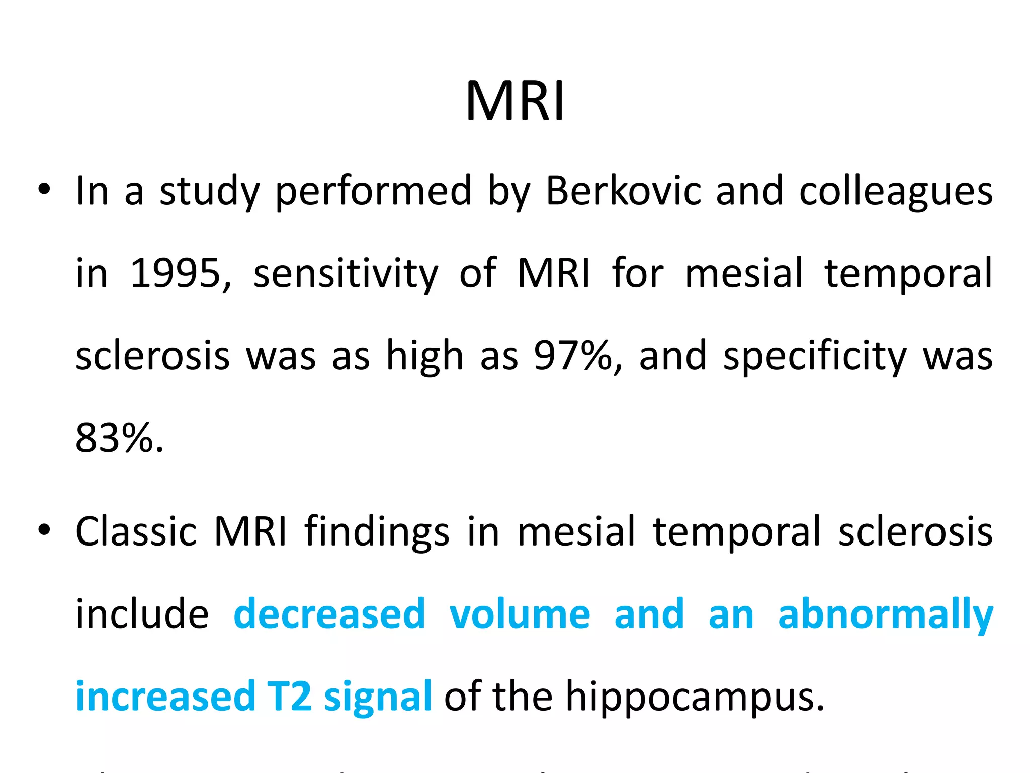
Mesial temporal lobe epilepsy
Anatomy of medial temporal lobe
• Consists of two interlocking
U shaped gray matter
structures
– Hippocampus proper
( Ammon horn)
Superolateral, upside-down U.
– Dentate gyrus
Inferomedial U
• Hippocampus is a curved
structure on the medial
aspect of temporal lobe
that bulges into floor of
temporal horn.
• Hippocampus – seahorse
• Cornu Ammonis (CA)
– CA1
– CA2
– CA3
– CA4
• Medially, the ambient cistern which separates
the hippocampus from brainstem.
• Choriodal fissure and temporal horn
superiorly.
• Parahippocampal gyrus inferiorly.
• Temporal horn of lateral ventricle laterally.
• The amygdala is an almond-
shaped mass that is located
deep within the brain. It is
between the temporal lobes,
hippocampus and
hypothalamus.
• plays a vital role in these
various parts of the brain and
with no surprise the amygdala
deals with our emotion,
particularly with fear.
History and evolution
• Bouchet and Cazauvieilh first described the association
between epilepsy and a sclerotic hippocampus in 1825.
• Hughlings Jackson recognized that partial seizures also
represented epileptic phenomena and made an
association between limbic-type seizures, which he
called “intellectual auras” or “dreamy states,” and
lesions in mesial temporal structures.
• Bailey and Gibbs were the first to perform anterior temporal
lobectomy on the basis of EEG evidence alone.
• Falconer in 1953
– en bloc resection including mesial temporal lobe structures.
– structural lesion was the cause, and not the effect, of recurrent
epileptic seizures.
– He also recognized the association between HS and both febrile
convulsions and a family history of epilepsy, which suggested the
existence of a specific syndrome.
• Crandall 1963 subsequently took advantage of the en bloc resection
and long-term depth-electrode recording to initiate basic research
on temporal lobe epilepsy.
Aetiology of HS
• still controversial and is likely to be multifactorial.
• widely considered as an acquired pathology, there is
even proven autosomal dominant inheritance.
• Alfred Meyer’s seminal hypothesis from the 1950s
proposed that an initiating event, injury or insult,
particularly prolonged febrile convulsions early in life,
primed the immature hippocampus for the subsequent
development of HS.
• TLE has been induced following prolonged febrile
seizures and as the FEBSTAT study have confirmed.
• ApoEε4 genotype has been associated with increased
risk of bilateral HS.
• More recently, HS and febrile seizures were linked by
common genetic variation around SCN1A gene.
Seizure-induced neuronal loss
• Necrotic or apoptotic neurones are seen in surgical
specimens of HS as evidence of ongoing neuronal
death.
• It is generally regarded that seizure induced neuronal
injury and subsequent loss results from excitotoxic,
glutamatergic neurotransmission, excessive Na+ and
Ca2+, resulting in osmolytic stress and cellular free-
radical production, culminating in necrosis of
neurones.
Inflammation
• In HS, neuronophagia or focal infiltrates of microglia are occasional
findings.
• There is evidence to support activation of both the innate and adaptive
immune system, for example IL-1β and IL-1 receptor upregulation, has
been noted in astrocytes, microglia and neurones in HS, and intercellular
adhesion molecule 1 (ICAM-1) and kallikrein expression in glia.
• Regarding underlying viral infection in HS, detection of human Herpes
virus 6 infection was frequently reported in one series but in another was
only identified in patients with a history of a prior episode of limbic
encephalitis.
Other aetiologies
• Infections- herpes encephalitis, bacterial meningitis,
neurocysticercosis
• Trauma - encephalomalacia or cortical scarring;
• Hamartomas
• Malignancies - meningiomas, gliomas, gangliomas
• Vascular malformations - arteriovenous
malformation, cavernous angioma)
• Idiopathic (genetic): rare, familial TLE
• Cryptogenic
Neuropathology
Mossy fibre sprouting
• Axonal sprouting, a common process in the developing
brain, is revived in adult tissues in response to seizures.
• Such plasticity may represent primarily a reparative
response to hippocampal neuronal loss, but may
ultimately be mal-adaptive and pro-epileptogenic.
– Selective loss of somatostatin and neuropeptide Y containing
hilar neurons.
• Abnormalities of the vasculature have been reported in
HS, with proliferation of micro-vessels, vascular
endothelial growth factor receptor expression and loss
of blood-brain barrier integrity .
• Vascular leakage of proteins, including IgG and albumin
may contribute to neuronal dysfunction in epilepsy.
• Gliosis is a striking component of HS with chronic,
fibrillary gliosis in CA1 and a radial gliosis in the
dentate gyrus; gliosis involving CA4 is a less specific
finding for HS.
Clinical features
• Temporal lobe epilepsy (TLE) is the most
common of the localization-related epilepsies.
• Most cases of TLE can be further localized to
the mesial temporal lobe (hippocampus,
amygdala, and parahippocampal gyrus).
• Focal seizures with impairment of
consciousness or awareness (previously
referred to as complex partial seizures) are the
most common manifestation of mesial TLE.
• About one-third of patients also have focal
seizures evolving to secondary generalized
seizures.
Initiating event e.g. complex febrile
convulsion/ status epilepticus or
other early insult
Latent period
Spontaneous seizure
Complete
remission Initial remission
Active seizure
disorder
Relapse, active seizure
disorder
One
year
4-5
years
12-13
years
14-18
years
Habitual
seizure,
respond well
to AED, do
well for
years
Silent period
Distinctive characteristics
• An aura (with sensory or psychic symptoms) occurs in
most patients.
• Common features include a rising epigastric sensation
(often likened to a "roller coaster" sensation), and
psychic or experiential phenomena, such as deja vu,
jamais vu, or fear.
• Auras of taste (gustatory hallucinations) and smell
(olfactory hallucinations) are less common.
• In either case, patients with mesial TLE usually
recall the seizure aura.
• Focal seizures with impairment of
consciousness manifest with a behavioral
arrest with staring and lasts between 30 and
120 seconds.
• The patients are generally unaware and
• Occasionally, such patients present with
amnestic attacks, but more detailed
questioning or observation of the seizures
reveal the presence of olfactory
hallucinations, other seizure auras, or ictal
automatisms.
• Automatisms are common, occurring in about
60 percent of focal seizures with impairment
of consciousness in patients with mesial TLE.
• These are repetitive, stereotyped, purposeless
movements.
• In TLE, they are typically mild, involving the
hands (picking, fidgeting, fumbling) and
• Lateralizing features can occur during or after
a focal seizure.
• Unilateral automatisms are usually ipsilateral
to the seizure focus, while dystonic posturing
almost invariably occurs on the contralateral
side.
• Head deviation at seizure onset is usually
ipsilateral to the seizure; when it occurs later,
it is contralateral.
• Contralateral clonic activity is relatively unusual.
• Lateralizing findings in the setting of mesial
temporal sclerosis should be interpreted with
some caution, as many of these patients have
bilateral, independent seizure foci.
• Less commonly observed behaviors associated
with a temporal lobe seizure include ictal speech
and vocalizations, affective behaviors (laughing,
crying or fear), hypermotor behaviors usually
Postictal phase
• Postictal confusion usually resolves within
minutes.
• If postictal psychosis occurs, it typically lasts
for days to weeks and often does not begin
immediately after the seizure.
• Postictal hemiparesis can occur contralateral
• Nose-wiping, performed by the hand
ipsilateral to the focus of seizure onset, is a
common postictal event in mesial TLE.
• Postictal wandering is not specific to TLE, but
is seen more often with temporal compared to
extratemporal seizures.
• Although some reports have noted that
tachycardia is more typical of seizures
originating from the right mesial temporal
lobe and bradycardia is more common with
left-sided seizures, most have not found this
to be a useful lateralizing feature.
Right vs left mTLE
• Lesion studies indicate that left and right temporomesial structures are
essential for verbal and visuospatial memory, respectively.
• Concordantly, patients with dominant medial temporal sclerosis (MTS)
have abnormalities of verbal memory, whereas those with nondominant
foci may have deficits of visuospatial memory.
• Visuospatial skills allow us to visually perceive objects and the spatial
relationships among objects.
• They allow us to retrace our way across the city because we have a visual
map in our memory.
Medial
Febrile seizure
Duration >2min
Aura : visceral,gustatory,
autonomic.
Motionless stare and
automatism
Autonomic phenomenon
Lateral
No
Lesser
Aura :auditory,hallucinations and
illusions.
Same
No
Electroencephalography
Interictal EEG
• Major part of temporal lobe is inaccessible to surface electrodes
• For a temporal spike to show up in scalp EEG, 6 cm of cortex needs to be
activated.
• Interictal phase shows– anterior temporal sharp waves, spikes and slow
waves.
• During auras – no EEG change.
• For scalp EEG to diagnose atleast 10cm2 of cortex should be involved.
• Additional electrode -10-10 system, true anterior temporal electrodes,
sphenoidal electrodes, zygomatic, cheek electrodes.
Postictal EEG Changes in mTLE
• The ictal onset on scalp recorded EEG can be obscured by
variety of artifacts.
• Therefore, sometimes postictal changes could provide
important information for lateralization of the seizure
onset.
• Postictal EEG changes in TLE could be in the form of
unilateral or bilateral slowing. Lateralized postictal slow
waves could be present in up to 70% of patients with mTLE.
Temporal Intermittent Rhythmic
Delta Activity (TIRDA)
• consists of trains of rhythmic delta activity lasting
4–20 seconds and is observed in up to 25% of
patients with TLE who are being evaluated for
surgery. TIRDA is more specific to TLE and often
associated with epileptiform discharges.
• In one study, TIRDA was found in up to 90% of
patients with MRI evidence of hippocampal
Intracranial Electrodes
• They include depth, subdural, epidural, and
foramen ovale electrodes.
• Depth electrodes are inserted through a burr
hole and can be removed at the bedside with
caution. They penetrate the brain and lie
within the cortex.
• Foramen ovale electrodes are similar to the
depth electrodes and are inserted under
fluoroscopy in radiology or in the operating
room.
• Subdural electrodes lie on the pial surface of
the brain. The strips have a single or double
row of contacts. They can be placed through a
• They record from gyral surface over multiple
areas and from the epidural or subdural spaces.
Subdural grids are larger in size and monitor a
larger area of cortex.
• They are also used for brain mapping. Subdural
strips are better tolerated than subdural grids and
can be removed at the bed side; however, grids
have to be removed in the operating room.
• The chronic intracranial electrodes have 1–4%
Subdural EEG
MRI
• In a study performed by Berkovic and colleagues
in 1995, sensitivity of MRI for mesial temporal
sclerosis was as high as 97%, and specificity was
83%.
• Classic MRI findings in mesial temporal sclerosis
include decreased volume and an abnormally
increased T2 signal of the hippocampus.
•Thin section angled coronal sequences at right angles to the longitudinal axis of
the hippocampus are required, to minimize volume averaging.
MR spectroscopy
• typically represent neuronal dysfunction
• decreased NAA and decreased NAA/Cho and
NAA/Cr ratios
• increased lipid and lactate soon after as
seizure.
Hippocampal sclerosis
Normal left hippocampus
Decreased NAA/Cho ratio.
Normal range of NAA/Cho
and NAA/Cr ratios
When severe and long standing, additional associated findings include :
1. atrophy of the ipsilateral fornix and mamillary body
2. increased signal and or atrophy of the anterior thalamic nucleus
3. atrophy of the cingulate gyrus
4. increased signal and/or reduction in the volume of the amygdala
5. reduction in the volume of the subiculum
6. dilatation of temporal horn and temporal lobe atrophy
7. collateral white matter and entorhinal cortex atrophy
8. thalamic and caudate atrophy
9. ipsilateral cerebral hypertrophy
10. contralateral cerebellar hemiatrophy
11. loss of grey-white matter interface in the anterior temporal lobe
12. reduced white matter volume in the parahippocampal gyrus.
FDG PET
• PET scans show glucose metabolism in the brain by
using a positron-emitting substance.
• Patients with temporal lobe epilepsy have decreased
glucose metabolism in the affected lobe during the
interictal period.
• Most sensitive interictal imaging technique for identifying
focal functional deficit.
• Region of PET hypometabolism is larger than epileptogenic
zone.
• Particularly useful in patients with normal MRI.
• EEG & PET + : can be taken for surgery without invasive
test.
Interictal FDG-PET (axial and coronal images) showing left
temporal hypometabolism in a 12-year-old child with TLE and
left HS
SPECT
• SPECT scans show the distribution of blood flow in
the brain at the time of the injection of a
radiotracer, which is injected ictally or interictally.
• If the radiotracer is injected ictally, focally increased
uptake is identified in the affected temporal lobe
(hot focus).
• If the radiotracer is injected interictally, the effected
Hyperperfusion on ictal SPECT (a) and hypoperfusion
on interictal SPECT (b) in a child with TLE.
• Sensitivity for detection of interictal seizure foci is
65-75% for both SPECT scans and PET scans.
• When the source of seizures is lateralized on PET
scans or SPECT scans, 94% of patients improve after
surgical resection.
Treatment
Medical management
– Carbamezapine,
– oxcarbazapine,
– lamotrigine,
– toperamate,
– levetiracetam.
Refractory epilepsy:
• 20% of the epilepsy patients are refractory to
medical management.
• Among them 25 to 50% cases have mTLE as
the diagnosis.
• Refractory epilepsy occurs when a person has
failed to become seizure free with adequate
doses of two AEDs, with atleast 2 episodes per
month for a disease duration of 2 years.
Candidates for Epilepsy Surgery
• Persistent seizures despite appropriate pharmacological treatment
– Usually at least two drugs, appropriate to seizure type, at adequate
doses, with adequate compliance
• Impairment of quality of life due to ongoing seizures
– Loss of driving privileges, employment opportunities, social/cultural
stigma, dependence on others, side effects of medications, under
achievement in school, memory deficit, attention deficit, injuries,
accidents
• Not a contraindication but rethink.
– Elderly
– Low IQ (intelligence quotient under 90)
– Psychiatric disease
– Other co morbidities
Presurgical predictors
fMRI
• Mapping of functional areas and
understanding lateralization of language and
memory help surgeons avoid removing critical
brain regions when they have to operate and
remove brain tissue.
• This is of particular importance in removing
tumors and in patients who have
aimed at localizing eloquent areas (e.g. speech, motor function)
Magnetoencephalography (MEG)
• Functional
neuroimaging technique for
mapping brain activity by
recording magnetic
fields produced by electrical
currents occurring naturally in
the brain, using very
sensitive magnetometers.
• Useful for neocortical
convexity lesions.
Electrocorticogrphy(ECoG)
• The ECoG recording is performed from
electrodes placed on the exposed cortex.
• Done under general anesthesia or under local
anesthesia if patient interaction is required for
functional cortical mapping.
• Success of the surgery depends on accurate
localization and removal of the epileptogenic
• Electrodes may either be
placed outside the dura
mater (epidural) or under
the dura mater
(subdural).
• Depth electrodes may
also be used to record
activity from deeper
structures such as
• Widely used for presurgical planning to guide
surgical resection of the lesion and epileptogenic
zone.
• ECoG data is assessed with regard to ictal spike
activity – “diffuse fast wave activity” recorded
during a seizure – and interictal epileptiform
activity (IEA), brief bursts of neuronal activity
recorded between epileptic events.
• Residual spikes on the ECoG, unaltered by the
resection, indicate poor seizure control, and
incomplete neutralization of the epileptogenic
cortical zone.
• Additional surgery may be necessary to
completely eradicate seizure activity.
Surgical management
• Temporal lobectomy.
– Most common surgical approach:
resection of anterior temporal lobe
with amygdala and hippocampus.
– Less extensive on left to save language
function.(superior temporal gyrus
spared )
Resection of the anterior temporal lobe (~4.5 cm on left side, ~5.5 cm on right side)
followed by resection of mesial structures (amygdala, hippocampus, parahippocampal
gyrus).
• Selective amygdalohippocampectomy
– Idea is to remove mesial structures (hippocampus, amygdala,
parahippocampal gyrus) leaving lateral temporal cortex intact
– Less risk for language function and equal outcome for seizure
control.
– Phonemic fluency better after transcortical than transsylvian
approach.
– Multiple subpial transections
• Disconnection of horizontal intracortical fibres and
preserving vertical connections. (ictal discharge spreads
horizontally and cortical functions vertically)
• Memory sparing procedure.
Engel Epilepsy Surgery Outcome Scale
• Class I: Free of disabling seizures
• Class II: Rare disabling seizures ("almost
seizure-free")
• Class III: Worthwhile improvement
• Class IV: No worthwhile improvement.
Prognosis
• With early surgical intervention, patients with mesial temporal lobe
epilepsy have an excellent chance of cure and a subsequent normal life.
• 60%--seizure free even after stopping all AED
• 20% reduced seizure frequency but need to continue with AED
• 10%--no benefit
• 10%--get worse
• Significant neurosurgical complication rarely occur
THANK YOU

MTLE

  • 1.
  • 2.
    Anatomy of medialtemporal lobe • Consists of two interlocking U shaped gray matter structures – Hippocampus proper ( Ammon horn) Superolateral, upside-down U. – Dentate gyrus Inferomedial U
  • 3.
    • Hippocampus isa curved structure on the medial aspect of temporal lobe that bulges into floor of temporal horn. • Hippocampus – seahorse
  • 4.
    • Cornu Ammonis(CA) – CA1 – CA2 – CA3 – CA4
  • 6.
    • Medially, theambient cistern which separates the hippocampus from brainstem. • Choriodal fissure and temporal horn superiorly. • Parahippocampal gyrus inferiorly. • Temporal horn of lateral ventricle laterally.
  • 8.
    • The amygdalais an almond- shaped mass that is located deep within the brain. It is between the temporal lobes, hippocampus and hypothalamus. • plays a vital role in these various parts of the brain and with no surprise the amygdala deals with our emotion, particularly with fear.
  • 10.
    History and evolution •Bouchet and Cazauvieilh first described the association between epilepsy and a sclerotic hippocampus in 1825. • Hughlings Jackson recognized that partial seizures also represented epileptic phenomena and made an association between limbic-type seizures, which he called “intellectual auras” or “dreamy states,” and lesions in mesial temporal structures.
  • 11.
    • Bailey andGibbs were the first to perform anterior temporal lobectomy on the basis of EEG evidence alone. • Falconer in 1953 – en bloc resection including mesial temporal lobe structures. – structural lesion was the cause, and not the effect, of recurrent epileptic seizures. – He also recognized the association between HS and both febrile convulsions and a family history of epilepsy, which suggested the existence of a specific syndrome. • Crandall 1963 subsequently took advantage of the en bloc resection and long-term depth-electrode recording to initiate basic research on temporal lobe epilepsy.
  • 12.
    Aetiology of HS •still controversial and is likely to be multifactorial. • widely considered as an acquired pathology, there is even proven autosomal dominant inheritance. • Alfred Meyer’s seminal hypothesis from the 1950s proposed that an initiating event, injury or insult, particularly prolonged febrile convulsions early in life, primed the immature hippocampus for the subsequent development of HS.
  • 13.
    • TLE hasbeen induced following prolonged febrile seizures and as the FEBSTAT study have confirmed. • ApoEε4 genotype has been associated with increased risk of bilateral HS. • More recently, HS and febrile seizures were linked by common genetic variation around SCN1A gene.
  • 14.
    Seizure-induced neuronal loss •Necrotic or apoptotic neurones are seen in surgical specimens of HS as evidence of ongoing neuronal death. • It is generally regarded that seizure induced neuronal injury and subsequent loss results from excitotoxic, glutamatergic neurotransmission, excessive Na+ and Ca2+, resulting in osmolytic stress and cellular free- radical production, culminating in necrosis of neurones.
  • 15.
    Inflammation • In HS,neuronophagia or focal infiltrates of microglia are occasional findings. • There is evidence to support activation of both the innate and adaptive immune system, for example IL-1β and IL-1 receptor upregulation, has been noted in astrocytes, microglia and neurones in HS, and intercellular adhesion molecule 1 (ICAM-1) and kallikrein expression in glia. • Regarding underlying viral infection in HS, detection of human Herpes virus 6 infection was frequently reported in one series but in another was only identified in patients with a history of a prior episode of limbic encephalitis.
  • 16.
    Other aetiologies • Infections-herpes encephalitis, bacterial meningitis, neurocysticercosis • Trauma - encephalomalacia or cortical scarring; • Hamartomas • Malignancies - meningiomas, gliomas, gangliomas • Vascular malformations - arteriovenous malformation, cavernous angioma) • Idiopathic (genetic): rare, familial TLE • Cryptogenic
  • 17.
    Neuropathology Mossy fibre sprouting •Axonal sprouting, a common process in the developing brain, is revived in adult tissues in response to seizures. • Such plasticity may represent primarily a reparative response to hippocampal neuronal loss, but may ultimately be mal-adaptive and pro-epileptogenic. – Selective loss of somatostatin and neuropeptide Y containing hilar neurons.
  • 18.
    • Abnormalities ofthe vasculature have been reported in HS, with proliferation of micro-vessels, vascular endothelial growth factor receptor expression and loss of blood-brain barrier integrity . • Vascular leakage of proteins, including IgG and albumin may contribute to neuronal dysfunction in epilepsy. • Gliosis is a striking component of HS with chronic, fibrillary gliosis in CA1 and a radial gliosis in the dentate gyrus; gliosis involving CA4 is a less specific finding for HS.
  • 19.
    Clinical features • Temporallobe epilepsy (TLE) is the most common of the localization-related epilepsies. • Most cases of TLE can be further localized to the mesial temporal lobe (hippocampus, amygdala, and parahippocampal gyrus).
  • 20.
    • Focal seizureswith impairment of consciousness or awareness (previously referred to as complex partial seizures) are the most common manifestation of mesial TLE. • About one-third of patients also have focal seizures evolving to secondary generalized seizures.
  • 21.
    Initiating event e.g.complex febrile convulsion/ status epilepticus or other early insult Latent period Spontaneous seizure Complete remission Initial remission Active seizure disorder Relapse, active seizure disorder One year 4-5 years 12-13 years 14-18 years Habitual seizure, respond well to AED, do well for years Silent period
  • 22.
    Distinctive characteristics • Anaura (with sensory or psychic symptoms) occurs in most patients. • Common features include a rising epigastric sensation (often likened to a "roller coaster" sensation), and psychic or experiential phenomena, such as deja vu, jamais vu, or fear. • Auras of taste (gustatory hallucinations) and smell (olfactory hallucinations) are less common.
  • 23.
    • In eithercase, patients with mesial TLE usually recall the seizure aura. • Focal seizures with impairment of consciousness manifest with a behavioral arrest with staring and lasts between 30 and 120 seconds. • The patients are generally unaware and
  • 24.
    • Occasionally, suchpatients present with amnestic attacks, but more detailed questioning or observation of the seizures reveal the presence of olfactory hallucinations, other seizure auras, or ictal automatisms.
  • 25.
    • Automatisms arecommon, occurring in about 60 percent of focal seizures with impairment of consciousness in patients with mesial TLE. • These are repetitive, stereotyped, purposeless movements. • In TLE, they are typically mild, involving the hands (picking, fidgeting, fumbling) and
  • 26.
    • Lateralizing featurescan occur during or after a focal seizure. • Unilateral automatisms are usually ipsilateral to the seizure focus, while dystonic posturing almost invariably occurs on the contralateral side. • Head deviation at seizure onset is usually ipsilateral to the seizure; when it occurs later, it is contralateral.
  • 27.
    • Contralateral clonicactivity is relatively unusual. • Lateralizing findings in the setting of mesial temporal sclerosis should be interpreted with some caution, as many of these patients have bilateral, independent seizure foci. • Less commonly observed behaviors associated with a temporal lobe seizure include ictal speech and vocalizations, affective behaviors (laughing, crying or fear), hypermotor behaviors usually
  • 28.
    Postictal phase • Postictalconfusion usually resolves within minutes. • If postictal psychosis occurs, it typically lasts for days to weeks and often does not begin immediately after the seizure. • Postictal hemiparesis can occur contralateral
  • 29.
    • Nose-wiping, performedby the hand ipsilateral to the focus of seizure onset, is a common postictal event in mesial TLE. • Postictal wandering is not specific to TLE, but is seen more often with temporal compared to extratemporal seizures.
  • 30.
    • Although somereports have noted that tachycardia is more typical of seizures originating from the right mesial temporal lobe and bradycardia is more common with left-sided seizures, most have not found this to be a useful lateralizing feature.
  • 31.
    Right vs leftmTLE • Lesion studies indicate that left and right temporomesial structures are essential for verbal and visuospatial memory, respectively. • Concordantly, patients with dominant medial temporal sclerosis (MTS) have abnormalities of verbal memory, whereas those with nondominant foci may have deficits of visuospatial memory. • Visuospatial skills allow us to visually perceive objects and the spatial relationships among objects. • They allow us to retrace our way across the city because we have a visual map in our memory.
  • 32.
    Medial Febrile seizure Duration >2min Aura: visceral,gustatory, autonomic. Motionless stare and automatism Autonomic phenomenon Lateral No Lesser Aura :auditory,hallucinations and illusions. Same No
  • 33.
  • 34.
    Interictal EEG • Majorpart of temporal lobe is inaccessible to surface electrodes • For a temporal spike to show up in scalp EEG, 6 cm of cortex needs to be activated. • Interictal phase shows– anterior temporal sharp waves, spikes and slow waves. • During auras – no EEG change. • For scalp EEG to diagnose atleast 10cm2 of cortex should be involved. • Additional electrode -10-10 system, true anterior temporal electrodes, sphenoidal electrodes, zygomatic, cheek electrodes.
  • 36.
    Postictal EEG Changesin mTLE • The ictal onset on scalp recorded EEG can be obscured by variety of artifacts. • Therefore, sometimes postictal changes could provide important information for lateralization of the seizure onset. • Postictal EEG changes in TLE could be in the form of unilateral or bilateral slowing. Lateralized postictal slow waves could be present in up to 70% of patients with mTLE.
  • 37.
    Temporal Intermittent Rhythmic DeltaActivity (TIRDA) • consists of trains of rhythmic delta activity lasting 4–20 seconds and is observed in up to 25% of patients with TLE who are being evaluated for surgery. TIRDA is more specific to TLE and often associated with epileptiform discharges. • In one study, TIRDA was found in up to 90% of patients with MRI evidence of hippocampal
  • 40.
    Intracranial Electrodes • Theyinclude depth, subdural, epidural, and foramen ovale electrodes. • Depth electrodes are inserted through a burr hole and can be removed at the bedside with caution. They penetrate the brain and lie within the cortex.
  • 42.
    • Foramen ovaleelectrodes are similar to the depth electrodes and are inserted under fluoroscopy in radiology or in the operating room. • Subdural electrodes lie on the pial surface of the brain. The strips have a single or double row of contacts. They can be placed through a
  • 43.
    • They recordfrom gyral surface over multiple areas and from the epidural or subdural spaces. Subdural grids are larger in size and monitor a larger area of cortex. • They are also used for brain mapping. Subdural strips are better tolerated than subdural grids and can be removed at the bed side; however, grids have to be removed in the operating room. • The chronic intracranial electrodes have 1–4%
  • 44.
  • 45.
    MRI • In astudy performed by Berkovic and colleagues in 1995, sensitivity of MRI for mesial temporal sclerosis was as high as 97%, and specificity was 83%. • Classic MRI findings in mesial temporal sclerosis include decreased volume and an abnormally increased T2 signal of the hippocampus.
  • 49.
    •Thin section angledcoronal sequences at right angles to the longitudinal axis of the hippocampus are required, to minimize volume averaging.
  • 50.
    MR spectroscopy • typicallyrepresent neuronal dysfunction • decreased NAA and decreased NAA/Cho and NAA/Cr ratios • increased lipid and lactate soon after as seizure.
  • 51.
    Hippocampal sclerosis Normal lefthippocampus Decreased NAA/Cho ratio. Normal range of NAA/Cho and NAA/Cr ratios
  • 52.
    When severe andlong standing, additional associated findings include : 1. atrophy of the ipsilateral fornix and mamillary body 2. increased signal and or atrophy of the anterior thalamic nucleus 3. atrophy of the cingulate gyrus 4. increased signal and/or reduction in the volume of the amygdala 5. reduction in the volume of the subiculum 6. dilatation of temporal horn and temporal lobe atrophy 7. collateral white matter and entorhinal cortex atrophy 8. thalamic and caudate atrophy 9. ipsilateral cerebral hypertrophy 10. contralateral cerebellar hemiatrophy 11. loss of grey-white matter interface in the anterior temporal lobe 12. reduced white matter volume in the parahippocampal gyrus.
  • 53.
    FDG PET • PETscans show glucose metabolism in the brain by using a positron-emitting substance. • Patients with temporal lobe epilepsy have decreased glucose metabolism in the affected lobe during the interictal period.
  • 54.
    • Most sensitiveinterictal imaging technique for identifying focal functional deficit. • Region of PET hypometabolism is larger than epileptogenic zone. • Particularly useful in patients with normal MRI. • EEG & PET + : can be taken for surgery without invasive test.
  • 55.
    Interictal FDG-PET (axialand coronal images) showing left temporal hypometabolism in a 12-year-old child with TLE and left HS
  • 56.
    SPECT • SPECT scansshow the distribution of blood flow in the brain at the time of the injection of a radiotracer, which is injected ictally or interictally. • If the radiotracer is injected ictally, focally increased uptake is identified in the affected temporal lobe (hot focus). • If the radiotracer is injected interictally, the effected
  • 57.
    Hyperperfusion on ictalSPECT (a) and hypoperfusion on interictal SPECT (b) in a child with TLE.
  • 58.
    • Sensitivity fordetection of interictal seizure foci is 65-75% for both SPECT scans and PET scans. • When the source of seizures is lateralized on PET scans or SPECT scans, 94% of patients improve after surgical resection.
  • 59.
  • 60.
    Medical management – Carbamezapine, –oxcarbazapine, – lamotrigine, – toperamate, – levetiracetam.
  • 61.
    Refractory epilepsy: • 20%of the epilepsy patients are refractory to medical management. • Among them 25 to 50% cases have mTLE as the diagnosis. • Refractory epilepsy occurs when a person has failed to become seizure free with adequate doses of two AEDs, with atleast 2 episodes per month for a disease duration of 2 years.
  • 62.
    Candidates for EpilepsySurgery • Persistent seizures despite appropriate pharmacological treatment – Usually at least two drugs, appropriate to seizure type, at adequate doses, with adequate compliance • Impairment of quality of life due to ongoing seizures – Loss of driving privileges, employment opportunities, social/cultural stigma, dependence on others, side effects of medications, under achievement in school, memory deficit, attention deficit, injuries, accidents • Not a contraindication but rethink. – Elderly – Low IQ (intelligence quotient under 90) – Psychiatric disease – Other co morbidities
  • 63.
  • 64.
    fMRI • Mapping offunctional areas and understanding lateralization of language and memory help surgeons avoid removing critical brain regions when they have to operate and remove brain tissue. • This is of particular importance in removing tumors and in patients who have
  • 65.
    aimed at localizingeloquent areas (e.g. speech, motor function)
  • 66.
    Magnetoencephalography (MEG) • Functional neuroimagingtechnique for mapping brain activity by recording magnetic fields produced by electrical currents occurring naturally in the brain, using very sensitive magnetometers. • Useful for neocortical convexity lesions.
  • 67.
    Electrocorticogrphy(ECoG) • The ECoGrecording is performed from electrodes placed on the exposed cortex. • Done under general anesthesia or under local anesthesia if patient interaction is required for functional cortical mapping. • Success of the surgery depends on accurate localization and removal of the epileptogenic
  • 68.
    • Electrodes mayeither be placed outside the dura mater (epidural) or under the dura mater (subdural). • Depth electrodes may also be used to record activity from deeper structures such as
  • 69.
    • Widely usedfor presurgical planning to guide surgical resection of the lesion and epileptogenic zone. • ECoG data is assessed with regard to ictal spike activity – “diffuse fast wave activity” recorded during a seizure – and interictal epileptiform activity (IEA), brief bursts of neuronal activity recorded between epileptic events.
  • 70.
    • Residual spikeson the ECoG, unaltered by the resection, indicate poor seizure control, and incomplete neutralization of the epileptogenic cortical zone. • Additional surgery may be necessary to completely eradicate seizure activity.
  • 71.
    Surgical management • Temporallobectomy. – Most common surgical approach: resection of anterior temporal lobe with amygdala and hippocampus. – Less extensive on left to save language function.(superior temporal gyrus spared ) Resection of the anterior temporal lobe (~4.5 cm on left side, ~5.5 cm on right side) followed by resection of mesial structures (amygdala, hippocampus, parahippocampal gyrus).
  • 72.
    • Selective amygdalohippocampectomy –Idea is to remove mesial structures (hippocampus, amygdala, parahippocampal gyrus) leaving lateral temporal cortex intact – Less risk for language function and equal outcome for seizure control. – Phonemic fluency better after transcortical than transsylvian approach.
  • 73.
    – Multiple subpialtransections • Disconnection of horizontal intracortical fibres and preserving vertical connections. (ictal discharge spreads horizontally and cortical functions vertically) • Memory sparing procedure.
  • 75.
    Engel Epilepsy SurgeryOutcome Scale • Class I: Free of disabling seizures • Class II: Rare disabling seizures ("almost seizure-free") • Class III: Worthwhile improvement • Class IV: No worthwhile improvement.
  • 76.
    Prognosis • With earlysurgical intervention, patients with mesial temporal lobe epilepsy have an excellent chance of cure and a subsequent normal life. • 60%--seizure free even after stopping all AED • 20% reduced seizure frequency but need to continue with AED • 10%--no benefit • 10%--get worse • Significant neurosurgical complication rarely occur
  • 78.