SlideShare a Scribd company logo
The entire purpose of electro diagnostic studies is to help you figure out whether there is a problem
in the nervous system and if so, where the problem is occurring.
NCS are done by placing electrodes on the skin and stimulating the nerves through electrical
impulses. To study motor nerves, electrodes are placed over a muscle that receives its innervation
from the nerve you want to test (stimulate). The electrical response of the muscle is then recorded
and you can determine both how fast and how well the nerve responded. This is very valuable
information and can help you to determine whether the patient’s condition is stemming from a
problem with the nerve or the muscle.
The electrodiagnostic process:
1. Evaluate the patient by doing a history and physical examination with the goal of developing
a differential diagnoses list.
2. Select the appropriate electrodiagnostic tests you want to perform in order to rule in or out
diagnoses on your list.
3. Explain to the patient what the test will feel like and why it is being done.
4. Perform the study in a technically competent fashion, usually starting with the nerve
conduction studies and then proceeding with the EMG.
5. Interpret the results in order to arrive at the correct diagnosis or to narrow your list of
differential diagnoses.
6. Communicate the test results to the referring physician in a timely and meaningful manner.
NCS are broken down into two categories: motor and sensory nerve conduction testing. The
autonomic nervous system can be tested, but rarely has clinical applications and is beyond the scope
of this text. NCS can be performed on any accessible nerve including peripheral nerves and cranial
nerves. The basic findings are generally twofold: 1. how fast is the impulse traveling? (e.g., how well
is the electrical impulse conducting?); and, 2. what does the electrical representation of the nerve
stimulation (action potential morphology) look like on the screen? (e.g., does there appear to be a
problem with the shape or height that might suggest an injury to some portion of the nerve such as
the axons or the myelin?)
The H-reflex is a monosynaptic or oligosynaptic spinal reflex involving both motor and sensory fibers.
It electrically tests some of the same fibers as are tested in the ankle jerk reflexes. In fact it is rare to
be unable to obtain an H-reflex in the presence of an ankle jerk reflex. If this occurs, technical factors
should be considered. In theory it is a sensitive measure in assessing radiculopathy because 1. it
helps to assess proximal lesions, 2. it becomes abnormal relatively early in the development of
radiculopathy, and 3. it incorporates sensory fiber function proximal to the dorsal root ganglion. The
H reflex primarily assesses afferent and efferent S1 fibers. Clinically, L5 and S1 radiculopathies may
appear similar on EMG due to the overlap of myotomes. H-reflexes are probably of greatest value in
distinguishing S1 from L5 radiculopathies. When assessing for S1 radiculopathy, the H-reflex latency
is recorded from the gastrocnemius-soleus muscle group upon stimulating the tibial nerve in the
popliteal fossa. The H-reflex is elicited with a submaximal stimulation with the cathode proximal to
the anode. As the intensity of the stimulation is gradually increased from peak H-amplitude, we
generally see a diminishment of the H-amplitude with a concurrent increase in the M wave
amplitude. With supra maximal stimulation, the H-reflex is usually absent. The H-reflex can also be
used in C6/C7 radiculopathy by recording over the flexor carpi radialis muscle and stimulating the
median nerve at the elbow. The median H-reflex is less commonly performed and clinically is less
likely to be helpful for radiculopathy than a lower extremity H-reflex. Generally, gastrocnemius-
soleus H-reflex latency side-toside differences of greater than 1.5 ms are suggestive of S1
radiculopathy. Although the H-reflex is sensitive, it has certain limitations: 1. patients with S1
radiculopathy can have a normal H-reflex; 2. an abnormal H-reflex is only suggestive, but not
definitive for radiculopathy because the abnormality may originate in other components of the long
pathway involved, such as the peripheral nerves, plexuses, or spinal cord; 3. once the H-reflex
becomes abnormal, it usually does not return to normal, even over time; and finally the H-reflex is
often absent in otherwise normal individuals over the age of 60 years. The reflexes therefore can be
considered a sensitive, but not specific indicator of pathology. Latency of the H-reflex is dependent
on the age and leg length of the patient. A side-to-side amplitude difference of 60% or more may
also indicate pathology. F-waves are low amplitude late responses thought to be due to antidromic
activation of motor neurons (anterior horn cells) following peripheral nerve stimulation, which then
cause orthodromic impulses to pass back along the involved motor axons. Some electromyographers
have called this a ‘backfiring’ of axons. It is called the F-wave because it was first noted in intrinsic
foot muscles. The F-wave has small amplitude, a variable configuration, and a variable latency.
Generally F-wave amplitudes are up to 5% of the orthodromically generated motor response (M-
response). The most widely used parameter is the latency of the shortest reproducible response. The
F-wave can be found in many muscles of the upper and lower extremities. Unfortunately F-waves
have not turned out to be as sensitive a test as initially hoped. The reasons for this are: 1. the
pathways involve only the motor fibers, 2. as with the H-reflex, it involves a long neuronal pathway
so that if there is a focal lesion it might be obscured, 3. if an abnormality is present, the F-wave will
not pinpoint the exact location because any lesion, from the anterior horn cell to the muscle being
tested, can affect the Fwave similarly, 4. since muscles have multiple root innervations, the shortest
latency may reflect the healthy fibers in the non-affected root, and 5. the latency and amplitude of
an F-wave is variable so that multiple stimulations must be performed to find the shortest latency. If
not enough stimulations are done (usually more than 10), the shortest latency may not be apparent.
Thus, use of Fwaves in evaluating for radiculopathy are extremely limited and should not be the sole
basis upon which the diagnosis is made.
F-wave Ratio Because errors can occur when measuring distances for F-wave conduction velocities,
an alternative F-wave technique was developed which did not require distance measurements. The
ratio is as follows:
(M = CMAP latency; this ratio may be rewritten as (F – M – 1)/2 M). The ratio assumes that the
distance from the elbow (or knee) to the hand (or foot) is approximately equal to the distance from
the elbow (or knee) to the spinal cord. Therefore, stimulation must be performed at the elbow or
knee. The normal F-wave ratio in the upper limb is approximately 1 ± 0.3 and in the lower limb the
normal F-ratio is 1.1 ± 0.3. A ratio higher than 1.3 indicates a proximal lesion, since the numerator of
the equation includes the proximal stimulation from the F-wave. A ratio below 0.7 indicates a distal
lesion, since a larger CMAP latency will decrease the numerator and increase the denominator.
Therefore, an F-ratio is not necessarily more sensitive than F-latency, but it does allow one to assess
whether the slowing is in the proximal or distal segment of the nerve. It should be noted that F-
waves are non-specific. Therefore, interpreting NCS results using F-waves must be done in
conjunction with other information.
Comparison of H-reflex and F-wave
Parameter H-reflex F-wave
Derivation of name Originally described Originally obtained in
by Hoffman foot muscles
Type of synapse Monosynaptic or oligosynaptic Polysynaptic
Pathway Sensory orthodromic Motor antidromic
Motor antidromic Motor orthodromic
Stimulus required Submaximal (stronger stimulation
produces inhibition secondary to
collision of orthodromic impulses by
antidromic conduction in motor
axons)
Supramaximal
Where they can be Soleus Most muscles (distal preferred)
elicited? (Normals) Flexor carpi radialis
Stimulation site Posterior tibial nerve in popliteal
fossa
Along peripheral nerve
Stimulus cathode Proximal Proximal
Size of response Amplification of motor Small (motor neurons are
(compared to M) response centrally activated infrequently with
(due to reflex activation of motor
neurons)
antidromic stimulation)
Facilitation Enhanced by maneuvers that increase
motor-neuron pool excitability
(contraction or
CNS lesion)
N/A
Uses S Radiculopathy
1
Demyelinating polyneuropathies
(sensitive but not Guillain–Barré
specific) Proximal nerve or root injury
Guillain–Barré syndrome (not test of choice – non-specific)
Latency, amplitude Reproducible latency Variable in amplitude, latency
and configuration and configuration (amplitude
dependent on stimulation)
and configuration
Side-to-side > 1.5 msec >2 msec from hand
difference >3 msec from calf
>4 msec from foot
Ratio N/A ––––––––– F – M – 1
2 M

More Related Content

What's hot

Nerve conduction study
Nerve conduction studyNerve conduction study
Nerve conduction studyGaraka Rabel
 
NERVE CONDUCTION STUDIES, ELECTROMYOGRAPHY
NERVE CONDUCTION STUDIES, ELECTROMYOGRAPHYNERVE CONDUCTION STUDIES, ELECTROMYOGRAPHY
NERVE CONDUCTION STUDIES, ELECTROMYOGRAPHY
PGIMER,DR.RML HOSPITAL
 
repetitive nerve stimulation
repetitive nerve stimulationrepetitive nerve stimulation
repetitive nerve stimulation
Neurology resident slides
 
Somatosensory evoked potentials
Somatosensory evoked potentialsSomatosensory evoked potentials
Somatosensory evoked potentials
Manideep Malaka
 
Lecture 10 electricalstimulation
Lecture 10 electricalstimulationLecture 10 electricalstimulation
Lecture 10 electricalstimulation
AVANIANBAN CHAKKARAPANI
 
S d curve
S d curveS d curve
S d curve
sanyal kumar
 
H reflex (Hoffmann's Reflex)
H reflex (Hoffmann's Reflex)H reflex (Hoffmann's Reflex)
H reflex (Hoffmann's Reflex)
Murtaza Syed
 
REPETITIVE NERVE STIMULATION (RNS)
REPETITIVE NERVE STIMULATION (RNS)REPETITIVE NERVE STIMULATION (RNS)
REPETITIVE NERVE STIMULATION (RNS)
Dhaval Modi
 
Interferential therapy
Interferential therapy Interferential therapy
Interferential therapy
Sreeraj S R
 
Electromyography (EMG)
Electromyography (EMG)Electromyography (EMG)
Electromyography (EMG)
Dr Usha (Physio)
 
Interpretation of NCS and EMG
Interpretation of NCS and EMG Interpretation of NCS and EMG
Interpretation of NCS and EMG
Mohamed E Elsebaey
 
Emg and ncs slides chapter 1 and 2
Emg and ncs slides chapter 1 and 2Emg and ncs slides chapter 1 and 2
Emg and ncs slides chapter 1 and 2
romegonzal
 
Repetitive Nerve Stimulation (RNS)
Repetitive Nerve Stimulation (RNS)Repetitive Nerve Stimulation (RNS)
Repetitive Nerve Stimulation (RNS)
Murtaza Syed
 
Electromyography (EMG) Basics
Electromyography (EMG) BasicsElectromyography (EMG) Basics
Electromyography (EMG) Basics
MUHAMMED AJMAL P
 
Interpretation of NCV.pptx
Interpretation of NCV.pptxInterpretation of NCV.pptx
Interpretation of NCV.pptx
KetakiPatani1
 
Ultrasound therapy
Ultrasound therapyUltrasound therapy
Ultrasound therapy
Muthuukaruppan
 
Fg test
Fg testFg test
Overview of Nerve Conduction Study
Overview of Nerve Conduction StudyOverview of Nerve Conduction Study
Overview of Nerve Conduction Study
Pramod Krishnan
 
Recent advances in electrotherapy
Recent advances in electrotherapyRecent advances in electrotherapy
Recent advances in electrotherapy
Radhika Chintamani
 
Therapeutic Ultrasound for Physiotherapy students
Therapeutic Ultrasound for Physiotherapy studentsTherapeutic Ultrasound for Physiotherapy students
Therapeutic Ultrasound for Physiotherapy students
Saurab Sharma
 

What's hot (20)

Nerve conduction study
Nerve conduction studyNerve conduction study
Nerve conduction study
 
NERVE CONDUCTION STUDIES, ELECTROMYOGRAPHY
NERVE CONDUCTION STUDIES, ELECTROMYOGRAPHYNERVE CONDUCTION STUDIES, ELECTROMYOGRAPHY
NERVE CONDUCTION STUDIES, ELECTROMYOGRAPHY
 
repetitive nerve stimulation
repetitive nerve stimulationrepetitive nerve stimulation
repetitive nerve stimulation
 
Somatosensory evoked potentials
Somatosensory evoked potentialsSomatosensory evoked potentials
Somatosensory evoked potentials
 
Lecture 10 electricalstimulation
Lecture 10 electricalstimulationLecture 10 electricalstimulation
Lecture 10 electricalstimulation
 
S d curve
S d curveS d curve
S d curve
 
H reflex (Hoffmann's Reflex)
H reflex (Hoffmann's Reflex)H reflex (Hoffmann's Reflex)
H reflex (Hoffmann's Reflex)
 
REPETITIVE NERVE STIMULATION (RNS)
REPETITIVE NERVE STIMULATION (RNS)REPETITIVE NERVE STIMULATION (RNS)
REPETITIVE NERVE STIMULATION (RNS)
 
Interferential therapy
Interferential therapy Interferential therapy
Interferential therapy
 
Electromyography (EMG)
Electromyography (EMG)Electromyography (EMG)
Electromyography (EMG)
 
Interpretation of NCS and EMG
Interpretation of NCS and EMG Interpretation of NCS and EMG
Interpretation of NCS and EMG
 
Emg and ncs slides chapter 1 and 2
Emg and ncs slides chapter 1 and 2Emg and ncs slides chapter 1 and 2
Emg and ncs slides chapter 1 and 2
 
Repetitive Nerve Stimulation (RNS)
Repetitive Nerve Stimulation (RNS)Repetitive Nerve Stimulation (RNS)
Repetitive Nerve Stimulation (RNS)
 
Electromyography (EMG) Basics
Electromyography (EMG) BasicsElectromyography (EMG) Basics
Electromyography (EMG) Basics
 
Interpretation of NCV.pptx
Interpretation of NCV.pptxInterpretation of NCV.pptx
Interpretation of NCV.pptx
 
Ultrasound therapy
Ultrasound therapyUltrasound therapy
Ultrasound therapy
 
Fg test
Fg testFg test
Fg test
 
Overview of Nerve Conduction Study
Overview of Nerve Conduction StudyOverview of Nerve Conduction Study
Overview of Nerve Conduction Study
 
Recent advances in electrotherapy
Recent advances in electrotherapyRecent advances in electrotherapy
Recent advances in electrotherapy
 
Therapeutic Ultrasound for Physiotherapy students
Therapeutic Ultrasound for Physiotherapy studentsTherapeutic Ultrasound for Physiotherapy students
Therapeutic Ultrasound for Physiotherapy students
 

Similar to Nerve Conduction Velocity: NCV

Presentation about late responses f wave h reflex.ppt
Presentation about late responses f wave h reflex.pptPresentation about late responses f wave h reflex.ppt
Presentation about late responses f wave h reflex.ppt
ArditTela1
 
Diagnostic tests in Physiotherapy.pptx
Diagnostic tests in Physiotherapy.pptxDiagnostic tests in Physiotherapy.pptx
Diagnostic tests in Physiotherapy.pptx
Dr.Aniruddha Barot (PT)
 
REFLEXS.pptx
REFLEXS.pptxREFLEXS.pptx
REFLEXS.pptx
IshfaqHamid5
 
late response# edited. .pptx
late response# edited.             .pptxlate response# edited.             .pptx
late response# edited. .pptx
azzaelnenaey
 
13- Electrodiagnosis in pedriatrics (DPT-9).pptx
13- Electrodiagnosis in pedriatrics (DPT-9).pptx13- Electrodiagnosis in pedriatrics (DPT-9).pptx
13- Electrodiagnosis in pedriatrics (DPT-9).pptx
cutefairy5
 
Advances in neuro anesthesia monitoring
Advances in neuro anesthesia monitoringAdvances in neuro anesthesia monitoring
Advances in neuro anesthesia monitoring
Wesam Mousa
 
Late Responses in Neurophysiology.pptx
Late Responses in Neurophysiology.pptxLate Responses in Neurophysiology.pptx
Late Responses in Neurophysiology.pptx
HouseXpanse
 
Ncs
NcsNcs
EEG & Evoked potentials
EEG & Evoked potentialsEEG & Evoked potentials
Amplitude - INTEGRATED( aEEG)
Amplitude - INTEGRATED( aEEG)Amplitude - INTEGRATED( aEEG)
Amplitude - INTEGRATED( aEEG)
Tarek Kotb
 
Evoked potentials and their clinical application
Evoked potentials and their clinical applicationEvoked potentials and their clinical application
Evoked potentials and their clinical applicationfizyoloji12345
 
Entrapment Neuropathies in Upper Limb.pptx
Entrapment Neuropathies in Upper Limb.pptxEntrapment Neuropathies in Upper Limb.pptx
Entrapment Neuropathies in Upper Limb.pptx
NeurologyKota
 
Clinical teaching on electroencephelography
Clinical teaching on electroencephelographyClinical teaching on electroencephelography
Clinical teaching on electroencephelography
Aquiflal KM
 
Neuro spinal monitoring
Neuro spinal monitoringNeuro spinal monitoring
Neuro spinal monitoring
PratikDhabalia
 
Medicine 5th year, 1st 2 lectures (Dr. Rasool)
Medicine 5th year, 1st 2 lectures (Dr. Rasool)Medicine 5th year, 1st 2 lectures (Dr. Rasool)
Medicine 5th year, 1st 2 lectures (Dr. Rasool)
College of Medicine, Sulaymaniyah
 
Eeg
EegEeg
Intraoperative Neurophysiological Monitoring Brain
Intraoperative Neurophysiological Monitoring BrainIntraoperative Neurophysiological Monitoring Brain
Intraoperative Neurophysiological Monitoring Brain
Farrukh Javeed
 

Similar to Nerve Conduction Velocity: NCV (20)

Presentation about late responses f wave h reflex.ppt
Presentation about late responses f wave h reflex.pptPresentation about late responses f wave h reflex.ppt
Presentation about late responses f wave h reflex.ppt
 
Diagnostic tests in Physiotherapy.pptx
Diagnostic tests in Physiotherapy.pptxDiagnostic tests in Physiotherapy.pptx
Diagnostic tests in Physiotherapy.pptx
 
REFLEXS.pptx
REFLEXS.pptxREFLEXS.pptx
REFLEXS.pptx
 
Electrodiagnosis 2
Electrodiagnosis 2Electrodiagnosis 2
Electrodiagnosis 2
 
late response# edited. .pptx
late response# edited.             .pptxlate response# edited.             .pptx
late response# edited. .pptx
 
13- Electrodiagnosis in pedriatrics (DPT-9).pptx
13- Electrodiagnosis in pedriatrics (DPT-9).pptx13- Electrodiagnosis in pedriatrics (DPT-9).pptx
13- Electrodiagnosis in pedriatrics (DPT-9).pptx
 
Advances in neuro anesthesia monitoring
Advances in neuro anesthesia monitoringAdvances in neuro anesthesia monitoring
Advances in neuro anesthesia monitoring
 
Late Responses in Neurophysiology.pptx
Late Responses in Neurophysiology.pptxLate Responses in Neurophysiology.pptx
Late Responses in Neurophysiology.pptx
 
Ncs
NcsNcs
Ncs
 
EEG & Evoked potentials
EEG & Evoked potentialsEEG & Evoked potentials
EEG & Evoked potentials
 
Ncs
NcsNcs
Ncs
 
Amplitude - INTEGRATED( aEEG)
Amplitude - INTEGRATED( aEEG)Amplitude - INTEGRATED( aEEG)
Amplitude - INTEGRATED( aEEG)
 
Evoked potentials and their clinical application
Evoked potentials and their clinical applicationEvoked potentials and their clinical application
Evoked potentials and their clinical application
 
Entrapment Neuropathies in Upper Limb.pptx
Entrapment Neuropathies in Upper Limb.pptxEntrapment Neuropathies in Upper Limb.pptx
Entrapment Neuropathies in Upper Limb.pptx
 
Clinical teaching on electroencephelography
Clinical teaching on electroencephelographyClinical teaching on electroencephelography
Clinical teaching on electroencephelography
 
Neuro spinal monitoring
Neuro spinal monitoringNeuro spinal monitoring
Neuro spinal monitoring
 
Medicine 5th year, 1st 2 lectures (Dr. Rasool)
Medicine 5th year, 1st 2 lectures (Dr. Rasool)Medicine 5th year, 1st 2 lectures (Dr. Rasool)
Medicine 5th year, 1st 2 lectures (Dr. Rasool)
 
Eeg
EegEeg
Eeg
 
Intraoperative Neurophysiological Monitoring Brain
Intraoperative Neurophysiological Monitoring BrainIntraoperative Neurophysiological Monitoring Brain
Intraoperative Neurophysiological Monitoring Brain
 
Protocols
ProtocolsProtocols
Protocols
 

More from Dr. Raj Maheshwari

Teres Major and Minor Pain
Teres Major and Minor PainTeres Major and Minor Pain
Teres Major and Minor Pain
Dr. Raj Maheshwari
 
Knee locking & Unlocking
Knee locking & UnlockingKnee locking & Unlocking
Knee locking & Unlocking
Dr. Raj Maheshwari
 
Anterior chest wall release
Anterior chest wall releaseAnterior chest wall release
Anterior chest wall release
Dr. Raj Maheshwari
 
Trigger points & Tender Points
Trigger points & Tender PointsTrigger points & Tender Points
Trigger points & Tender Points
Dr. Raj Maheshwari
 
Behaviour of tendons and ligaments towards Load
Behaviour of tendons and ligaments towards LoadBehaviour of tendons and ligaments towards Load
Behaviour of tendons and ligaments towards Load
Dr. Raj Maheshwari
 
Muscles of neck
Muscles of neckMuscles of neck
Muscles of neck
Dr. Raj Maheshwari
 
Mastectomy
MastectomyMastectomy
Mastectomy
Dr. Raj Maheshwari
 
Acute myocardial Infarction
Acute myocardial InfarctionAcute myocardial Infarction
Acute myocardial Infarction
Dr. Raj Maheshwari
 

More from Dr. Raj Maheshwari (8)

Teres Major and Minor Pain
Teres Major and Minor PainTeres Major and Minor Pain
Teres Major and Minor Pain
 
Knee locking & Unlocking
Knee locking & UnlockingKnee locking & Unlocking
Knee locking & Unlocking
 
Anterior chest wall release
Anterior chest wall releaseAnterior chest wall release
Anterior chest wall release
 
Trigger points & Tender Points
Trigger points & Tender PointsTrigger points & Tender Points
Trigger points & Tender Points
 
Behaviour of tendons and ligaments towards Load
Behaviour of tendons and ligaments towards LoadBehaviour of tendons and ligaments towards Load
Behaviour of tendons and ligaments towards Load
 
Muscles of neck
Muscles of neckMuscles of neck
Muscles of neck
 
Mastectomy
MastectomyMastectomy
Mastectomy
 
Acute myocardial Infarction
Acute myocardial InfarctionAcute myocardial Infarction
Acute myocardial Infarction
 

Recently uploaded

Surat @ℂall @Girls ꧁❤8527049040❤꧂@ℂall @Girls Service Vip Top Model Safe
Surat @ℂall @Girls ꧁❤8527049040❤꧂@ℂall @Girls Service Vip Top Model SafeSurat @ℂall @Girls ꧁❤8527049040❤꧂@ℂall @Girls Service Vip Top Model Safe
Surat @ℂall @Girls ꧁❤8527049040❤꧂@ℂall @Girls Service Vip Top Model Safe
Savita Shen $i11
 
Are There Any Natural Remedies To Treat Syphilis.pdf
Are There Any Natural Remedies To Treat Syphilis.pdfAre There Any Natural Remedies To Treat Syphilis.pdf
Are There Any Natural Remedies To Treat Syphilis.pdf
Little Cross Family Clinic
 
24 Upakrama.pptx class ppt useful in all
24 Upakrama.pptx class ppt useful in all24 Upakrama.pptx class ppt useful in all
24 Upakrama.pptx class ppt useful in all
DrSathishMS1
 
Non-respiratory Functions of the Lungs.pdf
Non-respiratory Functions of the Lungs.pdfNon-respiratory Functions of the Lungs.pdf
Non-respiratory Functions of the Lungs.pdf
MedicoseAcademics
 
The Normal Electrocardiogram - Part I of II
The Normal Electrocardiogram - Part I of IIThe Normal Electrocardiogram - Part I of II
The Normal Electrocardiogram - Part I of II
MedicoseAcademics
 
How to Give Better Lectures: Some Tips for Doctors
How to Give Better Lectures: Some Tips for DoctorsHow to Give Better Lectures: Some Tips for Doctors
How to Give Better Lectures: Some Tips for Doctors
LanceCatedral
 
BRACHYTHERAPY OVERVIEW AND APPLICATORS
BRACHYTHERAPY OVERVIEW  AND  APPLICATORSBRACHYTHERAPY OVERVIEW  AND  APPLICATORS
BRACHYTHERAPY OVERVIEW AND APPLICATORS
Krishan Murari
 
Physiology of Special Chemical Sensation of Taste
Physiology of Special Chemical Sensation of TastePhysiology of Special Chemical Sensation of Taste
Physiology of Special Chemical Sensation of Taste
MedicoseAcademics
 
For Better Surat #ℂall #Girl Service ❤85270-49040❤ Surat #ℂall #Girls
For Better Surat #ℂall #Girl Service ❤85270-49040❤ Surat #ℂall #GirlsFor Better Surat #ℂall #Girl Service ❤85270-49040❤ Surat #ℂall #Girls
For Better Surat #ℂall #Girl Service ❤85270-49040❤ Surat #ℂall #Girls
Savita Shen $i11
 
ACUTE SCROTUM.....pdf. ACUTE SCROTAL CONDITIOND
ACUTE SCROTUM.....pdf. ACUTE SCROTAL CONDITIONDACUTE SCROTUM.....pdf. ACUTE SCROTAL CONDITIOND
ACUTE SCROTUM.....pdf. ACUTE SCROTAL CONDITIOND
DR SETH JOTHAM
 
Triangles of Neck and Clinical Correlation by Dr. RIG.pptx
Triangles of Neck and Clinical Correlation by Dr. RIG.pptxTriangles of Neck and Clinical Correlation by Dr. RIG.pptx
Triangles of Neck and Clinical Correlation by Dr. RIG.pptx
Dr. Rabia Inam Gandapore
 
Report Back from SGO 2024: What’s the Latest in Cervical Cancer?
Report Back from SGO 2024: What’s the Latest in Cervical Cancer?Report Back from SGO 2024: What’s the Latest in Cervical Cancer?
Report Back from SGO 2024: What’s the Latest in Cervical Cancer?
bkling
 
basicmodesofventilation2022-220313203758.pdf
basicmodesofventilation2022-220313203758.pdfbasicmodesofventilation2022-220313203758.pdf
basicmodesofventilation2022-220313203758.pdf
aljamhori teaching hospital
 
Superficial & Deep Fascia of the NECK.pptx
Superficial & Deep Fascia of the NECK.pptxSuperficial & Deep Fascia of the NECK.pptx
Superficial & Deep Fascia of the NECK.pptx
Dr. Rabia Inam Gandapore
 
Charaka Samhita Sutra sthana Chapter 15 Upakalpaniyaadhyaya
Charaka Samhita Sutra sthana Chapter 15 UpakalpaniyaadhyayaCharaka Samhita Sutra sthana Chapter 15 Upakalpaniyaadhyaya
Charaka Samhita Sutra sthana Chapter 15 Upakalpaniyaadhyaya
Dr KHALID B.M
 
Hemodialysis: Chapter 3, Dialysis Water Unit - Dr.Gawad
Hemodialysis: Chapter 3, Dialysis Water Unit - Dr.GawadHemodialysis: Chapter 3, Dialysis Water Unit - Dr.Gawad
Hemodialysis: Chapter 3, Dialysis Water Unit - Dr.Gawad
NephroTube - Dr.Gawad
 
Lung Cancer: Artificial Intelligence, Synergetics, Complex System Analysis, S...
Lung Cancer: Artificial Intelligence, Synergetics, Complex System Analysis, S...Lung Cancer: Artificial Intelligence, Synergetics, Complex System Analysis, S...
Lung Cancer: Artificial Intelligence, Synergetics, Complex System Analysis, S...
Oleg Kshivets
 
Maxilla, Mandible & Hyoid Bone & Clinical Correlations by Dr. RIG.pptx
Maxilla, Mandible & Hyoid Bone & Clinical Correlations by Dr. RIG.pptxMaxilla, Mandible & Hyoid Bone & Clinical Correlations by Dr. RIG.pptx
Maxilla, Mandible & Hyoid Bone & Clinical Correlations by Dr. RIG.pptx
Dr. Rabia Inam Gandapore
 
heat stroke and heat exhaustion in children
heat stroke and heat exhaustion in childrenheat stroke and heat exhaustion in children
heat stroke and heat exhaustion in children
SumeraAhmad5
 
Physiology of Chemical Sensation of smell.pdf
Physiology of Chemical Sensation of smell.pdfPhysiology of Chemical Sensation of smell.pdf
Physiology of Chemical Sensation of smell.pdf
MedicoseAcademics
 

Recently uploaded (20)

Surat @ℂall @Girls ꧁❤8527049040❤꧂@ℂall @Girls Service Vip Top Model Safe
Surat @ℂall @Girls ꧁❤8527049040❤꧂@ℂall @Girls Service Vip Top Model SafeSurat @ℂall @Girls ꧁❤8527049040❤꧂@ℂall @Girls Service Vip Top Model Safe
Surat @ℂall @Girls ꧁❤8527049040❤꧂@ℂall @Girls Service Vip Top Model Safe
 
Are There Any Natural Remedies To Treat Syphilis.pdf
Are There Any Natural Remedies To Treat Syphilis.pdfAre There Any Natural Remedies To Treat Syphilis.pdf
Are There Any Natural Remedies To Treat Syphilis.pdf
 
24 Upakrama.pptx class ppt useful in all
24 Upakrama.pptx class ppt useful in all24 Upakrama.pptx class ppt useful in all
24 Upakrama.pptx class ppt useful in all
 
Non-respiratory Functions of the Lungs.pdf
Non-respiratory Functions of the Lungs.pdfNon-respiratory Functions of the Lungs.pdf
Non-respiratory Functions of the Lungs.pdf
 
The Normal Electrocardiogram - Part I of II
The Normal Electrocardiogram - Part I of IIThe Normal Electrocardiogram - Part I of II
The Normal Electrocardiogram - Part I of II
 
How to Give Better Lectures: Some Tips for Doctors
How to Give Better Lectures: Some Tips for DoctorsHow to Give Better Lectures: Some Tips for Doctors
How to Give Better Lectures: Some Tips for Doctors
 
BRACHYTHERAPY OVERVIEW AND APPLICATORS
BRACHYTHERAPY OVERVIEW  AND  APPLICATORSBRACHYTHERAPY OVERVIEW  AND  APPLICATORS
BRACHYTHERAPY OVERVIEW AND APPLICATORS
 
Physiology of Special Chemical Sensation of Taste
Physiology of Special Chemical Sensation of TastePhysiology of Special Chemical Sensation of Taste
Physiology of Special Chemical Sensation of Taste
 
For Better Surat #ℂall #Girl Service ❤85270-49040❤ Surat #ℂall #Girls
For Better Surat #ℂall #Girl Service ❤85270-49040❤ Surat #ℂall #GirlsFor Better Surat #ℂall #Girl Service ❤85270-49040❤ Surat #ℂall #Girls
For Better Surat #ℂall #Girl Service ❤85270-49040❤ Surat #ℂall #Girls
 
ACUTE SCROTUM.....pdf. ACUTE SCROTAL CONDITIOND
ACUTE SCROTUM.....pdf. ACUTE SCROTAL CONDITIONDACUTE SCROTUM.....pdf. ACUTE SCROTAL CONDITIOND
ACUTE SCROTUM.....pdf. ACUTE SCROTAL CONDITIOND
 
Triangles of Neck and Clinical Correlation by Dr. RIG.pptx
Triangles of Neck and Clinical Correlation by Dr. RIG.pptxTriangles of Neck and Clinical Correlation by Dr. RIG.pptx
Triangles of Neck and Clinical Correlation by Dr. RIG.pptx
 
Report Back from SGO 2024: What’s the Latest in Cervical Cancer?
Report Back from SGO 2024: What’s the Latest in Cervical Cancer?Report Back from SGO 2024: What’s the Latest in Cervical Cancer?
Report Back from SGO 2024: What’s the Latest in Cervical Cancer?
 
basicmodesofventilation2022-220313203758.pdf
basicmodesofventilation2022-220313203758.pdfbasicmodesofventilation2022-220313203758.pdf
basicmodesofventilation2022-220313203758.pdf
 
Superficial & Deep Fascia of the NECK.pptx
Superficial & Deep Fascia of the NECK.pptxSuperficial & Deep Fascia of the NECK.pptx
Superficial & Deep Fascia of the NECK.pptx
 
Charaka Samhita Sutra sthana Chapter 15 Upakalpaniyaadhyaya
Charaka Samhita Sutra sthana Chapter 15 UpakalpaniyaadhyayaCharaka Samhita Sutra sthana Chapter 15 Upakalpaniyaadhyaya
Charaka Samhita Sutra sthana Chapter 15 Upakalpaniyaadhyaya
 
Hemodialysis: Chapter 3, Dialysis Water Unit - Dr.Gawad
Hemodialysis: Chapter 3, Dialysis Water Unit - Dr.GawadHemodialysis: Chapter 3, Dialysis Water Unit - Dr.Gawad
Hemodialysis: Chapter 3, Dialysis Water Unit - Dr.Gawad
 
Lung Cancer: Artificial Intelligence, Synergetics, Complex System Analysis, S...
Lung Cancer: Artificial Intelligence, Synergetics, Complex System Analysis, S...Lung Cancer: Artificial Intelligence, Synergetics, Complex System Analysis, S...
Lung Cancer: Artificial Intelligence, Synergetics, Complex System Analysis, S...
 
Maxilla, Mandible & Hyoid Bone & Clinical Correlations by Dr. RIG.pptx
Maxilla, Mandible & Hyoid Bone & Clinical Correlations by Dr. RIG.pptxMaxilla, Mandible & Hyoid Bone & Clinical Correlations by Dr. RIG.pptx
Maxilla, Mandible & Hyoid Bone & Clinical Correlations by Dr. RIG.pptx
 
heat stroke and heat exhaustion in children
heat stroke and heat exhaustion in childrenheat stroke and heat exhaustion in children
heat stroke and heat exhaustion in children
 
Physiology of Chemical Sensation of smell.pdf
Physiology of Chemical Sensation of smell.pdfPhysiology of Chemical Sensation of smell.pdf
Physiology of Chemical Sensation of smell.pdf
 

Nerve Conduction Velocity: NCV

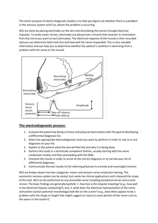
  • 1. The entire purpose of electro diagnostic studies is to help you figure out whether there is a problem in the nervous system and if so, where the problem is occurring. NCS are done by placing electrodes on the skin and stimulating the nerves through electrical impulses. To study motor nerves, electrodes are placed over a muscle that receives its innervation from the nerve you want to test (stimulate). The electrical response of the muscle is then recorded and you can determine both how fast and how well the nerve responded. This is very valuable information and can help you to determine whether the patient’s condition is stemming from a problem with the nerve or the muscle. The electrodiagnostic process: 1. Evaluate the patient by doing a history and physical examination with the goal of developing a differential diagnoses list. 2. Select the appropriate electrodiagnostic tests you want to perform in order to rule in or out diagnoses on your list. 3. Explain to the patient what the test will feel like and why it is being done. 4. Perform the study in a technically competent fashion, usually starting with the nerve conduction studies and then proceeding with the EMG. 5. Interpret the results in order to arrive at the correct diagnosis or to narrow your list of differential diagnoses. 6. Communicate the test results to the referring physician in a timely and meaningful manner. NCS are broken down into two categories: motor and sensory nerve conduction testing. The autonomic nervous system can be tested, but rarely has clinical applications and is beyond the scope of this text. NCS can be performed on any accessible nerve including peripheral nerves and cranial nerves. The basic findings are generally twofold: 1. how fast is the impulse traveling? (e.g., how well is the electrical impulse conducting?); and, 2. what does the electrical representation of the nerve stimulation (action potential morphology) look like on the screen? (e.g., does there appear to be a problem with the shape or height that might suggest an injury to some portion of the nerve such as the axons or the myelin?)
  • 2. The H-reflex is a monosynaptic or oligosynaptic spinal reflex involving both motor and sensory fibers. It electrically tests some of the same fibers as are tested in the ankle jerk reflexes. In fact it is rare to be unable to obtain an H-reflex in the presence of an ankle jerk reflex. If this occurs, technical factors should be considered. In theory it is a sensitive measure in assessing radiculopathy because 1. it helps to assess proximal lesions, 2. it becomes abnormal relatively early in the development of radiculopathy, and 3. it incorporates sensory fiber function proximal to the dorsal root ganglion. The H reflex primarily assesses afferent and efferent S1 fibers. Clinically, L5 and S1 radiculopathies may appear similar on EMG due to the overlap of myotomes. H-reflexes are probably of greatest value in distinguishing S1 from L5 radiculopathies. When assessing for S1 radiculopathy, the H-reflex latency is recorded from the gastrocnemius-soleus muscle group upon stimulating the tibial nerve in the popliteal fossa. The H-reflex is elicited with a submaximal stimulation with the cathode proximal to the anode. As the intensity of the stimulation is gradually increased from peak H-amplitude, we generally see a diminishment of the H-amplitude with a concurrent increase in the M wave amplitude. With supra maximal stimulation, the H-reflex is usually absent. The H-reflex can also be used in C6/C7 radiculopathy by recording over the flexor carpi radialis muscle and stimulating the median nerve at the elbow. The median H-reflex is less commonly performed and clinically is less likely to be helpful for radiculopathy than a lower extremity H-reflex. Generally, gastrocnemius- soleus H-reflex latency side-toside differences of greater than 1.5 ms are suggestive of S1 radiculopathy. Although the H-reflex is sensitive, it has certain limitations: 1. patients with S1 radiculopathy can have a normal H-reflex; 2. an abnormal H-reflex is only suggestive, but not definitive for radiculopathy because the abnormality may originate in other components of the long pathway involved, such as the peripheral nerves, plexuses, or spinal cord; 3. once the H-reflex becomes abnormal, it usually does not return to normal, even over time; and finally the H-reflex is often absent in otherwise normal individuals over the age of 60 years. The reflexes therefore can be considered a sensitive, but not specific indicator of pathology. Latency of the H-reflex is dependent on the age and leg length of the patient. A side-to-side amplitude difference of 60% or more may also indicate pathology. F-waves are low amplitude late responses thought to be due to antidromic activation of motor neurons (anterior horn cells) following peripheral nerve stimulation, which then cause orthodromic impulses to pass back along the involved motor axons. Some electromyographers have called this a ‘backfiring’ of axons. It is called the F-wave because it was first noted in intrinsic foot muscles. The F-wave has small amplitude, a variable configuration, and a variable latency. Generally F-wave amplitudes are up to 5% of the orthodromically generated motor response (M- response). The most widely used parameter is the latency of the shortest reproducible response. The F-wave can be found in many muscles of the upper and lower extremities. Unfortunately F-waves have not turned out to be as sensitive a test as initially hoped. The reasons for this are: 1. the pathways involve only the motor fibers, 2. as with the H-reflex, it involves a long neuronal pathway so that if there is a focal lesion it might be obscured, 3. if an abnormality is present, the F-wave will not pinpoint the exact location because any lesion, from the anterior horn cell to the muscle being tested, can affect the Fwave similarly, 4. since muscles have multiple root innervations, the shortest latency may reflect the healthy fibers in the non-affected root, and 5. the latency and amplitude of an F-wave is variable so that multiple stimulations must be performed to find the shortest latency. If not enough stimulations are done (usually more than 10), the shortest latency may not be apparent. Thus, use of Fwaves in evaluating for radiculopathy are extremely limited and should not be the sole basis upon which the diagnosis is made. F-wave Ratio Because errors can occur when measuring distances for F-wave conduction velocities, an alternative F-wave technique was developed which did not require distance measurements. The ratio is as follows:
  • 3. (M = CMAP latency; this ratio may be rewritten as (F – M – 1)/2 M). The ratio assumes that the distance from the elbow (or knee) to the hand (or foot) is approximately equal to the distance from the elbow (or knee) to the spinal cord. Therefore, stimulation must be performed at the elbow or knee. The normal F-wave ratio in the upper limb is approximately 1 ± 0.3 and in the lower limb the normal F-ratio is 1.1 ± 0.3. A ratio higher than 1.3 indicates a proximal lesion, since the numerator of the equation includes the proximal stimulation from the F-wave. A ratio below 0.7 indicates a distal lesion, since a larger CMAP latency will decrease the numerator and increase the denominator. Therefore, an F-ratio is not necessarily more sensitive than F-latency, but it does allow one to assess whether the slowing is in the proximal or distal segment of the nerve. It should be noted that F- waves are non-specific. Therefore, interpreting NCS results using F-waves must be done in conjunction with other information. Comparison of H-reflex and F-wave Parameter H-reflex F-wave Derivation of name Originally described Originally obtained in by Hoffman foot muscles Type of synapse Monosynaptic or oligosynaptic Polysynaptic Pathway Sensory orthodromic Motor antidromic Motor antidromic Motor orthodromic Stimulus required Submaximal (stronger stimulation produces inhibition secondary to collision of orthodromic impulses by antidromic conduction in motor axons) Supramaximal Where they can be Soleus Most muscles (distal preferred) elicited? (Normals) Flexor carpi radialis Stimulation site Posterior tibial nerve in popliteal fossa Along peripheral nerve Stimulus cathode Proximal Proximal Size of response Amplification of motor Small (motor neurons are (compared to M) response centrally activated infrequently with (due to reflex activation of motor neurons) antidromic stimulation) Facilitation Enhanced by maneuvers that increase motor-neuron pool excitability (contraction or CNS lesion) N/A Uses S Radiculopathy 1 Demyelinating polyneuropathies (sensitive but not Guillain–Barré specific) Proximal nerve or root injury Guillain–Barré syndrome (not test of choice – non-specific) Latency, amplitude Reproducible latency Variable in amplitude, latency
  • 4. and configuration and configuration (amplitude dependent on stimulation) and configuration Side-to-side > 1.5 msec >2 msec from hand difference >3 msec from calf >4 msec from foot Ratio N/A ––––––––– F – M – 1 2 M