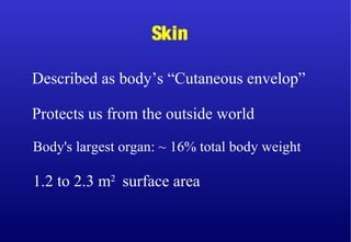
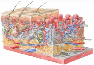
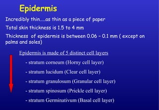
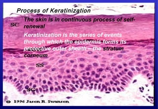
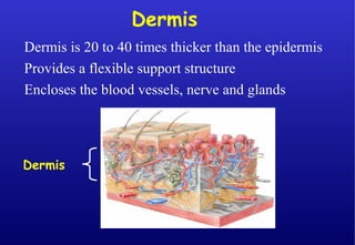
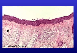
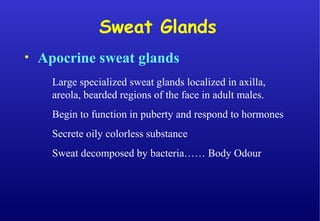
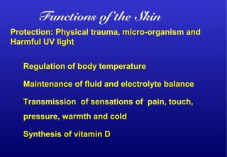
Skin
Body's largest organ: ~ 16% total body weight
1.2 to 2.3 m2
surface area
Described as body’s “Cutaneous envelop”
Protects us from the outside world
Epidermis
Incredibly thin…..as thin as a piece of paper
Total skin thickness is 1.5 to 4 mm
Thickness of epidermis is between 0.06 – 0.1 mm ( except on
palms and soles)
Epidermis is made of 5 distinct cell layers
- stratum corneum (Horny cell layer)
- stratum lucidum (Clear cell layer)
- stratum granulosum (Granular cell layer)
- stratum spinosum (Prickle cell layer)
- stratum Germinativum (Basal cell layer)
Process of Keratinization
The skin is in continuous process of self-
renewal
Keratinization is the series of events
through which the epidermis forms its
protective outer sheath – the stratum
corneum
Dermis
Dermis is 20 to 40 times thicker than the epidermis
Provides a flexible support structure
Encloses the blood vessels, nerve and glands
Dermis
• Apocrine sweat glands
Large specialized sweat glands localized in axilla,
areola, bearded regions of the face in adult males.
Begin to function in puberty and respond to hormones
Secrete oily colorless substance
Sweat decomposed by bacteria…… Body Odour
Sweat Glands
Protection: Physical trauma, micro-organism and
Harmful UV light
Regulation of body temperature
Maintenance of fluid and electrolyte balance
Transmission of sensations of pain, touch,
pressure, warmth and cold
Synthesis of vitamin D
Functions of the Skin
Skin Lesions
Small solid, elevated skin lesion,
usually 1 cm or less in diameter.
Seen in many skin diseases like
Acne
Papule
Elevated, flat-topped lesion
Usually greater than 1cm in diameter;
Described as being shaped like plateau
e. g.:
- psoriasis
Plaque
Solid deep seated mass in the dermis or
subcutaneous tissue
Produces elevation on the skin surface
Can be seen in gross inspection or only
on palpation.
Ex.:
- Nodulocystic acne
Nodule
Transient, solid, itchy, raised area of
cutaneous edema with irregular
shape, different diameter and
erythema.
e. g.:
- urticaria
- insect bites
Wheal
Elevated, thin-walled lesion;
filled with serous fluid, less than
1 cm in diameter.
Ex.:
- Herpes simplex
- Varicella
- Herpes Zoster
Vesicle
Thickened and rough
epidermis
Skin looks dry and leathery
e .g.:
- Atopic dermatitis
Lichenification
Change in skin’s appearance
following repair after skin
injury.
e. g.:
- healed wound
Scar
A linear and small split or
crack in the epidermis and
dermis.
e. g.:
- Cracked feet
Fissure
Depressed lesion that is wider
than a fissure but not as deep
Erosion
Deep erosion in which all
of epidermis and part of
dermis are eaten away
e. g.:
-wound ulcer
-Bed sore
Ulcers
Reduction of skin thickness occurring
at any skin layer.
e. g.:
- aged skin
Atrophy

Skin anatomy chc training 2012 [compatibility mode] [repaired]

  • 2.
    Skin Body's largest organ:~ 16% total body weight 1.2 to 2.3 m2 surface area Described as body’s “Cutaneous envelop” Protects us from the outside world
  • 7.
    Epidermis Incredibly thin…..as thinas a piece of paper Total skin thickness is 1.5 to 4 mm Thickness of epidermis is between 0.06 – 0.1 mm ( except on palms and soles) Epidermis is made of 5 distinct cell layers - stratum corneum (Horny cell layer) - stratum lucidum (Clear cell layer) - stratum granulosum (Granular cell layer) - stratum spinosum (Prickle cell layer) - stratum Germinativum (Basal cell layer)
  • 9.
    Process of Keratinization Theskin is in continuous process of self- renewal Keratinization is the series of events through which the epidermis forms its protective outer sheath – the stratum corneum
  • 11.
    Dermis Dermis is 20to 40 times thicker than the epidermis Provides a flexible support structure Encloses the blood vessels, nerve and glands Dermis
  • 15.
    • Apocrine sweatglands Large specialized sweat glands localized in axilla, areola, bearded regions of the face in adult males. Begin to function in puberty and respond to hormones Secrete oily colorless substance Sweat decomposed by bacteria…… Body Odour Sweat Glands
  • 20.
    Protection: Physical trauma,micro-organism and Harmful UV light Regulation of body temperature Maintenance of fluid and electrolyte balance Transmission of sensations of pain, touch, pressure, warmth and cold Synthesis of vitamin D Functions of the Skin
  • 21.
  • 23.
    Small solid, elevatedskin lesion, usually 1 cm or less in diameter. Seen in many skin diseases like Acne Papule
  • 24.
    Elevated, flat-topped lesion Usuallygreater than 1cm in diameter; Described as being shaped like plateau e. g.: - psoriasis Plaque
  • 25.
    Solid deep seatedmass in the dermis or subcutaneous tissue Produces elevation on the skin surface Can be seen in gross inspection or only on palpation. Ex.: - Nodulocystic acne Nodule
  • 26.
    Transient, solid, itchy,raised area of cutaneous edema with irregular shape, different diameter and erythema. e. g.: - urticaria - insect bites Wheal
  • 27.
    Elevated, thin-walled lesion; filledwith serous fluid, less than 1 cm in diameter. Ex.: - Herpes simplex - Varicella - Herpes Zoster Vesicle
  • 33.
    Thickened and rough epidermis Skinlooks dry and leathery e .g.: - Atopic dermatitis Lichenification
  • 34.
    Change in skin’sappearance following repair after skin injury. e. g.: - healed wound Scar
  • 36.
    A linear andsmall split or crack in the epidermis and dermis. e. g.: - Cracked feet Fissure
  • 37.
    Depressed lesion thatis wider than a fissure but not as deep Erosion
  • 38.
    Deep erosion inwhich all of epidermis and part of dermis are eaten away e. g.: -wound ulcer -Bed sore Ulcers
  • 39.
    Reduction of skinthickness occurring at any skin layer. e. g.: - aged skin Atrophy