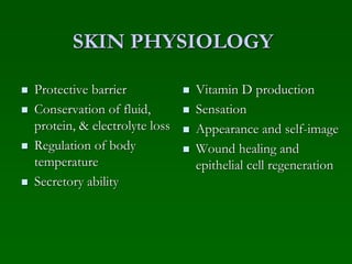
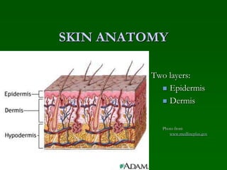
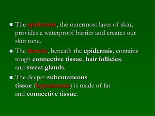
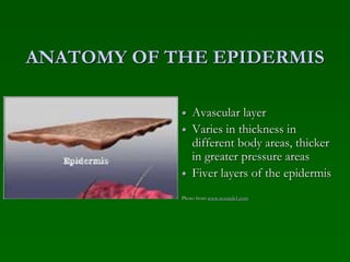
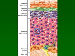
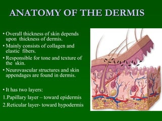
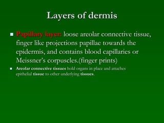
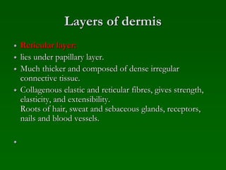
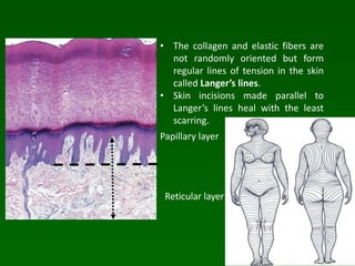
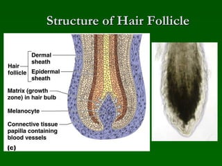
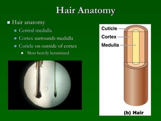
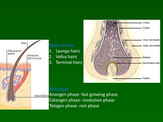
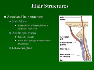
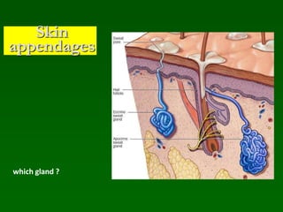
The skin has two main layers - the epidermis and dermis. The epidermis is the outermost layer and provides a protective barrier. It has five layers including the stratum corneum. The dermis lies beneath and contains tough connective tissue, hair follicles, and sweat glands. It consists of two layers - the papillary and reticular layers. Skin appendages like hair follicles, sebaceous glands, and sweat glands develop at the epidermal-dermal junction. Hair has a root that goes deep in the dermis and a shaft that projects out. Sweat glands secrete sweat which helps cool the body and remove waste.
The skin is the largest organ of the body, with a total area of about 20 square feet. ... Skin has three layers: The epidermis, the outermost layer of skin, provides a waterproof barrier and creates our skin tone. The dermis, beneath the epidermis, contains tough connective tissue, hair follicles, and sweat glands.
a brief ppt description about cartilage which may be usefull for teaching for first year mbbs, bds and paramedical students, hope it is helpfull to everyone
it describes the microanatomy of skin and its appendages in a concise format. it will give the overview of the integumentary system of our body and largest organ of our body.
Integumentary system
-The organs of the integumentary system include the skin and its accessory structures including hair, nails, and glands, as well as blood vessels, muscles and nerves.
-Dermatology is the medical specialty for the diagnosis and treatment of disorders of the integumentary system.
Structure Of The Skin
The skin (cutaneous membrane) covers the body and is the largest organ of the body by surface area and weight.
Its area is about 2 square meters (22 square feet) and weighs 4.5-5kg (10-11 lb), about 7% of body weight.
It is 0.5 – 4 mm thick, thinnest on the eyelids, thickest on the heels, the average thickness is 1 – 2 mm.
It consists of two major layers:
Outer, thinner layer called the epidermis, consists of epithelial tissue. Inner, thicker layer called the dermis.
Beneath the dermis is a subcutaneous layer (also called hypodermis) which attaches the skin to the underlying tissues and organs.
1. EPIDERMIS
-Covers, protects and waterproofs.
Contains five main layers:
- Stratum Basale
- Stratum Spinosum
- Stratum Granulosum
- Stratum lucidum
- Stratum corneum
The epidermis has a number of important characteristics:
The epidermis is composed of keratinized stratified squamous epithelium.
-It contains four major types of cells:
Keratinocytes (90% of the cells): It produce keratin which is a tough fibrous protein that provides protection.
-Melanocytes: which produce the pigment melanin that protects against damage by ultraviolet radiation.
-Langerhans cells: involved in immune responses, arise from red bone marrow.
-Merkel cells: which function in the sensation of touch along with the adjacent tactile discs.
2. DERMIS
-It is a deeper layer of skin, composed of connective tissue containing collagen and elastic fibers.
-It contains blood and lymph vessels, nerves, and other structures, such as hair follicles and sweat glands.
-The epidermis is avascular and cells of this layer get their oxygen and nutrients from capillaries in the dermis.
-The dermis can be divided into papillary layer and reticular layer.
Hypodermis :
- The hypodermis (also called the subcutaneous layer) is a layer directly below the dermis and serves to connect the skin to the underlying fascia (fibrous tissue) of the bones and muscles.
--Two types of glands are present in the skin over most of the body
These are sweat glands and sebaceous glands.
--Function Of Skin
Protection:
Sensation:
Heat regulation:
Storage and synthesis:
Synthesis of vitamin D:
Excretion and homeostasis:
Secretion:
Absorption:
Water resistance:
Colour :
Healing of wounds:
Aids in the diagnosis:
Skin = integument
Skin+ accessory organs = integumentary system.
The integumentary system comprises the skin and its appendages acting to protect the body from various kinds of damage, such as loss of water or damages from outside.
It includes hair, glands and nails.
It has a variety of additional functions; it may serve to waterproof, and protect the deeper tissues, excrete wastes, and regulate body temperature, and is the attachment site for sensory receptors to detect pain, sensation, pressure, and temperature,vitamin D synthesis.
skin and its layuer
Epidermis
Dermis
Hypodermis ( subcutaneous layer)
Epidermis
Most superficial layer of the skin
Approx 10 to 30 cells thick ( epithelial)
Cell types
Keratinocytes -90%
Melanocytes
Merkel cells 10%
Langerhens
stratum basale
Stratum germinativum
Deepest epidermal layer
Attaches to basal lamina
Cells bond to dermis via collagen fibres
Finger like projection called dermal papillae in dermis
Helps in stronger connection
Cells- cuboidal shaped keratinocytes
Grows constantly ,mitosis
Pushed up old cells
Applied aspects
Cells – merkel cell – function as receptor- stimulating sensory nerve fibres
Found in hairless skin.
Abundant on surface of hand and feet.
Melanocytes- produces pigment melanin
- gives hair and skin its color
-protect from u rays
Note: skin color influenced by ,melanin and carotene – carrot, oxygenated haemoglobin
Fingerprints-epidermal ridges
Stratum spinosum
Spiny in appearance
Desmosomsis protruding cell
Interlock between cells
Composed of 8to 10 layers of keratinocytes
Applied aspects
Langerhans cell –as a macrophage by engulfing bacteria and foreign body and damaged cell.
Keratinocytes synthesise keratin and prevent water loss
Stratum granulosam
Has a grainy appearance
Cells become flattened,and cell membrane thicken, and generate large amount of protein keratin and keratohyalin
After cell dies , keratin ,keratohyalin and cell membrane forms stratum lucidum
And accessory structures of hair and nail
Stratum lucidum
Smooth translucent layer
It is found only in thick skin of palms ,soles,digits
Cells are densely packed with eleidin , a clear protein rich in lipids,
Transparent appearance
Stratum corneam
Most superficial layer of epidermis
The increased keratinization of cell
It has 15 to 30 layers of cells
Water is lost from skin in 2 ways
Insensible perspiration
water diffuses from stratum corneum and evaporates from skin
-500 ml per day
Sensible perspiration
Water excreted by sweat glands.
clinical application
Some medications are toxic if swallowed, but safe if used topically (applied to surface of skin)
• Certain topical antibiotics are fairly toxic if taken by mouth, butcan be applied to skin with minimal risk of systemic absorption;they are polar molecules that cannot pass through epidermis toreach blood vessels in dermis; allows for local effect only
• Nonpolar substances cross epidermis much more easily;provides a c
The branch of science concerned with the bodily structure of humans, animals, and other living organisms, especially as revealed by dissection and the separation of parts.
The skin : هذا العرض يتحدث عن الجلد الذي يعتبر اكبر عضو بالجسم وشرح الطبقاة المكونة للجلد :
------------------------------------------
https://t.me/GoldenAlzaidy
------------------------------------------------------
youtube::: https://www.youtube.com/watch?v=Orumw-PyNjw
Deep Leg Vein Thrombosis (DVT): Meaning, Causes, Symptoms, Treatment, and Mor...The Lifesciences Magazine
Deep Leg Vein Thrombosis occurs when a blood clot forms in one or more of the deep veins in the legs. These clots can impede blood flow, leading to severe complications.
Defecation
Normal defecation begins with movement in the left colon, moving stool toward the anus. When stool reaches the rectum, the distention causes relaxation of the internal sphincter and an awareness of the need to defecate. At the time of defecation, the external sphincter relaxes, and abdominal muscles contract, increasing intrarectal pressure and forcing the stool out
The Valsalva maneuver exerts pressure to expel faeces through a voluntary contraction of the abdominal muscles while maintaining forced expiration against a closed airway. Patients with cardiovascular disease, glaucoma, increased intracranial pressure, or a new surgical wound are at greater risk for cardiac dysrhythmias and elevated blood pressure with the Valsalva maneuver and need to avoid straining to pass the stool.
Normal defecation is painless, resulting in passage of soft, formed stool
CONSTIPATION
Constipation is a symptom, not a disease. Improper diet, reduced fluid intake, lack of exercise, and certain medications can cause constipation. For example, patients receiving opiates for pain after surgery often require a stool softener or laxative to prevent constipation. The signs of constipation include infrequent bowel movements (less than every 3 days), difficulty passing stools, excessive straining, inability to defecate at will, and hard feaces
IMPACTION
Fecal impaction results from unrelieved constipation. It is a collection of hardened feces wedged in the rectum that a person cannot expel. In cases of severe impaction the mass extends up into the sigmoid colon.
DIARRHEA
Diarrhea is an increase in the number of stools and the passage of liquid, unformed feces. It is associated with disorders affecting digestion, absorption, and secretion in the GI tract. Intestinal contents pass through the small and large intestine too quickly to allow for the usual absorption of fluid and nutrients. Irritation within the colon results in increased mucus secretion. As a result, feces become watery, and the patient is unable to control the urge to defecate. Normally an anal bag is safe and effective in long-term treatment of patients with fecal incontinence at home, in hospice, or in the hospital. Fecal incontinence is expensive and a potentially dangerous condition in terms of contamination and risk of skin ulceration
HEMORRHOIDS
Hemorrhoids are dilated, engorged veins in the lining of the rectum. They are either external or internal.
FLATULENCE
As gas accumulates in the lumen of the intestines, the bowel wall stretches and distends (flatulence). It is a common cause of abdominal fullness, pain, and cramping. Normally intestinal gas escapes through the mouth (belching) or the anus (passing of flatus)
FECAL INCONTINENCE
Fecal incontinence is the inability to control passage of feces and gas from the anus. Incontinence harms a patient’s body image
PREPARATION AND GIVING OF LAXATIVESACCORDING TO POTTER AND PERRY,
An enema is the instillation of a solution into the rectum and sig
Explore our infographic on 'Essential Metrics for Palliative Care Management' which highlights key performance indicators crucial for enhancing the quality and efficiency of palliative care services.
This visual guide breaks down important metrics across four categories: Patient-Centered Metrics, Care Efficiency Metrics, Quality of Life Metrics, and Staff Metrics. Each section is designed to help healthcare professionals monitor and improve care delivery for patients facing serious illnesses. Understand how to implement these metrics in your palliative care practices for better outcomes and higher satisfaction levels.
Telehealth Psychology Building Trust with Clients.pptxThe Harvest Clinic
Telehealth psychology is a digital approach that offers psychological services and mental health care to clients remotely, using technologies like video conferencing, phone calls, text messaging, and mobile apps for communication.
Navigating Challenges: Mental Health, Legislation, and the Prison System in B...Guillermo Rivera
This conference will delve into the intricate intersections between mental health, legal frameworks, and the prison system in Bolivia. It aims to provide a comprehensive overview of the current challenges faced by mental health professionals working within the legislative and correctional landscapes. Topics of discussion will include the prevalence and impact of mental health issues among the incarcerated population, the effectiveness of existing mental health policies and legislation, and potential reforms to enhance the mental health support system within prisons.
Empowering ACOs: Leveraging Quality Management Tools for MIPS and BeyondHealth Catalyst
Join us as we delve into the crucial realm of quality reporting for MSSP (Medicare Shared Savings Program) Accountable Care Organizations (ACOs).
In this session, we will explore how a robust quality management solution can empower your organization to meet regulatory requirements and improve processes for MIPS reporting and internal quality programs. Learn how our MeasureAble application enables compliance and fosters continuous improvement.
CHAPTER 1 SEMESTER V PREVENTIVE-PEDIATRICS.pdfSachin Sharma
This content provides an overview of preventive pediatrics. It defines preventive pediatrics as preventing disease and promoting children's physical, mental, and social well-being to achieve positive health. It discusses antenatal, postnatal, and social preventive pediatrics. It also covers various child health programs like immunization, breastfeeding, ICDS, and the roles of organizations like WHO, UNICEF, and nurses in preventive pediatrics.
CHAPTER 1 SEMESTER V - ROLE OF PEADIATRIC NURSE.pdfSachin Sharma
Pediatric nurses play a vital role in the health and well-being of children. Their responsibilities are wide-ranging, and their objectives can be categorized into several key areas:
1. Direct Patient Care:
Objective: Provide comprehensive and compassionate care to infants, children, and adolescents in various healthcare settings (hospitals, clinics, etc.).
This includes tasks like:
Monitoring vital signs and physical condition.
Administering medications and treatments.
Performing procedures as directed by doctors.
Assisting with daily living activities (bathing, feeding).
Providing emotional support and pain management.
2. Health Promotion and Education:
Objective: Promote healthy behaviors and educate children, families, and communities about preventive healthcare.
This includes tasks like:
Administering vaccinations.
Providing education on nutrition, hygiene, and development.
Offering breastfeeding and childbirth support.
Counseling families on safety and injury prevention.
3. Collaboration and Advocacy:
Objective: Collaborate effectively with doctors, social workers, therapists, and other healthcare professionals to ensure coordinated care for children.
Objective: Advocate for the rights and best interests of their patients, especially when children cannot speak for themselves.
This includes tasks like:
Communicating effectively with healthcare teams.
Identifying and addressing potential risks to child welfare.
Educating families about their child's condition and treatment options.
4. Professional Development and Research:
Objective: Stay up-to-date on the latest advancements in pediatric healthcare through continuing education and research.
Objective: Contribute to improving the quality of care for children by participating in research initiatives.
This includes tasks like:
Attending workshops and conferences on pediatric nursing.
Participating in clinical trials related to child health.
Implementing evidence-based practices into their daily routines.
By fulfilling these objectives, pediatric nurses play a crucial role in ensuring the optimal health and well-being of children throughout all stages of their development.
Antibiotic Stewardship by Anushri Srivastava.pptxAnushriSrivastav
Stewardship is the act of taking good care of something.
Antimicrobial stewardship is a coordinated program that promotes the appropriate use of antimicrobials (including antibiotics), improves patient outcomes, reduces microbial resistance, and decreases the spread of infections caused by multidrug-resistant organisms.
WHO launched the Global Antimicrobial Resistance and Use Surveillance System (GLASS) in 2015 to fill knowledge gaps and inform strategies at all levels.
ACCORDING TO apic.org,
Antimicrobial stewardship is a coordinated program that promotes the appropriate use of antimicrobials (including antibiotics), improves patient outcomes, reduces microbial resistance, and decreases the spread of infections caused by multidrug-resistant organisms.
ACCORDING TO pewtrusts.org,
Antibiotic stewardship refers to efforts in doctors’ offices, hospitals, long term care facilities, and other health care settings to ensure that antibiotics are used only when necessary and appropriate
According to WHO,
Antimicrobial stewardship is a systematic approach to educate and support health care professionals to follow evidence-based guidelines for prescribing and administering antimicrobials
In 1996, John McGowan and Dale Gerding first applied the term antimicrobial stewardship, where they suggested a causal association between antimicrobial agent use and resistance. They also focused on the urgency of large-scale controlled trials of antimicrobial-use regulation employing sophisticated epidemiologic methods, molecular typing, and precise resistance mechanism analysis.
Antimicrobial Stewardship(AMS) refers to the optimal selection, dosing, and duration of antimicrobial treatment resulting in the best clinical outcome with minimal side effects to the patients and minimal impact on subsequent resistance.
According to the 2019 report, in the US, more than 2.8 million antibiotic-resistant infections occur each year, and more than 35000 people die. In addition to this, it also mentioned that 223,900 cases of Clostridoides difficile occurred in 2017, of which 12800 people died. The report did not include viruses or parasites
VISION
Being proactive
Supporting optimal animal and human health
Exploring ways to reduce overall use of antimicrobials
Using the drugs that prevent and treat disease by killing microscopic organisms in a responsible way
GOAL
to prevent the generation and spread of antimicrobial resistance (AMR). Doing so will preserve the effectiveness of these drugs in animals and humans for years to come.
being to preserve human and animal health and the effectiveness of antimicrobial medications.
to implement a multidisciplinary approach in assembling a stewardship team to include an infectious disease physician, a clinical pharmacist with infectious diseases training, infection preventionist, and a close collaboration with the staff in the clinical microbiology laboratory
to prevent antimicrobial overuse, misuse and abuse.
to minimize the developme
CRISPR-Cas9, a revolutionary gene-editing tool, holds immense potential to reshape medicine, agriculture, and our understanding of life. But like any powerful tool, it comes with ethical considerations.
Unveiling CRISPR: This naturally occurring bacterial defense system (crRNA & Cas9 protein) fights viruses. Scientists repurposed it for precise gene editing (correction, deletion, insertion) by targeting specific DNA sequences.
The Promise: CRISPR offers exciting possibilities:
Gene Therapy: Correcting genetic diseases like cystic fibrosis.
Agriculture: Engineering crops resistant to pests and harsh environments.
Research: Studying gene function to unlock new knowledge.
The Peril: Ethical concerns demand attention:
Off-target Effects: Unintended DNA edits can have unforeseen consequences.
Eugenics: Misusing CRISPR for designer babies raises social and ethical questions.
Equity: High costs could limit access to this potentially life-saving technology.
The Path Forward: Responsible development is crucial:
International Collaboration: Clear guidelines are needed for research and human trials.
Public Education: Open discussions ensure informed decisions about CRISPR.
Prioritize Safety and Ethics: Safety and ethical principles must be paramount.
CRISPR offers a powerful tool for a better future, but responsible development and addressing ethical concerns are essential. By prioritizing safety, fostering open dialogue, and ensuring equitable access, we can harness CRISPR's power for the benefit of all. (2998 characters)
1. Skin & Appendages
Dr. Shahbaz Ahmad PT
DPT[UIPT][UOL]
MS-MSK-PT [UIPT][UOL]*
Lecturer [LIHS][LCPS]
2. SKIN PHYSIOLOGY
Protective barrier
Conservation of fluid,
protein, & electrolyte loss
Regulation of body
temperature
Secretory ability
Vitamin D production
Sensation
Appearance and self-image
Wound healing and
epithelial cell regeneration
4. The epidermis, the outermost layer of skin,
provides a waterproof barrier and creates our
skin tone.
The dermis, beneath the epidermis, contains
tough connective tissue, hair follicles,
and sweat glands.
The deeper subcutaneous
tissue (hypodermis) is made of fat
and connective tissue.
5. ANATOMY OF THE EPIDERMIS
Avascular layer
Varies in thickness in
different body areas, thicker
in greater pressure areas
Fiver layers of the epidermis
Photo from www.wounds1.com
6. FIVER LAYERS OF THE EPIDERMIS
Stratum Corneum (waterproof; constantly being shed
Stratum Lucidium (only found in soles of feet and palms)
Stratum Granulosum (cells become more flattened)
Stratum Spinosum (active growing layer)
Stratum Germinativum (mitotic growth; makes melanin)
7.
8. EPIDERMIS
Four types of cells
1. Keratinocytes
2. Melanocytes
3. Langerhans cells
4. Merkel cells
9. EPIDERMIS
About 90% of the epidermal cells are
keratinocytes which produce keratin.
Keratin helps to waterproof and protect the skin
and underlying tissues from heat, light, microbes
and many chemicals.
Nanoparticles can diffuse and penetrate it.
(Phonophoresis Iontoporesis)
10. Epidermis
8% of the epidermal
cells are melanocytes
which produce the
pigment melanin.
Langerhans cells are
the third cell in the
epidermis, they interact
with helper T cells
(white blood cells) in
immune responses.
11. EPIDERMIS
The merkel cell is the fourth type of cell in
the epidermis and is found in the deepest layer
of hairless skin. These cells make contact with
the end of nerve cells and are thought to
function in the sensation of touch.
12. ANATOMY OF THE DERMIS
• Overall thickness of skin depends
upon thickness of dermis.
• Mainly consists of collagen and
elastic fibers.
• Responsible for tone and texture of
the skin.
• Neurovascular structures and skin
appendages are found in dermis.
• It has two layers:
1.Papillary layer – toward epidermis
2.Reticular layer- toward hypodermis
13. Layers of dermis
Papillary layer: loose areolar connective tissue,
finger like projections papillae towards the
epidermis, and contains blood capillaries or
Meissner’s corpuscles.(finger prints)
Areolar connective tissues hold organs in place and attaches
epithelial tissue to other underlying tissues.
14. Layers of dermis
Reticular layer:
lies under papillary layer.
Much thicker and composed of dense irregular
connective tissue.
Collagenous elastic and reticular fibres, gives strength,
elasticity, and extensibility.
Roots of hair, sweat and sebaceous glands, receptors,
nails and blood vessels.
15. Physiology of the Dermis
Supports epidermis through tensile strength and
elasticity
Nourishes epidermis through capillary loops
16. Reticular layer
• The collagen and elastic fibers are
not randomly oriented but form
regular lines of tension in the skin
called Langer’s lines.
• Skin incisions made parallel to
Langer’s lines heal with the least
scarring.
Papillary layer
17. Skin appendages
•These are developed as a result of down growth of
the epidermis of skin toward dermis and
hypodermis.
•It includes
1.Hair follicle: give rise to the hair
2.Sebaceous gland
3.Sweat gland
4.Nail
18. Appendages of the Skin
Hair
Produced by hair follicle
which are made of hard
keratinized epithelial cells
Melanocytes provide
pigment for hair color
22. Hair Structures
Associated hair structures
Hair follicle
Dermal and epidermal sheath
surround hair root
Arrector pili muscle
Smooth muscle
Pulls hairs upright when cold or
frightened
Sebaceous gland
23.
24. Nail structures
Nail Free edge
Body is the visible attached portion
Root of nail embedded in skin
Cuticle is the proximal nail fold that projects onto the nail body
25. Sweat and
Its Function
Composition
Mostly water
Salts and vitamin C
Some metabolic waste (urea and uric acid)
Fatty acids and proteins (apocrine only)
Function
Helps rid body of excess heat
Excretes waste products
Acidic nature inhibits bacteria growth
Odor is from associated bacteria