Downloaded 101 times
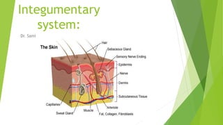


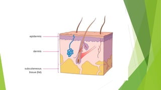
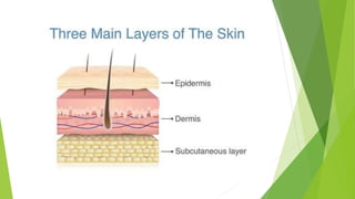
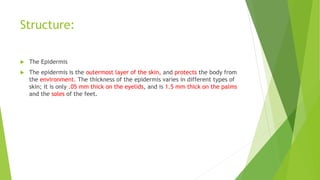
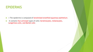

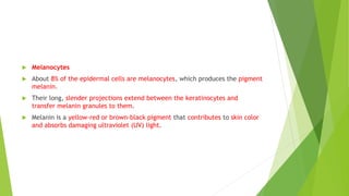

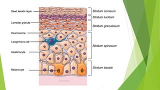


The integumentary system includes the skin, hair, nails, and glands. The skin is the body's largest organ and protects the body from pathogens, injury, heat, light, and helps regulate temperature and store vitamins. The skin has three layers - the epidermis, dermis, and subcutaneous tissue. The epidermis is the outer protective layer made of keratinocytes and contains melanocytes, Langerhans cells, and Merkel cells. The dermis lies below the epidermis and contains hair follicles, sweat and oil glands, blood vessels, nerves, and collagen and elastin for strength and flexibility.


