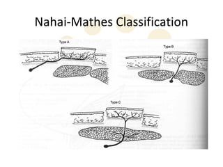
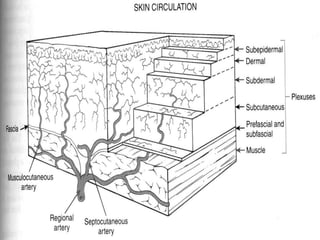
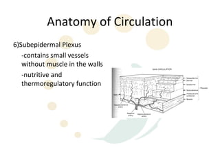
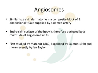
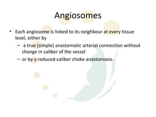
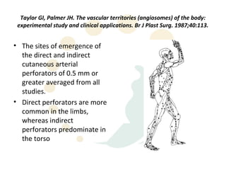
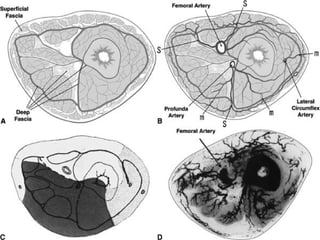
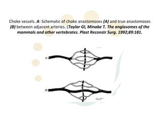
Blood Supply of the Skin
Anatomy of Circulation
• The blood reaching the skin originates from deep vessels

• These then feed interconnecting perforator vessels which
  supply the vascular plexus

• Thus skin fundamentally perfused by musculocutaneous or
  septocutaneous perforators
Nahai-Mathes Classification
Anatomy of Circulation


• The vascular plexuses of the fascia, subcutaneous tissue and
  skin are divided into 6 layers
Anatomy of Circulation
1)Subfascial plexus
   small plexus lying on the
   undersurface of the fascia
Anatomy of Circulation
2) Prefascial plexus
   -a larger plexus
   -particularly prominent on
   the limbs
   -fasciocutaneous vessels
Anatomy of Circulation
3)Subcutaneous Plexus
   -lies at the level of
   superficial fascia
   -Predominant on the torso
   -musculocutaneous vessels
Anatomy of Circulation
4)Subdermal Plexus
   -receives blood from
   underlying plexus
   -the main plexus supplying
   blood to the skin
   -represents the dermal
   bleed observed in incised
   skin
Anatomy of Circulation
5) Dermal Plexus
   -mainly arterioles
   -important in
   thermoregulation
Anatomy of Circulation
6)Subepidermal Plexus
   -contains small vessels
   without muscle in the walls
   -nutritive and
   thermoregulatory function
Angiosomes
• Similar to a skin dermatome is a composite block of 3
  dimensional tissue supplied by a named artery

• Entire skin surface of the body is therefore perfused by a
  multitude of angiosome units

• First studied by Marchot 1889, expanded by Salmon 1930 and
  more recently by Ian Taylor
Angiosomes
• Each angiosome is linked to its neighbour at every tissue
  level, either by
   – a true (simple) anastomotic arterial connection without
     change in caliber of the vessel
   – or by a reduced-caliber choke anastomosis.
Taylor GI, Palmer JH. The vascular territories (angiosomes) of the body:
 experimental study and clinical applications. Br J Plast Surg. 1987;40:113.


• The sites of emergence of
  the direct and indirect
  cutaneous arterial
  perforators of 0.5 mm or
  greater averaged from all
  studies.
• Direct perforators are more
  common in the limbs,
  whereas indirect
  perforators predominate in
  the torso
Choke vessels. A: Schematic of choke anastomoses (A) and true anastomoses
(B) between adjacent arteries. (Taylor GI, Minabe T. The angiosomes of the
    mammals and other vertebrates. Plast Reconstr Surg. 1992;89:181.
Choke Vessels
• Choke vessels play an important role in skin-flap survival, they
  provide an initial resistance to blood flow between the base
  and the tip of the flap.

• When a skin flap is delayed by the strategic division of
  cutaneous perforators along its length, these choke vessels
  dilate to the dimensions of true anastomoses thus enhancing
  the circulation to the distal flap
Delay Phenomenon
• Is a preliminary surgical intervention wherein a portion of the
  vascular supply to a flap is divided before definitive elevation
  and transfer of the flap

• Mechanism of this phenomenon is controversial
Delay Phenomenon
• Increased axiality of blood flow
   – Removal of blood flow from periphery of a random flap
     promotes development of axial flow
• Tolerance to ischaemia
   – Cells become accustomed to hypoxia
• Sympathectomy vasodilation theory
   – Thus leading to vasodilation
• Dilation of choke vessels
• Hyperadrenergic theory
The angiosome concept has important clinical implications

1) Each angiosome defines the safe anatomic
   boundary of tissue in each layer that can be
   transferred separately or combined on the
   underlying source vessels as a composite flap.
2) Because the junctional zone between adjacent
   angiosomes usually occurs within muscles of the
   deep tissue, rather than between them, these
   muscles provide an important anastomotic detour
   (bypass shunt) if the main source artery or vein is
   obstructed.
The angiosome concept has important clinical implications


3) Because most muscles span two or more angiosomes and are
    supplied from each territory, one is able to capture the skin
    island from one angiosome by muscle supplied in the
    adjacent territory.

Blood supply-of-the-skin

  • 2.
  • 3.
    Anatomy of Circulation •The blood reaching the skin originates from deep vessels • These then feed interconnecting perforator vessels which supply the vascular plexus • Thus skin fundamentally perfused by musculocutaneous or septocutaneous perforators
  • 4.
  • 5.
    Anatomy of Circulation •The vascular plexuses of the fascia, subcutaneous tissue and skin are divided into 6 layers
  • 7.
    Anatomy of Circulation 1)Subfascialplexus small plexus lying on the undersurface of the fascia
  • 8.
    Anatomy of Circulation 2)Prefascial plexus -a larger plexus -particularly prominent on the limbs -fasciocutaneous vessels
  • 9.
    Anatomy of Circulation 3)SubcutaneousPlexus -lies at the level of superficial fascia -Predominant on the torso -musculocutaneous vessels
  • 10.
    Anatomy of Circulation 4)SubdermalPlexus -receives blood from underlying plexus -the main plexus supplying blood to the skin -represents the dermal bleed observed in incised skin
  • 11.
    Anatomy of Circulation 5)Dermal Plexus -mainly arterioles -important in thermoregulation
  • 12.
    Anatomy of Circulation 6)SubepidermalPlexus -contains small vessels without muscle in the walls -nutritive and thermoregulatory function
  • 13.
    Angiosomes • Similar toa skin dermatome is a composite block of 3 dimensional tissue supplied by a named artery • Entire skin surface of the body is therefore perfused by a multitude of angiosome units • First studied by Marchot 1889, expanded by Salmon 1930 and more recently by Ian Taylor
  • 14.
    Angiosomes • Each angiosomeis linked to its neighbour at every tissue level, either by – a true (simple) anastomotic arterial connection without change in caliber of the vessel – or by a reduced-caliber choke anastomosis.
  • 15.
    Taylor GI, PalmerJH. The vascular territories (angiosomes) of the body: experimental study and clinical applications. Br J Plast Surg. 1987;40:113. • The sites of emergence of the direct and indirect cutaneous arterial perforators of 0.5 mm or greater averaged from all studies. • Direct perforators are more common in the limbs, whereas indirect perforators predominate in the torso
  • 17.
    Choke vessels. A:Schematic of choke anastomoses (A) and true anastomoses (B) between adjacent arteries. (Taylor GI, Minabe T. The angiosomes of the mammals and other vertebrates. Plast Reconstr Surg. 1992;89:181.
  • 18.
    Choke Vessels • Chokevessels play an important role in skin-flap survival, they provide an initial resistance to blood flow between the base and the tip of the flap. • When a skin flap is delayed by the strategic division of cutaneous perforators along its length, these choke vessels dilate to the dimensions of true anastomoses thus enhancing the circulation to the distal flap
  • 19.
    Delay Phenomenon • Isa preliminary surgical intervention wherein a portion of the vascular supply to a flap is divided before definitive elevation and transfer of the flap • Mechanism of this phenomenon is controversial
  • 20.
    Delay Phenomenon • Increasedaxiality of blood flow – Removal of blood flow from periphery of a random flap promotes development of axial flow • Tolerance to ischaemia – Cells become accustomed to hypoxia • Sympathectomy vasodilation theory – Thus leading to vasodilation • Dilation of choke vessels • Hyperadrenergic theory
  • 21.
    The angiosome concepthas important clinical implications 1) Each angiosome defines the safe anatomic boundary of tissue in each layer that can be transferred separately or combined on the underlying source vessels as a composite flap. 2) Because the junctional zone between adjacent angiosomes usually occurs within muscles of the deep tissue, rather than between them, these muscles provide an important anastomotic detour (bypass shunt) if the main source artery or vein is obstructed.
  • 22.
    The angiosome concepthas important clinical implications 3) Because most muscles span two or more angiosomes and are supplied from each territory, one is able to capture the skin island from one angiosome by muscle supplied in the adjacent territory.